Introduction
Over the past decades, there have been great advances in in vitro-production (IVP) systems with improved culture methods and new knowledge regarding embryo genetics, physiology, ultrastructure and morphology. At present, the IVP of bovine embryo has a successful commercial application. Nevertheless, the extensive use of this technology faces the hindrance of the IVP embryo high sensitivity to cryopreservation (Thompson, Reference Thompson1997; George et al., Reference George, Daniaux, Genicot, Verhaeghe, Lambert and Donnay2008).
Characteristics such as lipid accumulation, very electron-dense cytoplasm, loose blastomeres, buoyant density, slower growth, excessive metabolism of energy substrates, high thermal sensitivity, and apoptosis are indicators that the IVP embryos are more sensitive to cryopreservation than the ones produced in vivo (Gardner et al., Reference Gardner, Pool and Lane2000; Abe et al., Reference Abe, Yamashita, Satoh and Hoshi2002; Rizos et al., Reference Rizos, Ward, Duffy, Boland and Lonergan2002, Reference Rizos, Gutierrez-Adan, Perez-Garnelo, De La Fuente, Boland and Lonergan2003; De La Torre-Sanchez et al., Reference De La Torre-Sanchez, Prieis and Seidel2006; Mucci et al., Reference Mucci, Aller, Kaiser, Hozbor, Cabodevila and Alberio2006; Barceló-Fimbres & Seidel, Reference Barceló-Fimbres and Seidel2007a; Sudano et al., Reference Sudano, Paschoal, Rascado, Magalhães, Crocomo, Lima-Neto and Landim-Alvarenga2011). There is a consensus in the literature that the reduced cryotolerance of IVP embryos is frequently associated with high lipid content present in their cytoplasm (Abe et al., Reference Abe, Yamashita, Satoh and Hoshi2002; Rizos et al., Reference Rizos, Ward, Duffy, Boland and Lonergan2002, Reference Rizos, Gutierrez-Adan, Perez-Garnelo, De La Fuente, Boland and Lonergan2003; Mucci et al., Reference Mucci, Aller, Kaiser, Hozbor, Cabodevila and Alberio2006).
Nevertheless, the reasons for the accumulation of lipids and its effects upon embryo cryotolerance remain unclear until the present moment. Moreover, there are some controversial results in literature. While taurine embryos (Bos taurus taurus) presented greater lipid accumulation in the cytoplasm than zebuine embryos (Bos taurus indicus), the former showed higher cryotolerance (Visintin et al., Reference Visintin, Martins, Bevilacqua, Mello, Nicácio and Assumpção2002). Additionally, in a previous study of our group, the embryo quality assessed by apoptosis was a better parameter to predict embryo survival after vitrification than lipid content (Sudano et al., Reference Sudano, Paschoal, Rascado, Magalhães, Crocomo, Lima-Neto and Landim-Alvarenga2011). Both results and the literature call into question the actual influence of embryo lipid content on cryotolerance.
Nowadays, the most popular parameters analyzed before cryopreservation in commercial programs of IVP bovine embryos are development stage and quality degree score, which are extremely important despite the fact that their prediction of embryo cryotolerance still remains uncertain and subjective.
Therefore, the objective of the present study was to correlate the parameters of cleavage, blastocyst production, quality degree score, total cell number, apoptosis and lipid content of IVP bovine embryos with the post-cryopreservation survival.
Materials and methods
Experimental design
A total of 1727 IVP bovine blastocysts were used to establish the parameters of cleavage, blastocyst production, quality degree score, total cell number, apoptosis and lipid content. As for the determination of embryo survival after cryopreservation, 1316 blastocysts were vitrified. All materials were acquired from Sigma (Sigma-Aldrich Corp., St. Louis, MO, USA), except when specified.
In vitro production of embryos
Ovaries were recovered from slaughtered Nellore cows (Bos taurus indicus). Oocytes were aspirated from follicles ranging from 2 to 8 mm in diameter; only the oocytes with three or more compact layers of cumulus cells and homogeneous cytoplasm were used. Selected oocytes (n = 4320) were matured in vitro as described in Sudano et al. (Reference Sudano, Paschoal, Rascado, Magalhães, Crocomo, Lima-Neto and Landim-Alvarenga2011). At the end of the maturation period, oocytes were subjected to IVF with frozen semen from a single sample of a Nellore (Bos taurus indicus) bull with proven fertility. Sperm were selected using the Percoll method (Parrish et al., Reference Parrish, Krogenaes and Susko-Parrish1995), and the concentration was adjusted to 2 × 10⁶ sperm/ml. Fertilization occurred in HTF-FIV medium (Sudano et al., Reference Sudano, Paschoal, Rascado, Magalhães, Crocomo, Lima-Neto and Landim-Alvarenga2011). The day of fertilization was defined as day 0. At 18 h post-IVF, presumptive zygotes were denuded through repeated pipetting and transferred to culture plates in drops of 90 μl of culture media, covered with mineral oil, and cultured in an atmosphere of 5% CO2, 5% O2, and 90% N2. SOFaa was used as culture medium (Holm et al., Reference Holm, Booth, Schmidt, Greve and Callesen1999) with 2.7 mM myo-inosytol, 0.2 mM pyruvate, either 0, 2.5, 5, or 10% of FCS, 5 mg/ml BSA (fatty acid-free, Sigma A-8806), 100 μg/ml streptomycin sulfate, and 100 IU/ml of penicillin. On day 2.5 cleavage was checked, and on either this day or day 4, 0.3 μM of phenazine ethosulfate (PES) was supplemented or not to the culture medium resulting the study groups (Control, PES day 2.5 and PES day 4; Sudano et al., Reference Sudano, Paschoal, Rascado, Magalhães, Crocomo, Lima-Neto and Landim-Alvarenga2011). Embryos remained in these conditions until day 7, in which blastocyst production was checked and the blastocysts were either vitrified or assessed for quality degree score (Robertson & Nelson, Reference Robertson, Nelson, Stringfellow and Seidel1998), cell number, apoptosis and lipid content.
TUNEL (terminal deoxynucleotyl transferase uracil nick end labeling)
Samples of fresh and vitrified blastocysts (n = 60–134) were submitted to the TUNEL reaction (In Situ Cell Death Detection Kit with Fluorescein, Roche®, Mannheim, BW, Germany), according to the technique adapted by Paula-Lopes & Hansen (2002). In summary, embryos were fixed and permeabilized. All samples were incubated in an enzymatic and marking solution for 1 h. Samples were stained with Hoechst 33342. Nuclei with green fluorescence (FITC) were considered to be TUNEL positive (fragmented DNA). Hoechst 33342 stained all healthy and apoptotic cells, based on the presence of blue fluorescence. The Image J 1.41 software (Wayne Rasband National Institutes of Health, Bethesda, MD, USA) was used to count apoptotic and total number of cells.
Lipid quantification
A sample of blastocysts (n = 45–60) was selected randomly during the experimental replications and stained with Sudan Black B (Sigma S-0395), adapted from Abe et al. (Reference Abe, Yamashita, Satoh and Hoshi2002). The embryos were fixed previously; stained in drops of 1% Sudan Black B (w/v) in 70% ethanol, and then examined under a light microscope at ×600 magnification. To estimate the relative amount of lipid droplets in the cytoplasm in each embryo, a grid with five squares of 1600 μm2 each was designed with AutoCAD 2000 Software (Autodesk, Inc., San Francisco, CA, USA). Lipid droplets were classified as small, medium, and large (<2, 2–6 μm, and >6 μm, respectively). It was counted the number of droplets per category in the 1600 μm2 square, and calculated the average number of droplets from five squares for each embryo. Data regarding lipid accumulation are presented as number of lipid droplets per 1000 μm2. For the relationship between total lipid droplet value and apoptosis in vitrified blastocysts, small-sized and medium-sized droplets were converted to an equivalent number of large droplets; taking into account an average size of 1, 4 and 8 μm for small-, medium- and large-sized lipid droplets, respectively. As the comparison was done based on the volume, one large-sized droplet was equivalent to eight medium and 512 small droplets, respectively.
Vitrification
A two-step technique was used for the vitrification of blastocysts, as described by Campos-Chillòn et al. (Reference Campos-Chillòn, Walker, De La Torre-Sanchez and Seidel2006). Blastocysts were washed in holding solution (HS; Vigro®, Bioniche Co.) and transferred to vitrification solution VS1 (5 M of ethylene glycol in HS) in which they stayed for 3 min. Then, embryos were put in a 10 μl drop of vitrification solution VS2 (7 M ethylene glycol in HS + 0.5 M galactose + 18% (w/v) of Ficoll 70) for 45 s. Immediately afterwards, a 0.25 ml straw was mounted that contained 1 cm galactose solution (GS) (1 M of galactose in HS) + 0.5 cm air + 7 cm GS + 0.5 cm air + the drop of VS2 with the embryos + 0.5 cm air + GS until the straw was full. Straws remained for 1 min in liquid N2 vapour before being immersed in the liquid N2. Vitrification was done in groups of five to 10 blastocysts per straw.
Warming and subsequent culture of embryos
Vitrification straws were withdrawn from the liquid N2 and kept for 10 s in air, followed by 15 s in water at 35–37oC. Straws were slightly agitated to mix the columns of vitrification media. After 4 min, embryos were washed and cultured in drops of SOFaa supplemented with 10% FCS, 2.7 mM myoinositol, 0.2 mM pyruvate, 5 mg/ml BSA (fatty acid-free – Sigma A-8806), 100 μg/ml streptomycin sulfate, and 100 IU/ml penicillin under the same conditions of in vitro culture for 12 h to allow embryo re-expansion. Thereafter, embryos were evaluated to assess their re-expansion and submitted to the TUNEL reaction for determination of apoptosis in vitrified blastocysts. Post-vitrification embryo survival was defined as the re-expansion of the blastocoele associated with a reduced apoptosis rate.
Statistical analysis
Data were analyzed with analysis of variance (ANOVA) using the PROC GLIMMIX model of SAS statistical software package (SAS Inst. Inc., Cary, NC, USA). Embryo condition (fresh and vitrified blastocyst) was considered as fixed effect. The arcsine transformation was applied to percentage data. Data are reported as untransformed least square means and standard error. Pearson's correlation was calculated between the selected parameters (total cell number, quality degree score, total lipid droplets value and apoptosis in fresh blastocysts) and the cryotolerance (apoptosis in vitrified blastocysts) using the correlation procedure (PROC CORR) of the SAS. Differences in P-values < 0.05 were considered to be significant.
Results and Discussion
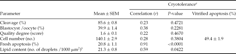
Main results are shown in Table 1 and Figs. 1, 2 and 3. Fresh apoptosis rate (20.8 ± 1.1) and total lipid droplets value (21.3 ± 0.8 droplets) were significantly correlated parameters (r = 0.94 and r = 0.64, respectively; P < 0.05) with post-cryopreservation embryo survival (49.4 ± 1.9). On the other hand, cleavage (85.6 ± 0.8), blastocyst production (39.9 ± 1.4), quality degree score (1.6 ± 0.1) and total cell number (140.1 ± 2.9) were not correlated with cryotolerance (r = 0.23, r = 0.38, r = 0.22 and r = 0.40, respectively; P > 0.05.
Relationship between embryo parameters and apoptosis of vitrified in vitro-produced bovine blastocysts

aEmbryo survival was defined as re-expansion of the blastocoele with a reduced apoptosis rate. bThe lipid content is expressed as an equivalent number of large lipid droplets.
Light micrographs of bovine embryos stained with Sudan Black B. Black dots indicate sudanophilic cytoplasmatic lipid droplets. Magnification ×600. Observations: 0 to 10% FCS: serum concentration used in the in vitro culture media. Phenazine ethosulfate (PES) D2.5: phenazine ethosulfate supplementation started on day 2.5 of in vitro culture. PES D4: phenazine ethosulfate supplementation started on day 4 of in vitro culture. (See online for a colour version of this figure.)
Fresh (A) and vitrified (B) bovine blastocysts submitted to the TUNEL reaction. Green nuclei fluorescence (FITC) was considered with fragmented DNA. Hoechst 33342 stained all healthy and apoptotic cells. The green and blue colors on cytoplasm were background. Magnification ×400. Observations: 0 to 10% FCS: serum concentration used in the in vitro culture media. PES D2.5: phenazine ethosulfate supplementation started on day 2.5 of in vitro culture. PES D4: phenazine ethosulfate supplementation started on day 4 of in vitro culture. (See online for a colour version of this figure.)
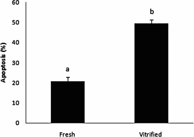
Fresh and vitrified apoptosis of in vitro-produced bovine blastocysts (mean ± SEM). a,bRefers to the difference (P < 0.0001) between fresh and vitrified apoptosis rate.
The ability of the embryo to survive after cryopreservation is a good indicator of its quality (George et al., Reference George, Daniaux, Genicot, Verhaeghe, Lambert and Donnay2008). On the other hand, a reduced cryotolerance is frequently associated with a series of morphological, metabolic and development kinetics disturbance characteristics in embryos (Abe et al., Reference Abe, Yamashita, Satoh and Hoshi2002; Rizos et al., Reference Rizos, Ward, Duffy, Boland and Lonergan2002, Reference Rizos, Gutierrez-Adan, Perez-Garnelo, De La Fuente, Boland and Lonergan2003; De La Torre-Sanchez et al., Reference De La Torre-Sanchez, Prieis and Seidel2006; Mucci et al., Reference Mucci, Aller, Kaiser, Hozbor, Cabodevila and Alberio2006; Barceló-Fimbres & Seidel, Reference Barceló-Fimbres and Seidel2007b). In the literature, the main characteristic that affects IVP embryo cryotolerance is the high lipid content present in their cytoplasm (Abe et al., Reference Abe, Yamashita, Satoh and Hoshi2002; Mucci et al., Reference Mucci, Aller, Kaiser, Hozbor, Cabodevila and Alberio2006; Barceló-Fimbres & Seidel, Reference Barceló-Fimbres and Seidel2007b). However, until the present moment it is not clear how and why lipid accumulation occurs in the IVP embryo cell's cytoplasm. However, it seems that the accumulation occurs as a result of FCS media supplementation (Abe et al., Reference Abe, Yamashita, Satoh and Hoshi2002; Rizos et al., Reference Rizos, Ward, Duffy, Boland and Lonergan2002; 2003; Mucci et al., Reference Mucci, Aller, Kaiser, Hozbor, Cabodevila and Alberio2006; Barceló-Fimbres & Seidel, Reference Barceló-Fimbres and Seidel2007a), or due to abnormalities in energy metabolism (Rieger, Reference Rieger1992; De La Torre-Sanchez et al., Reference De La Torre-Sanchez, Prieis and Seidel2006; Barceló-Fimbres & Seidel, Reference Barceló-Fimbres and Seidel2007a,Reference Barceló-Fimbres and Seidelb).
Indeed, the raise of FCS concentration increased the number of lipid droplets present in IVP embryo cytoplasm (Fig. 1) and was moderately correlated (r = 0.59; P = 0.0422) with the post-cryopreservation survival (Table 1). In the literature, the addition of serum to the culture medium has been associated with large offspring syndrome (Lazzari et al., Reference Lazzari, Wrenzycki, Herrmann, Duchi, Kruip, Niemann and Galli2002) and several other abnormalities, such as modifications of cell organelles (Abe et al., Reference Abe, Yamashita, Satoh and Hoshi2002), mitochondrial degeneration (Dorland et al., Reference Dorland, Gardner and Trounson1994), premature formation of the blastocoele, modification of gene expression pattern (Rizos et al., Reference Rizos, Ward, Duffy, Boland and Lonergan2002, Reference Rizos, Gutierrez-Adan, Perez-Garnelo, De La Fuente, Boland and Lonergan2003), and apoptosis (Fig. 2). Consequently, chemically-defined media (without FCS) were developed (Keskintepe & Brackett, Reference Keskintepe and Brackett1996); and more recently, the use of PES as a metabolic regulator has been indicated to reduce the serum deleterious effects and decrease the lipid accumulation (Barceló-Fimbres & Seidel, Reference Barceló-Fimbres and Seidel2007a,Reference Barceló-Fimbres and Seidelb; De La Torre-Sanchez et al., Reference De La Torre-Sanchez, Prieis and Seidel2006; Sudano et al., Reference Sudano, Paschoal, Rascado, Magalhães, Crocomo, Lima-Neto and Landim-Alvarenga2011).
The lipid content is not the only characteristic related with embryo cryotolerance. Several authors have described numerous points to improve the survival of cryopreserved embryos such as the choice of the optimal embryonic development stage (Sanchez-Osorio et al., Reference Sanchez-Osorio, Cuello, Gil, Almiñana, Parrilla, Caballero, Garcia, Vazquez, Roca and Martinez2008; Abdalla et al., Reference Abdalla, Shimoda, Hara, Morita, Kuwayama, Hirabayashi and Hochi2010; Shirazi et al., Reference Shirazi, Soleimani, Karimi, Nazari, Ahmadi and Heidari2010), embryo age (Abdalla et al., Reference Abdalla, Shimoda, Hara, Morita, Kuwayama, Hirabayashi and Hochi2010), the embryo donor (Sanchez-Osorio et al., Reference Sanchez-Osorio, Cuello, Gil, Almiñana, Parrilla, Caballero, Garcia, Vazquez, Roca and Martinez2008), cryopreservation method (Shirazi et al., Reference Shirazi, Soleimani, Karimi, Nazari, Ahmadi and Heidari2010; Nicacio et al., Reference Nicacio, Simões, Paula-Lopes, de Barros, Peres, Assumpção and Visintin2011), culture conditions (Rizos et al., Reference Rizos, Ward, Duffy, Boland and Lonergan2002, Reference Rizos, Gutierrez-Adan, Perez-Garnelo, De La Fuente, Boland and Lonergan2003, Reference Rizos, Clemente, Bermejo-Alvarez, de La Fuente, Lonergan and Gutiérrez-Adán2008), season of the year (Marquez et al., Reference Márquez, Galina, Moreno, Ruiz, Ruiz and Merchant2005), as well as interactions between these factors (Morató et al., Reference Morató, Izquierdo, Paramio and Mogas2010).
Despite the indication to use the quality degree score and embryo development stage to analyze embryos prior to cryopreservation, their prediction of embryo cryotolerance remains uncertain. In the literature, there is still no consensus as to the optimal development stage for embryo cryotolerance (Saragusty & Arav, Reference Saragusty and Arav2011). Moreover, embryos classified as good quality, according experienced technicians following established criteria (Lindner & Wright, Reference Lindner and Wright1983) showed degenerative stages features under electron microscopy in 30% of the cases (Aguilar et al., Reference Aguilar, Galina, Merchant, Montiel, Canseco and Márquez2002). Farin et al. (Reference Farin, Britt, Shaw and Sleining1995) have proved the subjectivity of the stereoscopic evaluation by showing that embryos that were classified as being of high quality by the time they were frozen, were in fact in due process of degeneration when assessed by more accurate techniques. In the present study, parameters such as cleavage, blastocyst production, quality degree score and total cell number were not correlated (P > 0.05) with embryo post-cryopreservation survival (Table 1).
In contrast, increased apoptosis in fresh blastocysts was strongly correlated (r = 0.91; P < 0.0001) with apoptosis in vitrified blastocysts (Table 1). Apoptosis is a highly conserved form of cell death and it acts as a mechanism of cell quality control by removing damaged cells (Jacobson et al., Reference Jacobson, Weil and Raff1997; Paula-Lopes & Hansen, Reference Paula-Lopes and Hansen2002). However, a disproportionate incidence of this kind of cell death is associated with the reduction of embryo viability (Byrne et al., Reference Byrne, Southgate, Brison and Leese1999). In this study, the majority of apoptosis in fresh blastocysts occurred in the inner cell mass (ICM), when compared with the trophoblast (Fig. 2a), on the very same fashion that was described in both murine (Hardy & Handyside, Reference Hardy and Handyside1996) and bovine (Byrne et al., Reference Byrne, Southgate, Brison and Leese1999) embryos. The higher incidence of apoptosis in ICM might be explained by the removal of damaged or excessive cells, which are not needed or competent for the embryo development. In this case, apoptosis exerts a rigorous control over the quality of the ICM, which is the cell lineage from which the fetus originates (Byrne et al., Reference Byrne, Southgate, Brison and Leese1999).
Such events like thermal stress (Paula-Lopes and Hansen, Reference Paula-Lopes and Hansen2002; Hansen, Reference Hansen2007), oxidative stress (Guerin et al., Reference Guerin, Mouatassim and Ménézo2001), unfavorable conditions in culture media composition (Devreker and Hardy, Reference Devreker and Hardy1997; Moley et al., Reference Moley, Chi, Knudson, Korsmeyer and Mueckler1998), and cryopreservation (Sudano et al., Reference Sudano, Paschoal, Rascado, Magalhães, Crocomo, Lima-Neto and Landim-Alvarenga2011) are potential causes of embryo apoptosis. The freezing and thawing processes decrease embryo viability, which is attributed to physical and chemical damage induced during cryopreservation (Overstrom, Reference Overstrom1996; Baguisi et al., Reference Baguisi, Lonergan, Overstrom and Boland2000).
In the present study, the stress generated by cryopreservation corresponded to an increase of 2.4 fold in the apoptosis rate when fresh and vitrified (20.8 ± 1.1 vs. 49.4 ± 1.9; P < 0.0001) blastocysts were compared (Figs. 2 and 3). Moreover, there was a random distribution of apoptosis incidence in both ICM and trophoblast cells in vitrified blastocysts, which corroborated to the reduction in embryo quality (Fig. 2b). The same results were described by Park et al. (Reference Park, Kim, Cui, Tae, Lee, Kim, Park and Lim2006) and Márquez-Alvarado et al. (Reference Márquez-Alvarado, Galina, Castilla, León and Moreno-Mendoza2004), in which the apoptosis rate in cryopreserved embryos increased in 3.7- and 1.7-fold compared with apoptosis in fresh embryos, respectively. This damage may occur due to the use of high concentrations of cryoprotectant solutions employed in the current vitrification technique to generate a glass-like physical state (Campos-Chillòn et al., Reference Campos-Chillòn, Walker, De La Torre-Sanchez and Seidel2006), which increases the risk of osmotic and toxic damage (Barceló-Fimbres & Seidel, Reference Barceló-Fimbres and Seidel2007b). Conversely, Morató et al. (Reference Morató, Izquierdo, Paramio and Mogas2010) did not observe any difference in apoptosis rates between fresh and vitrified blastocysts. However, the authors justify that the period between warming and DNA fragmentation examination (3 h) was not enough to identify the extention of damage caused by the vitrification-warming procedure.
In conclusion, the increased lipid content was moderately correlated with apoptosis in vitrified blastocysts. On the other hand, increased apoptosis in fresh blastocysts was strongly correlated with apoptosis in vitrified blastocysts, indicating that fresh apoptosis rate was better than lipid content to predict post-vitrification embryo survival.
Acknowledgements
We thank the Fundação de Amparo à Pesquisa do Estado de São Paulo (São Paulo Research Foundation – FAPESP) for the financial support (grants 07/57766-4, 08/51378-5, 09/54513-3 and 10/09922-0).