





During pregnancy, maternal nutrition plays a pivotal role in shaping both immediate and long-term health outcomes for the fetus and mother(Reference Chouli, Bothou and Kyrkou1). Adequate dietary patterns and appropriate macronutrient and micronutrient intake significantly influence birth weight, a key indicator of intrauterine fetal growth and a well-recognised predictor of cardio-metabolic health in life(Reference Chouli, Bothou and Kyrkou1,Reference Kennedy, Reynolds and O’Malley2) . This is particularly noticeable in cases of extreme birth weights, such as small- and large-for-gestational-age newborns, which further increase the likelihood of non-communicable diseases in adult life(Reference de Mendonça, de Lima Macêna and Bueno3,Reference Xue, Qin and Xi4) .
Unhealthy dietary habits – characterised by high fat and low micronutrient intake – are increasingly prevalent worldwide and contribute to maternal inflammation, placental dysfunction and adverse pregnancy outcomes(Reference Gernand, Schulze and Stewart5,Reference Anelli, Parisi and Sarno6) . Consequently, systematic assessment of maternal nutrition and lifestyle should be considered an essential component of prenatal care. In 2015, the International Federation of Gynecology and Obstetrics proposed a rapid and reproducible nutrition checklist to facilitate early identification of nutritional risk in pregnancy(7). Several studies have demonstrated the high reproducibility and acceptability of the Federation of Gynecology and Obstetrics nutrition checklist, showing strong agreement with the gold-standard FFQ in identifying inadequate dietary patterns(Reference Killeen, Callaghan and Jacob8–Reference Tsoi, Chan and Li10). Based on Italian recommendations on maternal nutrition(11), our group adapted the Federation of Gynecology and Obstetrics nutrition checklist by incorporating a specific item on iodised salt use and by aligning fruit and vegetable intake with the recommendation of five daily servings (≥ 400 g/d) (SIMPLE nutrition checklist). Adequate iodine intake in pregnancy is essential for maternal thyroid hormone production and fetal neurodevelopment, and mild-to-moderate deficiency remains a public health concern in many settings(Reference Cetin, Devlieger and Isolauri12). In the Italian context, consensus recommendations explicitly emphasise ensuring adequate iodine intake during pregnancy, including via iodised salt as a practical dietary strategy(11). By integrating this item and refining dietary thresholds, the SIMPLE checklist increases sensitivity to common and clinically actionable micronutrient gap. In a pilot study, we demonstrated that the SIMPLE score was associated with early markers of placental development and selected pregnancy outcomes, supporting its potential utility as an early screening tool for maternal nutritional inadequacies(Reference Parisi, Savasi and Di Bartolo13). These findings were subsequently evaluated in a large multicentre Italian prospective cohort study (SIMPLE study) of healthy singleton pregnancies(Reference Parisi, Coco and Cetin14). In this population, women with a SIMPLE score ≥ 6 had higher first trimester serum pregnancy-associated plasma protein-A (PAPP-a) concentrations and lower rates of emergency cesarean section compared with women with a SIMPLE score lower than 6. Additionally, the first trimester Hg item (higher than 110 g/l) was independently associated with early placental markers, as well as with birth and placental weights in multi-adjusted models(Reference Parisi, Coco and Esposito15). Importantly, PAPP-A is a recognised marker of early placental function and trophoblastic development(Reference Smith, Stenhouse and Crossley16). Therefore, the observed association between higher SIMPLE scores and increased PAPP-A concentrations suggests that early maternal nutritional status may influence placental biology. The concurrent association with delivery outcomes further supports the hypothesis that early nutritional exposures may have downstream implications for fetal development and pregnancy course. However, longitudinal fetal growth trajectories, which represent a dynamic proxy of intrauterine development and a potential intermediate pathway between early placental function and birth outcomes, have not yet been addressed.
The present study was designed to address this gap. Specifically, we aimed to evaluate the associations between maternal nutritional status, as assessed by the first trimester SIMPLE score and pregestational BMI, and longitudinal fetal growth trajectories. We examined serial measurements of abdominal circumference (AC), estimated fetal weight (EFW), growth velocity and birth weight to determine whether early maternal nutritional status is associated not only with early placental biomarkers, but also with subsequent patterns of fetal growth across gestation. By extending prior findings from static early markers to dynamic growth parameters, this study seeks to clarify the potential continuum linking maternal nutrition, placental function and intrauterine development and to inform the clinical utility of early nutritional screening in routine obstetric care.
Materials and methods
The SIMPLE study is an ongoing multicentre, prospective observational cohort currently involving twenty-four Italian maternity units(Reference Parisi, Coco and Cetin14). The present work reports a subgroup analysis of women assessed for longitudinal ultrasound fetal growth measurements between January 2019 and September 2023.
This study was conducted in accordance with the Declaration of Helsinki, and all procedures involving human patients were approved by the Medical Ethical Committee and Institutional Review Board at the ‘Vittore Buzzi’ Children Hospital, Milan (promoting centre, reference number 46091/2018, 7/11/2018) and subsequently by the local ethical committees of all recruiting centres. Written informed consent was obtained from all subjects.
The study was reported in accordance with the STROBE-Nut (Strengthening the Reporting of Observational Studies in Epidemiology – Nutritional Epidemiology) guidelines.
All participants provided signed informed consent to collect personal and clinical data. Healthy women, without comorbidities or ongoing chronic therapies, carrying a singleton gestation, were enrolled between 11+0 and 13+6 gestational weeks during the ultrasound scan of the first trimester combined screening test for aneuploidies. Gestational age was based on the crown-rump length measurement at enrollment (range 45–84 mm). Exclusion criteria included: age under 18 years, language barrier, any pre-existing chronic medical or psychiatric condition requiring ongoing medical follow-up and/or pharmacological treatment (including, but not limited to, pregestational diabetes, chronic hypertension, autoimmune, renal or CVD and psychiatric disorder), oocyte donation pregnancy and fetal congenital anomalies or aneuploidies. At enrollment, maternal demographic (age, ethnicity and education level) and anthropometric data (weight, height and pregestational BMI), smoking habits and obstetric and medical history were collected. As exposure variable, each participant completed the SIMPLE nutrition checklist to assess the adherence to a healthy diet and lifestyle during pregnancy through the SIMPLE score calculation. The SIMPLE checklist includes ten binary (yes/no) items evaluating consumption of meat (two to three times per week), fruits and vegetables (five portions per day), fish (once to twice weekly), dairy products (daily), whole grains (daily), sweets and snacks (less than five times per week), folic acid supplementation (during the preconception period and the first trimester), use of iodised salt, sun exposure (at least 10 to 15 min per day) and first trimester Hg concentrations (greater than 110 g/l). Every affirmative answer was scored as one point, yielding a total SIMPLE score ranging from 0 to 10, with higher scores indicating greater adherence to a balanced diet. Based on the cut-off of six identified in prior SIMPLE studies as the threshold associated with early placental markers (including PAPP-A) and adverse obstetric outcomes(Reference Parisi, Savasi and Di Bartolo13,Reference Parisi, Coco and Esposito15) , the same cut-off score was selected to classify participants into low- (< 6) and high- (≥ 6) adherence groups.
As outcome measures, all women underwent two ultrasound fetal growth assessment during second (first assessment, 18–22 gestational weeks) and third trimester (second assessment, 28–34 gestational weeks) with collection of biparietal diameter, head circumference, AC, femur length and estimated fetal weight (EFW) according to the International Society of Ultrasound in Obstetrics and Gynecology guidelines. Mid-pregnancy EFW was calculated using the Hadlock formula: 10 × (1·3596 − 0·00386 × AC × femur length + 0·0064 × head circumference + 0·00061 × BPD × AC + 0·0424 × AC + 0·174 × femur length)(Reference Hadlock, Harrist and Sharman17).
At birth, delivery and pregnancy outcomes – including gestational age, mode of delivery, birth weight and neonatal sex – were collected.
Statistical analysis
Descriptive statistics for quantitative variables were presented as median and range or mean and standard deviation, as appropriate, while categorical variables were expressed as absolute and relative frequencies. χ 2 and Mann–Whitney U tests were used to compare baseline characteristics in the two groups, as appropriate.
To investigate associations between SIMPLE score groups and intrauterine longitudinal growth trajectories, linear mixed models were estimated to account for correlations between repeated measurements within the same pregnancy. First, an unconditional individual growth model over time (gestational age (days) at each assessment) was fitted using linear mixed models with a random intercept, with serial ultrasound measurements (AC, EFW) as response variables and gestational age as predictor. Both linear and quadratic terms of gestational age were tested. Gestational age was centred at 183 d, the value closest to the mean across the dataset, by subtracting 183 from each participant’s gestational age at each assessment. In a second step, the SIMPLE score was included in the model, coded as 0 if higher or equal to 6 (high adherence, reference group) or 1 if lower than 6 (low adherence). The multivariable model further included the following covariates selected a priori based on biological plausibility and association with intrauterine growth and birth outcomes: maternal age, pregestational BMI, parity, educational level, smoking status, ethnicity and neonatal sex. All selected covariates were entered simultaneously into a single multivariable model to obtain multi-adjusted estimates. The SIMPLE score was additionally tested as a continuous variable in sensitivity analyses. In addition, the analysis was stratified by fetal sex, given its known association with intrauterine growth trajectories(Reference Parisi, Coco and Esposito15). To assess intrauterine growth velocity, absolute differences in AC and EFW between third and second trimester measurements were computed as follows: (measurement at third trimester – measurement at second trimester)/(gestational age at third trimester scan – gestational age at second trimester scan). Linear mixed models with random intercepts were then fitted using these differences as response variables, with the SIMPLE score and its single items as predictors.
Finally, to fully investigate the association between maternal nutritional status and intrauterine longitudinal growth trajectories, the independent role of maternal pregestational BMI (treated as a continuous variable) was additionally tested including in the multi-adjusted model the previously defined terms.
All analyses were performed using R version 4.2.2 and SAS statistical software version 9.4 (SAS Institute, Inc.). Statistical significance was set at P ≤ 0·05.
Results
The study population comprised 938 women with complete first trimester SIMPLE score assessment, second and third trimester ultrasound fetal growth measurements and available neonatal data. Based on first trimester SIMPLE score values, 829 (88·4 %) women were classified in the high-adherence group (SIMPLE score ≥ 6) and 109 (11·6 %) in the low-adherence group (SIMPLE score < 6). Among the individual SIMPLE components, the highest proportions of negative responses were observed for whole-grain consumption (47·3 %) and adequate sun exposure (44·7 %), with 93·4 % of participants presenting at least one nutritional risk factor (i.e. at least one negative response).
Table 1 shows maternal baseline characteristics, ultrasound fetal growth measurements and neonatal and pregnancy outcomes according to SIMPLE score group. Compared with the low-adherence group, women in the high adherence group were older, had higher educational levels and showed lower rates of pregestational obesity. No differences were detected between groups in fetal ultrasound measurements, neonatal outcomes or pregnancy complications, although gestational age at delivery showed a difference approaching statistical significance.
Maternal baseline characteristics, ultrasound fetal growth measurements and neonatal and pregnancy outcomes according to the SIMPLE score groups

Table 1. Long description
The table presents data on maternal baseline characteristics, ultrasound fetal growth measurements, and neonatal and pregnancy outcomes, categorized by SIMPLE score groups. It includes two groups: high adherence (829 participants) and low adherence (109 participants). The table has 21 rows and 10 columns, with headers for each column. Key columns include maternal characteristics such as age, body mass index (BMI), overweight, obesity, ethnicity, education, nulliparity, and smoking habit. Ultrasound fetal measurements include second and third trimester estimated fetal weight (EFW), abdominal circumference (AC), and head circumference to abdominal circumference ratio (HC/AC). Neonatal and pregnancy outcomes include sex, gestational age at delivery, birth weight, birth weight percentiles, gestational diabetes mellitus (GDM), and fetal growth restriction (FGR). Notable trends include older age, higher educational levels, and lower rates of pregestational obesity in the high adherence group compared to the low adherence group. No significant differences were detected in fetal ultrasound measurements, neonatal outcomes, or pregnancy complications, although gestational age at delivery showed a difference approaching statistical significance.
Fisher test or χ 2 test was performed for categorical variables, as appropriate, Mann–Whitney U test for continuous variables. Obesity was defined as pregestational BMI ≥ 30 kg/m2. Birth weight (BW) centiles were calculated based on the Intergrowth21 tables (available at https://intergrowth21.com/tools-resources/newborn-size).
Bold type highlights significant results. AC, abdominal circumference; EFW, estimated fetal weight; FGR, fetal growth restriction; GA, gestational age; GDM, gestational diabetes mellitus; HC, head circumference. †4 missing values among the high adherence group; ‡3 missing values among the high adherence group and two missing values among the low-adherence group.
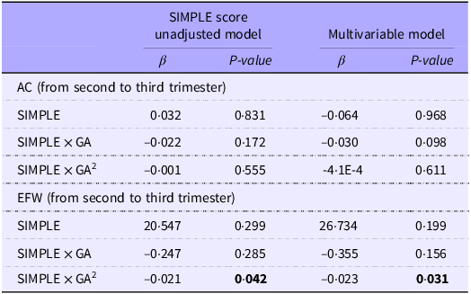
For the selected outcome measures, unconditional growth models demonstrated a significant positive effect of gestational age and a significant quadratic term, confirming a non-linear growth trajectory over time. Figures 1 and 2 illustrate the estimated growth curves for AC and EFW/birth weight, respectively, derived from linear quadratic models. Table 2 provides the effect estimates from linear mixed models evaluating the associations between SIMPLE score group and longitudinal fetal/neonatal measurements. The linear interaction between SIMPLE score group and time was not significant for either AC or EFW, suggesting that group differences did not follow a strictly linear pattern over gestation. However, fetal weight gain (g/d) from the second to the third trimester was significantly lower in the low-adherence group (P < 0·05), reflecting a reduced growth velocity compared with the high-adherence group (Figure 3). Sex-stratified analyses are shown in Table 3.
Longitudinal abdominal circumference measurements during second and third trimester in the study population.

Figure 1. Long description
A scatter plot showing the relationship between gestational age in days and abdominal circumference in millimeters. The x-axis represents gestational age in days, ranging from 120 to 280 days. The y-axis represents abdominal circumference in millimeters, ranging from 150 to 350 millimeters. The plot contains hundreds of data points, each representing a measurement. A clear positive correlation is visible, with data points forming a dense cluster along a diagonal line. A blue regression line is present, indicating the trend of the data. All values are approximated.
Longitudinal second and third trimester estimated fetal weight and birth weight measurements in the study population.

Figure 2. Long description
A scatter plot illustrates the relationship between estimated fetal weight and gestational age. The plot contains hundreds of data points, each representing an estimated fetal weight measurement. The x-axis represents gestational age in days, ranging from approximately 100 to 300 days. The y-axis represents estimated fetal weight in grams, ranging from 0 to 5000 grams. The data points show a positive correlation, indicating that as gestational age increases, estimated fetal weight also increases. A blue regression line is present, highlighting the overall trend. The data points are densely clustered around the regression line, with some variability. All values are approximated.
Effect estimates from linear mixed models for the associations between SIMPLE score group and longitudinal fetal/neonatal measurements

Table 2. Long description
The table presents effect estimates from linear mixed models evaluating the associations between SIMPLE score group and longitudinal fetal/neonatal measurements. It includes data for abdominal circumference (AC) and estimated fetal weight (EFW) from the second to the third trimester. The table has four rows and six columns, with column headers labeled SIMPLE score unadjusted model beta, SIMPLE score unadjusted model P, Multivariable model beta, and Multivariable model P. Row labels include SIMPLE, SIMPLE x GA, and SIMPLE x GA squared for both AC and EFW. Notable values include a significant P value of 0.042 for SIMPLE x GA squared under the unadjusted model for EFW and a significant P value of 0.031 for the same interaction under the multivariable model. The data suggests non-linear growth trajectories and significant effects of gestational age.
AC, abdominal circumference; EFW, estimated fetal weight; GA, gestational age.
The SIMPLE score was coded as 0 if ≥ 6 (high adherence, reference category) or 1 if < 6 (low adherence). The multivariable model further included terms for maternal age, pregestational BMI, parity, educational level, smoking status, ethnicity and neonatal sex. Bold type highlights significant results.
Longitudinal fetal estimated weight measurements according to the SIMPLE score groups.

Figure 3. Long description
A scatter plot illustrates the relationship between estimated fetal weight and gestational age. The x-axis represents gestational age in days, ranging from 120 to 280 days, while the y-axis represents estimated fetal weight in grams, ranging from 0 to 4000 grams. The plot includes hundreds of data points, each representing an estimated fetal weight measurement. Two trend lines are present: a green line for the high adherence group and a red line for the low adherence group. The data points form a positive correlation, indicating that as gestational age increases, estimated fetal weight also increases. The high adherence group generally shows higher estimated fetal weights compared to the low adherence group. All values are approximated.
Effect estimates from multi-adjusted linear mixed models for the associations between the SIMPLE score group and longitudinal fetal/neonatal measurements according to neonatal sex

Table 3. Long description
The table presents data on the effect estimates from multi-adjusted linear mixed models for the associations between the SIMPLE score group and longitudinal fetal/neonatal measurements according to neonatal sex. It includes measurements for male and female fetuses, with columns for beta coefficients and P-values. The table has two main sections: abdominal circumference and estimated fetal weight, each with three rows for SIMPLE, SIMPLE multiplied by gestational age, and SIMPLE multiplied by gestational age squared. Notable trends include significant P-values for SIMPLE multiplied by gestational age in male fetuses for abdominal circumference and estimated fetal weight, indicating a non-linear growth trajectory over time. The data highlights differences in growth patterns between male and female fetuses.
GA, gestational age.
The SIMPLE score was coded as 0 if ≥ 6 (high adherence, reference category) or 1 if < 6 (low adherence). Bold type highlights significant results.
No significant associations were observed among female fetuses. In contrast, among male fetuses, the interaction between SIMPLE score group and squared time was significant, indicating a slower EFW growth trajectory from the second to the third trimester in the low adherence group. Additionally, for AC, a significant interaction between SIMPLE score and time was detected in male fetuses (β = −0·053, P = 0·032), consistent with lower AC growth trajectories in the low adherence compared to the high adherence group.
Table 4 shows the effect estimates from linear mixed models assessing associations between individual SIMPLE checklist items and absolute daily changes in fetal growth parameters, including adjustment for the previously defined covariates. Among the ten SIMPLE score components, only first trimester Hg concentrations higher than 110 g/l were negatively associated with the absolute daily change in EFW from the second to the third trimester (mean difference: −0·70 g/d). The SIMPLE score was additionally tested as a continuous variable in sensitivity analyses. When modeled continuously, each one-point increase in the score was not significantly associated with fetal growth trajectories. Finally, linear mixed models analysing the independent contribution of maternal pregestational BMI showed significant positive associations with absolute daily changes in both AC and EFW. Furthermore, pregestational obesity was significantly associated with increased birth weights by 167 g (95 % IC: 64·5, 269·6, P = 0·001) compared with non-obese women.
Effect estimates from the fully-adjusted linear mixed models for the associations between the SIMPLE score individual components, pregestational BMI and absolute differences in selected longitudinal fetal measurements (i.e. abdominal circumference, estimated fetal weight)

Table 4. Long description
The table presents effect estimates from linear mixed models, focusing on the associations between individual components of the SIMPLE score, pregestational BMI, and changes in fetal measurements such as abdominal circumference and estimated fetal weight. The table includes ten SIMPLE score components and their respective beta values and P-values for both abdominal circumference and estimated fetal weight. Notable findings include a negative association between first trimester hemoglobin concentrations higher than 110 grams per liter and the absolute daily change in estimated fetal weight from the second to the third trimester. Pregestational BMI shows significant positive associations with both abdominal circumference and estimated fetal weight. The table consists of three main columns: SIMPLE score component, abdominal circumference with beta and P-values, and estimated fetal weight with beta and P-values. Each row lists a different SIMPLE score component along with its corresponding beta values and P-values for the two fetal measurements.
FA, folic acid.
* Absolute differences as response variables and BMI as a predictor.
Discussion
The present study extends previous findings from the SIMPLE project by demonstrating that early maternal nutritional status – assessed through both pregestational BMI and first trimester SIMPLE score – is associated not only with early placental markers but also with longitudinal fetal growth trajectories and velocity. While previous studies have largely focused on birth weight as a static endpoint, our data highlight the importance of examining longitudinal growth trajectories, which may more sensitively capture subtle intrauterine adaptations. Specifically, the low-adherence group (SIMPLE score lower than 6) was associated with decreased EFW growth acceleration from the second to the third trimester of pregnancy compared with the high-adherence group, with sex-specific effects observed predominantly among male fetuses. Overall, these findings build upon our prior evidence linking higher SIMPLE scores to increased first-trimester PAPP-A concentrations and more favourable obstetric outcomes(11,Reference Parisi, Savasi and Di Bartolo13) . Given that PAPP-A is a recognised marker of placental development(Reference Smith, Stenhouse and Crossley16,Reference Tzanaki, Makrigiannakis and Lymperopoulou18) , the present results support a potential biological continuum linking early maternal nutritional status, placental development and subsequent fetal growth dynamics. To further disentangle the associations of individual nutritional exposures, a significant negative association between first trimester Hg concentrations higher than 110 g/l and EFW growth velocity was shown.
This study also revealed alarmingly high rates of nutritional risk among pregnant women in the first trimester: 93·4 % of the study population presented at least one nutritional risk (i.e. negative response to at least one SIMPLE checklist item) and 7 % were classified as obese, in line with national reports for women of reproductive age (available at https://osservatoriosullasalute.it/osservasalute/rapporto-osservasalute-2022). The most prevalent areas of deficiency were whole grain consumption and sun exposure, with approximately half of participants providing negative responses. As expected, the low-adherence group included younger, less educated and more frequently obese women compared with the high-adherence group.
Importantly, no differences emerged in absolute ultrasound measurements at isolated time points, underscoring that fetal size alone may not fully reflect growth impairment. Instead, the observed differences in EFW growth acceleration emphasise the relevance of dynamic growth patterns. Although fetal size below fixed thresholds (third, fifth or tenth percentile) is commonly used as a proxy for impaired intrauterine growth and a predictor of adverse pregnancy outcomes, recent evidence emphasises the importance of deranged EFW growth velocity as a more sensitive marker of pathological fetal growth. Pacora and colleagues demonstrated that longitudinal growth velocity doubled the sensitivity for predicting antepartum fetal death compared with a single size assessment, irrespective of EFW percentile(Reference Pacora, Romero and Jung19). Similarly, Gardosi and Hugh proposed the projected optimal weight range method to identify non-small for gestational age fetuses with slow growth at increased risk of stillbirth(Reference Gardosi and Hugh20,Reference Hugh, Cowan and Butler21) . A secondary analysis of the TRUFFLE-2 study further showed that reduced fetal growth velocity is associated with adverse perinatal outcomes, independent of signs of cerebral blood flow redistribution(Reference Stampalija, Wolf and Mylrea-Foley22). Additionally, Mac Donald and colleagues demonstrated that reduced growth velocity between 28 and 36 gestational weeks is associated with antenatal (cerebro-placental ratio), intrapartum (arterial cord blood pH) and neonatal (neonatal body fat percentage) markers of placental insufficiency, regardless of adequate birth weight(Reference MacDonald, Hui and Tong23). Overall, these data underscore the clinical relevance of identifying reduced fetal growth velocity as an early indicator of growth impairment warranting further investigation. Reference growth velocity curves have been recently proposed within the INTERGROWTH-21st Project(Reference Ohuma, Villar and Feng24). To our knowledge, the present study is the first to evaluate maternal nutritional habits in relation to fetal growth rate, highlighting that maternal first trimester low adherence to a healthy diet and lifestyle – as measured by the SIMPLE score – is associated with slower fetal growth trajectories compared with fetuses of well-nourished mothers.
Notably, sex-specific differences emerged in the observed associations. Sexually dimorphic responses during intrauterine life have been increasingly reported, with substantial differences in both short- and long-term maternal and offspring outcomes according to fetal sex(Reference Alur, Holla and Hussain25,Reference Sheiner, Levy and Katz26) . To explain the higher perinatal mortality rate observed among males, the concept of a ‘more dangerous life in the womb’ has been proposed(Reference Eriksson, Kajantie and Osmond27). According to this hypothesis, male fetuses prioritise feto-placental growth pathways, whereas female fetuses enhance placental cellular adaptation and vasculogenesis, thereby increasing resilience to adverse environmental conditions(Reference Meakin, Cuffe and Darby28). Our results are consistent with this paradigm and suggest that suboptimal maternal nutritional patterns in early pregnancy may disproportionately affect male fetal growth trajectories.
The positive association between maternal pregestational BMI and intrauterine fetal growth aligns with robust evidence showing higher birth weights and increased neonatal adiposity among offspring of overweight and obese mothers, with maternal pregestational BMI and gestational weight being the key predictors of neonatal macrosomia(Reference Sewell, Huston-Presley and Super29–Reference Lewandowska31).
Conversely, the inverse association between first trimester Hg concentration above 110 g/l and EFW growth velocity is consistent with a U-shaped relationship previously described between maternal Hg levels and birth weight, suggesting that both Fe deficiency and Fe excess may adversely affect placental perfusion and intrauterine growth(Reference Evanchuk, Kozyrskyj and Vaghef-Mehrabani32,Reference Rooney, Conway and O’Keeffe33) .
The present study has several strengths. First, the longitudinal, multicentre, nationwide design provides a reliable picture of the pregnant population in Italy, enhancing the external validity of the findings. Furthermore, the multi-adjusted models accounted for most known determinants of prenatal growth, increasing the robustness of the results. All ultrasound measurements were performed by trained clinicians according to International Society of Ultrasound in Obstetrics and Gynecology guidelines, ensuring consistency across recruiting centres. Lastly, the Federation of Gynecology and Obstetrics Nutrition checklist has been recently validated against the gold standard food FFQ, supporting its use as an effective screening tool to identify suboptimal dietary quality in early pregnancy(Reference Tsoi, Chan and Li10).
On the other hand, several limitations should be acknowledged. First, the observational design precludes any inference of causality. Consequently, future research should include randomised controlled trials to determine whether interventions guided by the SIMPLE score are effective in improving pregnancy outcomes and fetal growth trajectories. Additionally, the SIMPLE score was assessed only once, in the late first trimester, when nausea, vomiting, food aversions or increased health awareness may have temporarily influenced maternal dietary behaviors. Therefore, reliance on a single dietary assessment at a time of potential behavioural modification represents a limitation and changes in nutritional habits later in pregnancy cannot be excluded. However, available evidence suggests that dietary patterns during pregnancy are relatively stable over time, supporting the validity of early pregnancy dietary assessment as a reasonable proxy for subsequent nutritional behaviours(Reference Crozier, Robinson and Godfrey34). Moreover, nutritional exposures in early pregnancy are biologically plausible determinants of placental development and are more likely to influence fetal growth trajectories in the second and early third trimester than dietary behaviors assessed later in gestation. Another limitation relates to the dichotomisation of the SIMPLE score using a predefined cut-off (≥ 6 v. < 6). Although this threshold was identified in our previous studies as clinically relevant and facilitates interpretability in routine practice, dichotomisation inevitably results in some loss of information and may oversimplify the underlying variability in dietary behaviours. For example, women with very similar scores (e.g. 5 v. 6) may be classified into different categories despite minimal differences in overall nutritional profile. Moreover, when the SIMPLE score was modelled as a continuous variable, no significant linear association was observed with fetal growth trajectories, suggesting that the relationship may not follow a strictly dose–response pattern across the entire score range. Therefore, while the use of a clinically oriented cut-off improves applicability, the possibility of residual misclassification and reduced statistical sensitivity should be acknowledged. Another limitation is the lack of information on physical activity, a key component of overall metabolic health and lifestyle, as well as on gestational weight gain at delivery, which was unavailable for more than half of the study population. Ultrasound assessments were limited to the second and early third trimesters, with no evaluations performed in late gestation. Therefore, potential late-pregnancy fetal growth decelerations or accelerations may not have been captured. Additionally, maternal nutritional blood biomarkers were not available to further validate the SIMPLE score and checklist as a measure of maternal nutritional status. Finally, the absence of neonatal and infant follow-up data precluded the assessment of long-term growth outcomes.
Conclusions
The present study shows that early maternal nutritional status, assessed by the SIMPLE score and pregestational BMI, is associated with longitudinal fetal growth trajectories and velocity, with sex-specific effects predominantly observed among male fetuses. These findings extend previous evidence linking the SIMPLE score to early placental biomarkers and suggest that early maternal nutritional patterns may influence subsequent intrauterine growth dynamics. However, given the observational design and the absence of intervention data, our results should be interpreted as hypothesis-generating. Further studies are needed to confirm these associations, explore underlying mechanisms – particularly regarding sex-specific vulnerability – and evaluate whether nutritional screening and targeted interventions based on the SIMPLE score can improve maternal and fetal outcomes.
Acknowledgements
Promoted by the Italian Association of Academic Gynecologists (AGUI).
The authors are thankful to all the midwives and nurses for their expertise and cooperation and to all pregnant women for participating in the study. We thank Prof. Chiara Mandò for the scientific support in the ‘ONFOODS Project’.
The authors are very grateful to Prof. Giovanni Scambia† (Full Professor of Gynaecology and Obstetrics at Università Cattolica del Sacro Cuore and Scientific Director of UOC of Gynaecologic Oncology and Fondazione Policlinico Universitario Agostino Gemelli IRCCS) for having strongly supported the start of this collaboration among Italian university centres.
This work was partially supported by the Italian Ministry of Health. The funding of Prof.ssa Irene Cetin and Dr. Gaia Maria Anelli comes from a project funded under the National Recovery and Resilience Plan (PNRR), Mission 4, Component 2, Investment 1·3 – Call for Proposal No. 341 of 15 March 2022 by the Italian Ministry of University and Research, funded by the European Union – NextGenerationEU. Project code PE00000003, Concession Decree No. 1550 of 11 October 2022, adopted by the Italian Ministry of University and Research, CUP D93C22000890001. Project title: ‘ON Foods – Research and Innovation Network on Food and Nutrition Sustainability, Safety, and Security – Working ON Foods’.
Conceptualisation: F. Parisi., I. C.; Data curation: F. Parisi., G. E.; Formal analysis: F. Parisi., G. E.; Investigation: F.Parisi., G. A., A. S., G. C., M. B., F. Petraglia., A. C., A. L., P. G., F. O., N. C., G. B., M. V., M. P.; Methodology: F. Parisi., G. A.; Supervision: V. S., I. C.; Validation: F. Parisi.; Visualisation: F. Parisi., A. S, G. C., M. B., F. Petraglia., A. C., A. L., P. G., F. O., N. C., G. B., M. V., M. P., I. C.; Writing – original draft: F. Parisi., C. L.; Writing – review & editing: F. Parisi., G. A., C. L., I. C., V. S.
The authors declare no conflicts of interest.
CONTRIBUTION
What are the novel findings of this work?
The present work confirms the clinical utility of the SIMPLE score, showing significant associations with intrauterine growth trajectory and velocity, independent of other markers of nutritional status (e.g. pregestational BMI). To our knowledge, this is the first study evaluating maternal nutritional habits using a fast, reproducible and validated checklist, in association with fetal growth rate, highlighting that low adherence to a healthy diet is associated with slower fetal growth trajectories. Additionally, a slower estimated fetal weight trajectory from the second to the third trimester was observed in non-anaemic compared with anaemic women in the first trimester. The positive association between maternal pregestational BMI and intrauterine fetal growth aligns with robust evidence showing that infants of overweight or obese mothers tend to have higher birth weight and increased adiposity.
What are the clinical implications of this work?
The evidence from this cohort provides the foundation for the use of the SIMPLE score and its single components in clinical settings, where assessing maternal nutritional habits is often time-consuming, challenging and requires specialised personnel. Future research should evaluate the applicability of the SIMPLE score among high-risk pregnancies, while randomised controlled trials are warranted to clarify the effects of targeted interventions in women at high nutritional risk.




 Open access

