Introduction
The cost of drug development has increased dramatically during the last few decades due to the inefficiency of current preclinical drug evaluation models and the decreasing number of newly developed drugs.Reference Munos1 Major disadvantages of conventional preclinical models include a phylogenetic gap between human and animal models and a lack of similarity between two-dimensional (2D) cell culture systems and in vivo systems. Microengineering technologies combined with cell biology have been expected to solve this inefficiency by bridging the gap between human and preclinical models. The concept of “organs-on-chips” was proposed recently to establish in vitro models that precisely recreate in vivo characteristics as well as drug responses.Reference Esch, King and Shuler2,Reference Huh, Torisawa, Hamilton, Kim and Ingber3
Vascularization, a construction of the microvascular niche, is essential to mimic in vivo organ characteristics, as microvessels and capillaries cover organs almost entirely. In tissue engineering, vascularization has been emphasized to perfuse large-scale engineered tissue and supply oxygen and nutrients to interior cells, particularly for in vivo transplantation.Reference Auger, Gibot and Lacroix4,Reference Rouwkema, Rivron and van Blitterswijk5 In contrast, supplementation with oxygen and nutrients is not the main purpose of vascularization for organs-on-chips, as the entire system is typically small enough (a few hundred microns in dimension) to be supplied by oxygen diffusion in culture media. Instead, vascularization of organs-on-chips can establish in vivo-like target organ microenvironments, as microvascular endothelial cells (ECs, a cell type that composes the interior surface of the blood vessels) play critical roles in organ function in cooperation with the cell types specific to each organ.Reference Nolan, Ginsberg, Israely, Palikuqi, Poulos, James, Ding, Schachterle, Liu, Rosenwaks, Butler, Xiang, Rafii, Shido, Rabbany, Elemento and Rafii6
In this review, we focus on the importance of vascularization for in vitro organ models. Beginning with the pathophysiological importance of the endothelium, subsequent sections will introduce specific features of chip-level in vitro microvascular mimicking systems. Recent efforts to vascularize organs-on-chips and material considerations for establishing organs-on-chips are also covered.
Organ-specific endothelial physiology
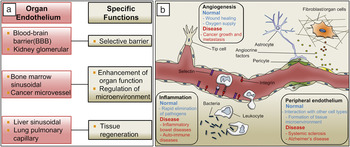
The microvascular system is the infrastructure of the body that not only supplies oxygen and nutrients to tissues but is also related to numerous pathophysiological conditions. The microvascular endothelium, which is the internal surface of blood vessels consisting of ECs, plays diverse roles in organ function (Figure 1a). As the specific characteristics of each organ are determined by the endothelium,Reference Nolan, Ginsberg, Israely, Palikuqi, Poulos, James, Ding, Schachterle, Liu, Rosenwaks, Butler, Xiang, Rafii, Shido, Rabbany, Elemento and Rafii6,Reference Nachman7 understanding endothelial organ specificity is necessary for reconstituting functional organs in vitro.
(a) The physiological roles of the microvascular system in various organs. In the brain and kidney, the endothelium forms selective barriers through an increased number of tight junctions in cooperation with supporting cell types. In bone marrow and cancer, the microvascular endothelium secretes angiocrine factors to provide an optimum organ-specific microenvironment to enhance their innate functions. Sinusoidal endothelium of the liver and lung assists in the regeneration of organs by distinct molecular mechanisms. (b) Representative physiological phenomena present in a microvascular system and their dual role in normal and pathological conditions. Although angiogenesis is necessary to recover wounded or low-oxygen tissues, it can also provide nutrients and oxygen for cancer. Inflammation is the first mechanism of protection from pathogens, but an uncontrolled immune response can damage normal tissue and cause auto-immune disease. Peripheral endothelial cells that compose the microvascular system play critical roles in organs by interacting with various cell types, while secreting toxic factors that damage native tissue.

Selective barrier
Although the selective barrier function is a natural and basic function of the endothelium, some organs have highly specialized endothelial barriers. ECs form the blood–brain barrier (BBB) in cooperation with astrocytes (a non-neural cell type that is most abundant in the human brain)Reference Abbott, Rönnbäck and Hansson8 and pericytes (a contractile cell type that wraps around the endothelial cells of capillaries),Reference Armulik, Genové, Mäe, Nisancioglu, Wallgard, Niaudet, He, Norlin, Lindblom, Strittmatter, Johansson and Betsholtz9 which have tight cell-cell junctions and lower permeability to protect the central nervous system from various potentially harmful external factors.
The kidney microvascular endothelium also functions as a specialized selective barrier and is closely related to numerous kidney-related diseases.Reference Liu10,Reference Long, Norman and Fine11 ECs form the glomerular filtration barrier in the kidney, which resists the high pressure needed to filter huge volumes of water into the urinary space while retaining 99.9% of the large proteins. Podocytes, specialized supporting cells that are similar to astrocytes in the BBB, interact tightly with ECs to precisely control barrier function.Reference Welsh and Saleem12
Tissue-specific microenvironments
Sinusoidal ECs (a special type of fenestrated capillaries that have larger openings) in bone marrow are continuously remodeling the sinusoidal microenvironment by secreting various paracrine factors to maintain bone marrow functions, such as hematopoiesis (formation of cellular components in the blood) and hematopoietic stem and progenitor cell (HSPC) maintenance.Reference Hooper, Butler, Nolan, Kranz, Iida, Kobayashi, Kopp, Shido, Petit, Yanger, James, Witte, Zhu, Wu, Pytowski, Rosenwaks, Mittal, Sato and Rafii13–Reference Ding, Saunders, Enikolopov and Morrison15 Trafficking and homing of lymphocytesReference Streeter, Berg, Rouse, Bargatze and Butcher16 and HSPCsReference Naiyer, Jo, Ahn, Mohle, Peichev, Lam, Silverstein, Moore and Rafii17,Reference Sipkins, Wei, Wu, Runnels, Côté, Means, Luster, Scadden and Lin18 are also regulated by organ-specific chemokines (low molecular weight soluble proteins or peptides that induce homing of cells) and adhesion molecules on ECs in bone marrow.
Abnormal morphology (tortuous and leaky), functionally disrupted pericytes,Reference Greenberg, Shields, Barillas, Acevedo, Murphy, Huang, Scheppke, Stockmann, Johnson, Angle and Cheresh19 and a different extracellular matrix (ECM) composition are the main characteristics of tumor vessels.Reference Ruoslahti20,Reference Tozer, Kanthou and Baguley21 ECs that compose the tumor microenvironment also enhance tumor progression by secreting various growth and other factors.Reference Pirtskhalaishvili and Nelson22,Reference Butler, Kobayashi and Rafii23 Recent studies have demonstrated that the microvascular niche in tumor sites also plays essential roles in inducing breast cancer quiescence.Reference Ghajar, Peinado, Mori, Matei, Evason, Brazier, Almeida, Koller, Hajjar, Stainier, Chen, Lyden and Bissell24
Regeneration of injured organs
ECs play critical roles in the regeneration of organs, such as the liver and lungs, with distinct underlying mechanisms. Ding et al. reported that the liver sinusoidal endothelium activates hepatocyte proliferation by secreting Wnt2 and hepatocyte growth factor.Reference Ding, Nolan, Butler, James, Babazadeh, Rosenwaks, Mittal, Kobayashi, Shido, Lyden, Sato, Rabbany and Rafii25 The same group demonstrated that lung pulmonary capillary ECs induce lung regeneration through matrix metalloproteinase 14 expression, which leads to epithelial progenitor cell proliferation via epidermal growth factor receptors.Reference Ding, Nolan, Guo, Babazadeh, Cao, Rosenwaks, Crystal, Simons, Sato, Worgall, Shido, Rabbany and Rafii26
The microvascular system in pathogenic conditions
General physiological events in the microvascular system, such as angiogenesis, inflammation, and EC interactions with perivascular cells, are important for maintaining the stability of our body. Notably, these events also play key roles in various disease states (Figure 1b).
Angiogenesis: Cancer growth and metastasis
Angiogenesis, a physiological process that forms new blood vessels from pre-existing vessels, is one of the most representative phenomena that occur in the microvascular system and is necessary to maintain homeostasis. Angiogenesis is involved in many biological processes such as wound healing, regulation of acute inflammation, and remodeling of vessels to supply oxygen.
However, angiogenesis also occurs under various pathological conditions. A large amount of clinical evidence indicates that angiogenesis from microvascular beds near cancer sites provide oxygen and nutrients for growth and channels for metastasis.Reference Folkman27–Reference Weidner29 Strategies to wither cancer by blocking angiogenesis have been rigorously studied during the last two decades;Reference Carmeliet30,Reference Ferrara and Kerbel31 however, clinical trials have shown that strategies depending solely on anti-angiogenesis are not as effective as expected. In many cases, cancers develop in alternative ways to avoid therapies, such as by developing resistance to drug treatment.Reference Shojaei32 To overcome these limitations, many complementary strategies are emerging, such as tumor vessel normalizationReference Jain33,Reference Goel, Fukumura and Jain34 and cancer immunotherapy.Reference Mellman, Coukos and Dranoff35,Reference Palucka and Banchereau36
Inflammation: Inflammatory diseases
Inflammation is a rapid immune response by injured or infected tissues to recruit leukocytes (white blood cells) to eliminate pathogens. During development of peripheral inflammation, microvessels synthesize and secrete pro-inflammatory cytokines, including various interleukins (a substance extracted from white blood cells that stimulates the function of the immune system), to attract immune cells to sites of inflammation.
However, failure to regulate inflammation often leads to inflammatory disease. Several molecules and mechanisms are involved in the regulation of inflammatory responsesReference Tak and Firestein37–Reference Deban, Correale, Vetrano, Malesci and Danese39 and are being investigated as potential therapeutic targets for immune-related disorders.Reference Luster, Alon and von Andrian40 For example, tumor necrosis factor (TNF) is the main molecular regulator of inflammation and is related to various inflammatory diseases. TNF levels are increased in patients with inflammatory bowel diseases, and the symptoms are attenuated by blocking TNF.Reference Bradley41 TNF also induces chronic inflammation in cooperation with various cytokines and growth factors in patients with rheumatoid arthritis.Reference McInnes and Schett42 Furthermore, a claim that auto-inflammation causes obesity-related Type 2 diabetes is supported by various studies.Reference Donath and Shoelson43
Peripheral endothelium: Complications of microangiopathy
ECs, which compose microvessels, dynamically regulate the microenvironment by interacting with other cell types, including immune cells, pericytes, and fibroblasts (a cell type that synthesizes the framework of animal tissues). Microangiopathy, a general term that indicates dysfunction of microvessels, is mainly caused by abnormal behavior of ECs. Microangiopathy can lead to various diseases, such as systemic sclerosis,Reference Cutolo, Sulli and Smith44 sepsis, thrombosis,Reference Semeraro, Ammollo, Semeraro and Colucci45 obesity,Reference de Jongh46 and complications of diabetes.Reference Tooke47 In particular, the complications of diabetes are followed by various diseases such as retinal microvessel malfunction (retinopathy) and kidney glomerular dysfunction (nephropathy).Reference Tooke47,Reference Cracowski, Minson, Salvat-Melis and Halliwill48 Furthermore, the EC-derived neurotoxic thrombin increases significantly in the brains of patients with Alzheimer’s disease.Reference Yin, Wright, Wall and Grammas49 Other neurodegenerative diseases, such as Parkinson’s disease and multiple sclerosis, are caused by dysfunction in the brain endothelium.Reference Grammas, Martinez and Miller50
In vitro microvascular models
Many in vitro studies have contributed to a deeper understanding of vascular biology. The conventional blood vessel model starts with a scheme of confluent EC cultures on a Petri dish,Reference Kramer and Nicolson51 Transwell membrane (a cell-culture platform that is composed of two chambers separated by a porous membrane),Reference Li and Zhu52 or hydrogel.Reference Voura, Sandig, Kalnins and Siu53 Assuming that the confluent monolayer of ECs is a blood vessel wall, many studies of the physiology of blood vessels have been conducted, including trans-endothelial migration of cancer or immune cells,Reference Kramer and Nicolson51–Reference Voura, Sandig, Kalnins and Siu53 the effects of shear stress on blood vessels,Reference Levesque and Nerem54,Reference Chien55 vascular permeability studies,Reference Kazakoff, McGuire, Hoie, Cano and Iversen56,Reference Friedl, Puhlmann, Bartlett, Libutti, Turner, Gnant and Alexander57 and angiogenesis from pre-existing blood vessels.Reference Davis, Black and Bayless58
Two-dimensional monolayer models have several limitations. They poorly represent the structural characteristics of the body, as they replicate only a portion of the blood vessel wall without any circular or lumenized (hollowed) structures. Additionally, 2D monolayer models cannot be used to study luminal flow with 3D tubular microvessel structures; thus, replicating the effect of physiological blood flow and the interactions with other cell types remains challenging.
To overcome these disadvantages, much effort has been devoted to recreating 3D structural characteristics of blood vessels in vitro. Soft lithography and polydimethylsiloxane (PDMS)-based microfluidic devices have opened a new era of in vitro models for biological research.Reference Anderson, Chiu, Wu, Schueller and Whitesides59 The use of these microchips has several advantages over conventional macroscale platform-based in vitro models.Reference Chung, Sudo, Vickerman, Zervantonakis and Kamm60 Substrates with micro-nano patterns or microstructures that mimic the in vivo cellular microenvironment can be incorporated in vitro. Additionally, complex environmental factors can be precisely integrated and controlled, such as chemical gradients or interstitial flow that provides an in vivo-like environment. As a microchip uses a much smaller volume of reagent and number of cells compared to conventional models, performing biological research using a microchip also lowers research costs.
We introduce in vitro microvascular models using microfluidic techniques, which are potentially applicable to organ-on-chip vascularization. The models are grouped into two main types: (1) Attaching ECs to the wall of a micropatterned hydrogel structure, and (2) using natural morphogenic processes for microvessel formation inside a microfluidic device by spatially controlling the patterning of the hydrogel.
Attaching ECs to the walls of hydrogel channels
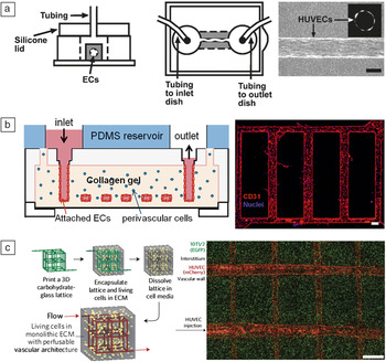
This scheme uses micropatterned hydrogels and ECs that attach to the wall to create microvessels on a chip. Chrobak et al. created a lumenized microvessel by constructing a collagen gel with a long, hollow structure using a silicon housing and a microneedle (Figure 2a).Reference Chrobak, Potter and Tien61 The microneedle-containing silicone housing is filled with collagen, and the microneedle is removed after collagen polymerization to leave a hollow microchannel structure. By attaching a confluent culture of ECs onto the wall of the hollow collagen structure, a 3D microvessel that has tubular structure was engineered in vitro. Vascular permeability was investigated using this microvessel, and the interaction between ECs and other cell types such as leukocytes or pericytes was examined by perfusion of media inside the microvessel lumen.
Various methods for recreating in vitro microvessels by attachment to micropatterned hydrogel walls. (a) Schematic of microvessel construction by using silicon housing and microneedles as molds. Endothelial cells (ECs) were attached to the hydrogel wall to form a microvessel structure. Micrograph on right side shows top and sectioned view (inset) of the formed microvessel. Scale bar, 100 μm. (Reproduced with permission from Reference 61. © 2006 Elsevier.) (b) A micropatterned silicon stamp was used as a mold to construct a microvessel network shape inside the hydrogel. The micropatterned hydrogel was coated with ECs to model the microvessel network. The micrograph of the sample immunostained with CD31 shows robust microvessel network formation. Scale bar, 100 μm. (Reproduced with permission from Reference 64. © 2012 National Academy of Sciences, USA.) (c) A three-dimensional (3D) microvessel network was formed by rapid casting of carbohydrate glass with a 3D printer. The lattice of the carbohydrate glass was covered with a perivascular cell-containing hydrogel and melted with cell media to form the hollowed network inside the hydrogel tissue. The micrograph (right) shows HUVECs expressing mCherry (red fluorescent protein) that are attached to the hydrogel wall to generate the microvessel network. 10T1/2 cells (mouse fibroblast cell line) expressing EGFP (green fluorescent marker) are embedded with hydrogel to visualize interstitium. Scale bar, 1 mm. (Reproduced with permission from Reference 65. © 2012 Nature Publishing Group.) Note: HUVECs, human umbilical vein endothelial cells; PDMS, polydimethylsiloxane; ECM, extracellular matrix; EGFP, enhanced green fluorescence protein.

Bischel et al. developed a simpler technique to generate hollow structures inside a hydrogel using “viscous finger patterning.”Reference Bischel, Young, Mader and Beebe62 During polymerization of a hydrogel inside the microchannel, the partially polymerized hydrogel in the central part of the channel is washed away by passive pumping-based media flow,Reference Walker and Beebe63 leaving a hollow lumen-like structure inside the hydrogel. ECs are attached inside the wall of the hollow structure to complete the microvessels with a 3D tubular structure on the chip. Multiple, parallel microvessels are created to observe angiogenesis toward a vascular endothelial growth factor (VEGF) gradient as well as the effect of smooth muscle cells on the angiogenic process. Consequently, this device can be used for various angiogenesis assays using different growth factors and drug candidates.
Zheng et al. replicated a network of microvessels rather than a single tube (Figure 2b).Reference Zheng, Chen, Craven, Choi, Totorica, Diaz-Santana, Kermani, Hempstead, Fischbach-Teschl and López64 The microvascular structure was patterned on a collagen Type I gel using a microstructured silicone stamp. First, the patterned collagen is housed between two flex-glass pieces, followed by attaching ECs to the collagen wall. Several aspects of vascular biology were observed, such as interactions between pericytes and ECs, endothelial barrier function under different media conditions or co-cultured cells, and interactions between whole blood components and endothelium under a perfused flow condition.
Miller et al. established a novel method of creating a microvessel network in a hydrogel structure (Figure 2c).Reference Miller, Stevens, Yang, Baker, Nguyen, Cohen, Toro, Chen, Galie, Yu, Chaturvedi, Bhatia and Chen65 Pertinent levels of carbohydrate glass—formed by mixing sucrose, glucose, and dextran in water and boiling off the solvent—were extruded to model microvessels by optimizing the nozzle velocity and diameter of a 3D printer. After thermally extruding the carbohydrate glass network utilizing the 3D printer, a fibrin gel was used to cover the microvessels, similar to interstitial ECM. The carbohydrate glass is dissolved with cell media, leaving the network structure inside the fibrin. ECs are attached to the fibrin gel walls to form a microvessel network in the tissue. The fibrin gel can be encapsulated with fibroblasts and co-cultured with ECs to form a robust microvascular network. The researchers clearly showed that hepatocytes (liver cells) in the gel exhibit increased metabolic functions by establishing a microvascular structure inside the hydrogel.
These in vitro microvascular models share several features. They commonly use molds and housings to construct the hydrogel structure, which resemble the luminal structure of blood vessels, followed by attaching EC monolayers to the walls of the structure to recreate microvessels. Although these in vitro microvessels have properties similar to the Transwell membrane model, their structural properties are much closer to in vivo microvessels and can be easily perfused with cell culture media. An advantage of this method is that microvessel geometry can be controlled easily through the use of microfabricated mold structures according to the experimental purpose. Furthermore, as the shape of the mold determines microvessel structure, applying physiological levels of media using an external pump is easy, as the applied shear stress can be calculated from the engineered microvessel diameter and pump flow rate. However, the microvessels developed in these models are not formed by a vasculogenic (formation of new blood vessels in the absence of pre-existing vessels) or angiogenic process, which is the main biological process that living organisms use to establish the circulatory system. That is, the physiological relevance of these engineered microvessels with actual in vivo blood vessels is attenuated, as the process for engineering the microvascular structure is based mainly on artificially depositing ECs on hydrogel walls.
Utilizing the angiogenic/vasculogenic properties of ECs to recreate microvessels on a chip
Numerous studies in vascular biology have shown that ECs have morphogenic features that differentiate into a lumenized microvessel structure by stimulation with several growth factors (e.g., VEGF and basic fibroblast growth factor [bFGF])Reference Koolwijk, Van Erck, De Vree, Vermeer, Weich, Hanemaaijer and Van Hinsbergh66 or by co-culture with other cell types such as fibroblastsReference Montesano, Pepper and Orci67 in ECM-like hydrogels, including collagen and fibrin. Naturally lumenized microvessels can be engineered on a chip by combining the angiogenic/vasculogenic properties of ECs with construction of microstructures.
Raghavan et al. introduced a method that geometrically controls the shape of microvessels formed by the vasculogenic process.Reference Raghavan, Nelson, Baranski, Lim and Chen68 A mixture of collagen gel and ECs was added to micropatterned PDMS channels, and the cells were stimulated with bFGF and VEGF to promote vasculogenesis. The microvessels formed by the ECs were geometrically aligned in various shapes by the geometrical confinement of the PDMS. Various shapes and radial sizes of microvessels could be achieved by modifying the collagen concentration and design of the PDMS microchannels. Although media perfusion in these microvessels is not possible because external pumps or other pumping systems cannot be connected to a defined microvessel inlet or outlet, this dimensionally confining method for engineering various microvessel shapes has the potential for use in many tissue engineering applications.
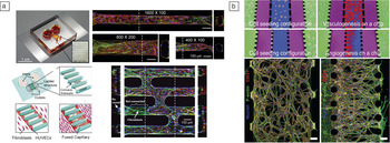
Yeon et al. established an array of perfusable microvessels on a single microfluidic chip.Reference Yeon, Ryu, Chung, Hu and Jeon69 After the fibrin gel was selectively patterned in the middle of a PDMS channel array as a provisional matrix for angiogenic sprouts, ECs were attached to both sides of the fibrin gel wall (Figure 3a). The ECs were stimulated to form sprouts by co-culture with fibroblasts. After 3–4 days, the endothelial sprouts from both sides of the middle channel connected to form lumenized perfusable microvessels. Various dimensions or networks of microvessels can be engineered by fabricating different PDMS geometries.
Various methods for recreating in vitro microvessels utilizing morphogenic properties of endothelial cells (ECs). (a) An array of microvessels was formed inside the microfluidic chip by utilizing the angiogenic properties of ECs (top left). HUVECs were attached on both sides of the fibrin gel patterned inside the microchannel (bottom left). HUVECs were co-cultured with lung fibroblasts to stimulate the angiogenic process. The HUVEC sprouts from both sides were fused to form perfusable microvessels. Various shapes and network of the microvessel were formed by controlling the geometry of the microchannel (top and bottom right). Cross-sectional views at the dotted lines are shown next to respective micrographs. (Reproduced with permission from Reference 69. © 2012 Royal Society of Chemistry.) Scale bars, except where indicated otherwise, are 100 μm. (b) A microvascular network was formed by utilizing the vasculogenic or angiogenic process (upper and lower part of schematic, respectively) of HUVECs. HUVECs were either encapsulated in or attached to the fibrin gel (blue channels) and co-cultured with lung fibroblasts (green channels) to stimulate microvessel formation. The microvessel network was opened to both media channels (purple channels) for perfusion by media. Immuno-fluorescent images show the engineered microvessel network formed by the vasculogenic and angiogenic (left and right in micrographs, respectively) process. Scale bars, 100 μm. (Reproduced with permission from Reference 70. © 2013 Royal Society of Chemistry.) Note: PDMS, polydimethylsiloxane; HUVECs, human umbilical vein endothelial cells.

Kim et al. used two approaches to generate a perfusable microvessel network:Reference Kim, Lee, Chung and Jeon70 filling the middle channel with a fibrin gel and EC mixture for the vasculogenic process, and filling the middle channel with fibrin gel and attaching ECs on the gel wall for the angiogenic process (Figure 3b). Fibroblasts were co-cultured, and ECs were stimulated to form a blood vessel network by the secreted factors of the fibroblasts. After the perfusable microvessel network is formed in 4–5 days, various aspects of vascular biology were investigated, including vascular permeability, alignment of F-actin, expression of EC nitric oxide by stimulation from media perfusion, and interactions with other cell types, such as pericytes and immune cells.
Hsu et al. demonstrated another microfluidic system in which an array of vascular networks was engineered.Reference Hsu, Moya, Hughes, George and Lee71 Based on an analogy between an electrical circuit and a microfluidic network, this system applied a physiological level of media flow into EC/fibroblast co-cultured microvessel chambers. ECs were mixed with fibroblasts in the fibrin gel to stimulate the vasculogenic process, and interstitial flow by a pressure drop across the vessel chamber applied a physiological level of interstitial flow, mimicking the in vivo microenvironment.
In contrast to methods that attach ECs to hydrogel walls, the main advantage of these methods is that they are based on a more in vivo-like vessel formation process by utilizing the angiogenic/vasculogenic properties of ECs. Moreover, these schemes, particularly those for the vasculogenesis methods, can be applied to tissue engineering. As the degree of biological similarity between the microvessels produced using this scheme and the in vivo condition has not been fully realized, future work should concentrate on investigating the detailed biological relevance of vessels produced using this scheme compared to the endothelial-cell-attachment scheme. Moreover, the shape and dimensions of these microvessels cannot be precisely controlled, unlike the EC-attachment scheme on a micropatterned hydrogel. Refining the scheme for engineering defined microvessel structure is required for future studies that use a media perfusion system to apply a physiological level of flow to the microvessels.
Vascularized organs on a chip
Numerous in vitro models that mimic certain characteristics of human organs have been established. However, as blood vessels closely interact with other organ systems, vascularizing in vitro organ models will greatly improve their physiological relevance. This will lead to better prediction of actual organ responses in the preclinical stage of the drug discovery process. We next summarize recent efforts aimed at establishing vascularized in vitro organ models on a chip and their applications.
Heart
Chiu et al. engineered in vitro functional cardiac tissue by culturing vascularized cardiomyocytes (heart muscle cells) on a PDMS chip (Figure 4a).Reference Chiu, Montgomery, Liang, Liu and Radisic72 Arterial and venous explants (removal of living tissue from the natural site of growth and placement in a medium for culture) from mice were cultured on a PDMS structure with a collagen-chitosan hydrogel coating for vascularization. The hydrogel was encapsulated with Tβ4, which is an angiogenic and cardioprotective peptide, and the PDMS was micropatterned to promote vascularization by the explants. Cardiomyocytes were cultured after the hydrogel-coated PDMS substrate was vascularized for three weeks. Vascularized cardiac tissue was successfully modeled by culturing the cardiomyocytes for an additional seven days. The vascularized cardiac tissue showed closer characteristics with the actual heart in terms of excitation threshold, sarcomere organization, and cell–cell junctions compared to cardiomyocytes without vascularization. This work represents the importance of vascularization for engineering in vitro organ models.
Recent representative in vitro vascularized organ models. (a) Cardiomyocytes were cultured on vascularized polydimethylsiloxane (PDMS) substrates to model cardiac tissue. Hydrogel-coated PDMS was vascularized with arterial and venous explants for three weeks. Cardiomyocytes were cultured on the vascularized substrates for an additional seven days to model the cardiac tissue. The micrograph shows vascularized cardiomyocytes (purple regions) cultured on the PDMS substrate. The capillary outgrowths from the vascular explant (asterisk) are indicated by arrows. Scale bar, 200 μm. (Reproduced with permission from Reference 72. © 2012 National Academy of Sciences, USA.) (b) In vitro vascularized lung model by co-culturing epithelial cells (cells that cover the side of the lung in contact with air) with endothelial cells (ECs) on a PDMS membrane to generate the alveolar-capillary interface (left). Improved lung function was modeled by applying cyclic mechanical strain via vacuum (middle) to the cells with air-liquid interface culture. Scale bar, 25 μm. (Reproduced with permission from Reference 73. © 2010 The American Association for the Advancement of Science.) (c) Cancer angiogenesis was modeled on a microfluidic chip. ECs were coated on a fibrin gel wall, and cancer cells were loaded at the opposite side of the EC attached region to stimulate cancer angiogenesis. Micrographs show the angiogenic sprouts with branching tip cells (white arrowhead) and convoluted tubules (yellow arrowhead) induced by secretion of cancer on days 2 and 4, respectively. Scale bars, 20 μm. (Reproduced with permission from Reference 70. © 2013 Royal Society of Chemistry.) (d) A cancer intravasation model was established using EC-attached hydrogel and cancer cells. The cancer cells were cultured in the red region, and they migrated toward the EC monolayer on the hydrogel wall in the green region to intravasate through the ECs (left). The black arrow indicates a y-junction between the two regions, which is required for precise control of chemical gradient in the channels. The micrograph shows intravasating cancer cells (red) through the ECs (green). Blue indicates nuclei of the cells. Scale bars, 2 mm (left) and 30 μm (right), respectively. (Reproduced with permission from Reference 76. © 2012 National Academy of Sciences, USA.) Note: Tβ4, thymosin β4; VE cadherin, vascular endothelial cadherin; CD31, also known as PECAM-1 (platelet endothelial cell adhesion molecule); F-actin, a protein that constitutes actin filaments of the cells.

Lung
Huh et al. reconstituted a functional alveolar-capillary interface of the human lung on a chip (Figure 4b).Reference Huh, Matthews, Mammoto, Montoya-Zavala, Hsin and Ingber73 The chip consists of three PDMS layers—an upper and lower PDMS layer sandwiching a porous PDMS membrane. The side channels were pumped with PDMS etchant, which is composed of tetrabutyl-ammonium fluoride and N-methylpyrrolidinone, to etch a redundant PDMS membrane. A monolayer of ECs is cultured on the bottom layer of the membrane for vascularization. Additionally, epithelial cells are cultured on the upper side of the PDMS membrane in the middle chamber to mimic the alveolar-capillary interface. An air–liquid interface culture was performed to mimic the microenvironment of the in vivo alveolar-capillary interface, which pumps media into only the endothelial layer; the epithelial layer (outer surface layer throughout the body) is exposed to air. The thin layer of surfactant that covers and stabilizes the epithelium is successfully produced by the environment, which closely resembles the in vivo alveolar condition. Furthermore, an in vivo-like inflammatory response and nanotoxicology were successfully modeled by this system using cyclic mechanical strain by applying a vacuum to the side chambers. Among the numerous in vitro models, this system is representative of pertinent combinations of materials (PDMS membrane, PDMS layers and etchant), mechanical elements (mechanical strain by the vacuum and media flow), and cell types (ECs and epithelial cells) to establish a vascularized in vivo-like organ system on a single chip.
Cancer
Cancer progression and metastasis has multiple stages. After they recruit surrounding blood vessels to supply additional nutrients and oxygen, cancer cells intravasate (penetrate) into a blood vessel, circulate in the blood or lymph vessels, and extravasate (escape) into a secondary site.Reference Bergers and Benjamin74,Reference Sahai75 Several microfluidic chip models have been developed to mimic vascularized cancer sites and to study detailed biological aspects of each stage during cancer progression.
Kim et al. developed a microfluidic chip to investigate cancer angiogenesis (Figure 4c).Reference Kim, Lee, Chung and Jeon70 Fibrin gel was injected into the middle channel as a provisional matrix for endothelial sprouting. Cancer cells were mixed with fibrin gel and introduced into the side channel, and ECs were attached to the fibrin gel wall in the middle channel. ECs that attach on the fibrin gel wall are regarded as a pre-existing vessel wall, which could be a model for a vascularized cancer site during the angiogenic process in cancer progression. The endothelial sprouts from the “pre-existing vessel” promoted by the secretion of cancer cells had aberrant morphology compared to fibroblast sprouts, which is a general characteristic of cancer vasculature.
Zervantonakis et al. developed a microfluidic chip that uses hydrogel and ECs to mimic the blood vessel wall to study cancer intravasation (Figure 4d).Reference Zervantonakis, Hughes-Alford, Charest, Condeelis, Gertler and Kamm76 After attaching a monolayer of ECs to the hydrogel, cancer cells were introduced at the basal region of the ECs. The cancer cells migrated toward the endothelial region until they intravasated into the endothelial monolayer. Furthermore, the effect of tumor-associated macrophages and TNF-α (tumor necrosis factor α, a protein involved in the inflammatory response) treatment on cancer intravasation was also demonstrated. This model shows simple mimicry of a vascularized cancer site during intravasation by using an EC monolayer co-cultured with cancer cells.
Blood-brain barrier
The blood-brain barrier (BBB) is a highly regulated barrier of brain capillaries with a large number of tight junctions that block molecules entering from the blood circulation to protect the central nervous system. Astrocytes are one of the major elements of the BBB and perform their functions through their interactions with brain ECs.Reference Lai and Kuo77 A microfluidic chip was developed to functionally model the BBB.Reference Booth and Kim78 The chip consists of two PDMS channel layers (upper and lower), separated by a polycarbonate membrane and two electrodes for measuring vascular permeability. An endothelial monolayer was cultured on the upper side of the membrane to mimic blood vessel structure. Astrocytes were cultured on the lower side of the membrane to interact with ECs and form the functional BBB. The upper layer was constantly perfused with media to mimic blood flow. The results showed that the permeability of the endothelial monolayer increased significantly due to media perfusion and the astrocyte culture, indicating the functional characteristics of the BBB. They also showed real-time permeability recovery after treatment with histamine, which is a vascular permeability factor. This system is a robust vascularized in vitro organ model that uses several materials, including PDMS and polycarbonate membrane, with a co-culture of astrocytes and media perfusion.
Material considerations for establishing organs-on-chips
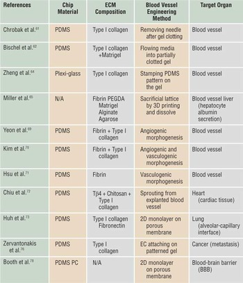
Until now, numerous materials (e.g., silicon, glass, elastomers, thermoplastics) have been used to fabricate microfluidic devices.Reference Ren, Zhou and Wu79 Among these, we need to select those materials that meet several essential requirements for constructing organs-on-chips: (1) The materials should not be toxic to cells for long-term culture in the chips. Any toxic reagents or materials should be completely removed before introducing cells inside the chip. (2) The materials should be gas permeable enough to supply a sufficient amount of oxygen to the cells inside the chip. If less permeable materials such as glass or thermoplastics are used, the oxygen supplement issue should also be solved. Opening the cell region into air or partially using gas permeable materials can be solutions. (3) The materials should be optically transparent for microscopic imaging. (4) The main purpose of establishing organs-on-chips is for in vitro high-throughput drug screening. Therefore, costs of the materials and the chip fabrication process are important issues for material selection of organs-on-chips. (5) Properties of the materials should be carefully considered to model specific properties of organs. For example, Huh et al. used an elastomer (PDMS), which has a comparably low Young’s modulus, to model the stretching motion of lungs during the respiratory process.Reference Huh, Matthews, Mammoto, Montoya-Zavala, Hsin and Ingber73 As shown in Table I, most organs-on-chips are made of PDMS, as it is biocompatible, oxygen permeable, optically transparent, easy to use, and inexpensive.Reference McDonald and Whitesides80 Although PDMS satisfies most essential requirements of organ-on-chip materials, it also has several limitations.Reference Mukhopadhyay81 First, the surface of PDMS typically absorbs biomolecules, which could lead to inaccurate results of dose-dependent chemical experiments. Furthermore, compared to plastic based materials, it is difficult to produce a PDMS-based chip with conventional mass-productive systems, which can hamper the commercialization and standardization of the chips as an assay platform. Therefore, research needs to be focused on finding new materials that can cover all the requirements of organ-on-chip materials.
Materials and characteristics of various in vitro blood vessel & organ models.

Note: PDMS, polydimethylsiloxane; PC, polycarbonate; PEGDA, polyethylene glycol diacrylate; ECM, extracellular matrix; Tβ4, thymosin β4; EC, endothelial cell; BBB, blood-brain barrier.
In the human body, cells are exposed to a 3D microenvironment with various components of the ECM. Mimicry of the ECM composition in the target organ is essential to achieve higher biological similarity of organs-on-chips, as the microenvironment of cells can intensely affect the behavior of cells.Reference Griffith and Swartz82 As shown in Table I, natural ECMs (collagen or fibrin) are the most commonly used materials to mimic the 3D microenvironment of target organs. These natural ECMs are commercially available and provide numerous types of chemical and physical cues that present in vivo. However, as naturally derived ECMs have various unknown factors that can affect cell behavior, we cannot selectively isolate the effects of specific factors. Additionally, the use of components that closely mimic the specific ECM properties of target organs is essential for designing organs-on-chips with higher biological similarity. Therefore, synthetic ECMs can be a promising solution, as they can be tailored to mimic specific ECM properties in target organs.Reference Lutolf and Hubbell83 To date, synthetic ECMs have not fully been established enough to be commonly used in organ-on-chip systems. Future work in biomaterial science should be concentrated on tailoring synthetic ECMs enough to substitute natural ECMs for more specific and precise mimicry of target organs.
Conclusion
The endothelium plays essential roles in cancer progression, inflammation, and complications of microangiopathy. Furthermore, the endothelium has specific functions in organs, such as a selective barrier in the brain and kidney, provision of tissue-specific microenvironments in bone marrow and tumors, and regeneration in the liver and lungs. Therefore, vascularization of in vitro organ models is crucial to mimic in vivo organ conditions.
Methods for engineering in vitro microvessels can be categorized into (1) attaching endothelial cells (ECs) to micropatterned hydrogel walls and (2) utilizing the morphogenic properties of ECs. The geometric characteristics of microvessels engineered by attaching ECs can be controlled precisely. Microvessels formed by utilizing the natural morphogenic properties of ECs are expected to have more in vivo-like properties, while their geometric characteristics cannot be controlled precisely. Therefore, establishing more precise and geometrically controlled microvessel engineering methods and illuminating the biological similarities with actual blood vessels in vivo is required in future studies.
Recent studies have used various methods to establish vascularized in vitro organ models. These models combine various elements (e.g., mechanical stress, various materials, geometrical structures) with vascularization to mimic target organ characteristics. The vascularized models reveal more physiological characteristics, indicating the importance of vascularization for in vitro organ models.
We have reviewed the importance of microvasculature for organs-on-chips, with methods for engineering microvessel and vascularized organs-on-chips, and material considerations for establishing organs-on-chips. We postulate that one of the main considerations for engineering more physiologically relevant organs-on-chips is selecting pertinent materials to construct the extracellular matrix (ECM). Most of the methods introduced in this review used naturally derived ECM components, such as collagen or fibrin, as the basic material for constructing the ECM. However, utilizing synthetic materials to recreate the ECM has another advantage, as it can be engineered to recapitulate target organ-specific characteristics in vitro. Therefore, establishing organs-on-chips with greater biological relevance will lead to further biological research if combined with vascularization and selection of pertinent materials for construction of the ECM.
Acknowledgment
This work was supported by the Pioneer Project (2013–041859) and the global PhD Fellowship Program (NRF-2012-Global Ph.D. Fellowship Program) through the National Research Foundation funded by the Ministry of Education, Science and Technology of Korea.