
I. INTRODUCTION
The development of efficient and scalable biotechnological methods of preparation of highly nanostructured materials represents a hot topic of current nanotechnology. In fact, many natural structures can be viewed as three dimensional (3-D) organic and inorganic nanoarchitectures having reproducible and complex hierarchical morphologies that are still difficult to achieve with human technologies. Actually, many living organisms have evolved their metabolism to optimize the structure of some components, from the nano- to the microscale, to confer upon them multifunctional properties that are fundamental for their growth and survival. Among these organisms, diatoms have attracted special attention.
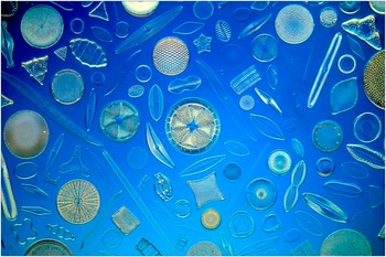
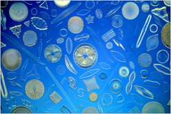
Diatoms are unicellular eukaryotic microalgae (Fig. 1) ubiquitously distributed in oceans, freshwater habitats, soils, and inhospitable surfaces, and thus have catched the attention of many classes of scientists thanks to the beautiful ornate nanoarchitecture of their silica shell walls. Since the end of 19th century, microscopy has disclosed the extraordinary features of such small organisms, and nowadays materials scientists are exploiting their intriguing potentialities as nanomaterials. Reference Werner1,Reference Wetherbee2
Artistic assembly of different species of diatoms (Klaus Kemp, master of the Victorian art of diatom arrangement).

Diatoms are essential to all life on earth: their origin dates back to the Jurassic period and, for this reason, their microfossils (diatomaceous earth or diatomite) are useful indicators for monitoring paleoceanographic events and environmental changes in lakes or oceans. Reference Smol and Stoermer3 As other photosynthetic algae, they use light and carbon dioxide as an energy source to produce their own carbohydrates.
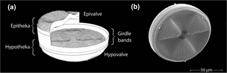
Diatoms are unique in the unicellular algae world and appealing for the materials research community since they bear nanostructured rigid cell walls, called frustules, made of amorphous hydrated silica. The mesoporous highly ordered structure of frustule has been optimized over millions years of evolution to constrain and enclose the eukaryotic protoplast, protecting it from unfavorable environmental conditions and from dangerous light wavelengths and, at the same time, to promote nutrient uptake and secretion of cellular products through an intricate pattern of pores and slits on the frustule surface. The biosilica shell has a pill-box like structure composed of two valves (the epitheca and the hypotheca), connected by lateral rings (girdles) that confer flexibility to the structure (Fig. 2). Reference Riedrichs, Maier and Hamm4
Structure of a diatom cell wall. (a) Schematic cross-section of a box-like diatom frustule in a centric species. (b) SEM picture of a centric diatom. Reproduced with permission from Wiley. Reference Riedrichs, Maier and Hamm4

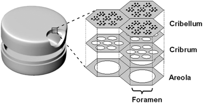
Both valves and girdles have a species-specific “layer by layer” hierarchical architecture in which every layer has a periodic nanotexturized topography with an ordered disposition of pores (areolae) whose diameter increases passing from the internal to the external layers. In spite of the fluctuation in higher taxonomic categories, a classification of diatoms can be made on the basis of frustule shape and symmetry of pore patterns: centric are all diatoms whose frustule has radial symmetry, while pennate are those in which valves are bilaterally symmetric. Reference Williams and Kociolek5 A schematic 3-D cross-section of a frustule of centric diatoms is shown in Fig. 3: the hierarchical organization of porous plates (foramen, cribrum, and cribellum) involves pore diameters with different patterns that range from nanometers to micrometers.
Hierarchical organization of biosilica layers in a centric diatom valve. The honeycomb-like vertical chambers in the inner layer are called areolae. The outer siliceous thin layer membrane (cribellum) consists of very small pores. Reproduced with permission from Royal Society of Chemistry. Reference Losic, Mitchell and Voelcker58

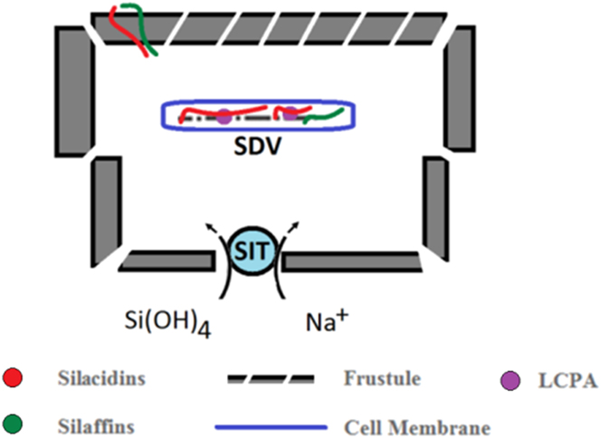
Hence diatoms can be regarded as microscopic biofactories of nanostructured biosilica, that is generated in vivo by a complex biomineralization process Reference Hildebrand and Buerlein6 occurring in specialized intracellular compartments, named the silica deposition vesicles (SDVs), Reference Thamatrakoln, Alverson and Hildebrand7 which act as cellular reaction vessels (Fig. 4). Silicon is uptaken by diatoms in the form of orthosilicic acid Si(OH)4, which is abundant in almost every water habitat. Si(OH)4 penetrates into the cell cytoplasm by specific silicic acid transporter proteins (SITs). SITs are membrane proteins containing 10 transmembrane domains and they directly bind Si(OH)4 via glutamine residues present in two adjacent domains, allowing the Si(OH)4 transport into cytoplasm and, more specifically, into the SDV. Reference Hildebrand, Dahlin and Volcani8 Inside the SDV, Si(OH)4 is converted into a silica network (SiO2)n in the presence of long chain polyamines (LCPAs), specific proteins (silaffins), and polyanionic peptides (silacidins) (Fig. 4). Reference Kröger, Deutzmann, Bergsdorf and Sumper9–Reference Parmisky and Golokhvast13 Understanding the processes involved in biomineralisation can eventually enable production of nanostructured silica by bio-mimetic methods with minimal environmental impact. Actually, silaffins and silacidins have been extracted from the silica walls and used to induce in vitro precipitation from silica acid solution of silica nanostructures such as spheres, rods, or platelets. Reference Kröger, Deutzmann and Sumper14–Reference Jin and Levi17 The study of the mechanisms of the in vivo construction of 3D hierarchically nanostructured silica in diatoms can offer useful inspiration in the synthesis of novel magnetic, electronic, pharmaceutical, or adhesive materials on a nanometer scale.
Schematic representation of intimate interaction between organic diatom machinery and silica components.

A very high number of diatom species has been estimated worldwide thus far, being of the order of 2 × 10⁵. Despite the fact that their classification and taxonomy are still under debate, Reference Mann18,Reference Sims, Mann and Medlin19 mesoporous silica shells of these microalgae are attractive materials thanks to their biocompatibility, high surface area, tunable pore size, and versatile surface chemistry, enabling their possible applications in various fields such as sensing, drug delivery, optics, and photonics. Reference Drum and Gordon20–Reference Fuhrmann, Landwehr, El Rharbi-Kucki and Sumper22 The study of properties and nanotechnological potentialities of monodisperse nanostructured frustules actually represents a very intriguing research field, going beyond the long time and well-known practical use of morphologically heterogeneous diatomaceous earth in chemistry, agriculture, and industry. Reference Martin23–Reference Bennett, Yee, Rhee and Cheng27
II. DIATOM FRUSTULES AS PHOTONIC CRYSTALS
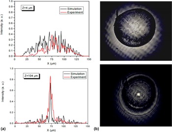
Diatoms’ biosilica shells can be regarded as “nanostructured microboxes” acting as photonic crystals, since the regular disposition of pores in superimposed layers of biosilica induces a hierarchically ordered periodic pattern of high and low refractive index. Reference Fuhrmann, Landwehr, El Rharbi-Kucki and Sumper22 Periodicity as well as morphology and size of these pores strictly depend on the algal species and enable potential application of frustules as natural micro-lenses, optical filters, polarizers, waveguides, beam splitters, couplers, and laser cavities. Indeed, as a common feature of regularly patterned systems, light propagation through the valve not only depends on the interaction with matter but also on the spatial order of the periodic structure. The light focusing ability of valves of the centric Coscinodiscus wailesii diatoms was first investigated by De Stefano et al. Reference De Stefano, Rea, Rendina, De Stefano and Moretti28 In particular, a radial symmetry of pores with almost perfect hexagonal pattern was observed, this resembling the symmetry of some man-made photonic crystal fibers and phase-locked arrays of optical fibers and lasers. Illuminating the valve by a red laser beam with 100 μm spot size, a coherent superimposition of the light scattered by pores is responsible for focusing the beam to less than a 10 μm spot size at a distance of 104 μm after light transmission through the valve (Fig. 5).
(a) Transmitted light intensity distribution at the distance (z) of 4 μm (top plot) and 104 μm (bottom plot): the red curve is the experimental recorded one, the black one is the numerical estimated. (b) Diatom surface images recorded by a CCD camera at the two distances. Reproduced with permission from OSA publishing. Reference De Stefano, Rea, Rendina, De Stefano and Moretti28

A large scale and low cost production can be envisaged for such natural micro lenses ensuring a precise and reproducible nanostructure simply controlled by the biological metabolism of the selected diatom species. Moreover, thanks to their high mechanical resistance, the valves can be easily handled by standard micro-needles or micro-tips, and mounted on top of optical fibers or in the vertical cavity surface of an emitting laser source, thus affording lensed fibers or focusing laser beams.
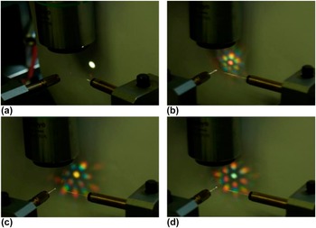
For instance, white light of a supercontinuum broadband (400–1700 nm) laser source was used to irradiate spot regions of ∼20 μm diameter on the surface of a single Coscinodiscus wailesii diatom valve attached to the end of a supporting optical fiber. In this way, white light diffraction and transmission were investigated as functions of the spacing of pores in different regions of the valve. As shown in Fig. 6, the periodic pore structure of the valve is responsible for the generation of colorful patterns due to the diffraction of different applied wavelengths at different angles. In particular, while a white light circular spot is observed when the valve is not in the light path [Fig. 6(a)], the hexagonal light scattering pattern of the beam crossing the valve results from the hexagonal periodic pore valve structure and the color of the central spot changes by changing the irradiated spot region on the valve surface [Figs. 6(b)–6(d)]. Reference Noyes, Sumper and Vukusic29,Reference Kieu, Li, Fang, Cohoon, Herrera, Hildebrand, Sandhage and Norwood30
Optical images of the diffraction patterns obtained from the different regions of a Coscinodiscus wailesii valve: (a) no diatom is present in the beam path, (b–d) the frustule valve is in the beam path. Reproduced with permission from OSA publishing. Reference Kieu, Li, Fang, Cohoon, Herrera, Hildebrand, Sandhage and Norwood30

The biosilica valves have also been investigated for their potential application as optical filters. Indeed, the transmission of incident light by their nanostructure is strongly wavelength dependent, being more pronounced for the red light rather than for the blue and green wavelengths Reference Yamanaka, Yano, Usami, Hayashida, Ohguchi, Takeda and Yoshino31 ; this is likely due to the living diatoms that evolved their ordered biosilica shells to reflect the noxious UV and blue lights, thus protecting the inner protoplasm.
A recent study carried out in 2014 also demonstrated that single valves of Arachnoidiscus diatoms are able to confine monochromatic radiations of the visible light in a series of hotspots whose position along the optical axis is strictly dependent on wave length. Reference Ferrara, Dardano, De Stefano, Rea, Coppola, Rendina, Congestri, Antonucci, De Stefano and De Tommasi32 This feature may disclose new frontiers in optics, due to the possibility to employ diatom valves as micro-monochromator elements.
III. IN VITRO AND IN VIVO CHEMICAL MODIFICATION OF DIATOM BIOSILICA
Integration of functional molecules suitable for various applications into diatom biosilica shells can be pursued by two main approaches, the first based on in vivo stable incorporation of molecules into the bulk of the biosilica nanostructured architecture, while the second approach consists in affixing, by condensation reactions, functional molecules on the surface of silica shells after their purification by removal of the organic matrix of the cell. Both approaches have been initially used to stain frustules with dyes and to study diatom biosilycification mechanism and frustules morphology.
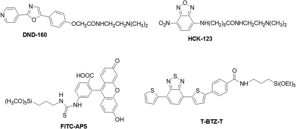
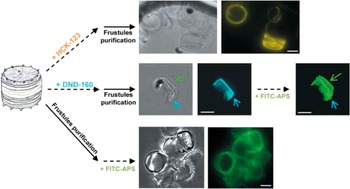
For instance, Desclés et al. reported a selective in vivo incorporation of the blue DND-160 and the yellow HCK-123 dyes (Fig. 7) into the biosilica shells of various diatoms and demonstrated both dyes’ resistance to harsh acid oxidative treatment with potassium permanganate and sulfuric acid used to purify the fluorescent frustules from the organic autotrophic matter (Fig. 8). Reference Desclés, Vartanian, Harrak, Quinet, Bremond, Sapriel, Bibette and Lopez33
Chemical structures of some dyes used for frustule staining.

Frustules staining with DND-160 (cyan), HCK-123 (yellow) and FITC-APS (green). Bars, 5 μm. Reproduced with permission from Wiley. Reference Wachter, Lechleitner, Artner-Dworzak, Hausen, Jarosch, Widner, Patsch, Pfeiffer and Fuchs24

The same authors also demonstrated that using a trimethoxysilyl substituted green dye (FITC-APS, Fig. 7) it is possible to stain in vitro previously isolated natural or pre-stained frustules by a condensation reaction of the terminal silyloxy moiety of the dye with the silanolic groups on the silica surface (Fig. 8).
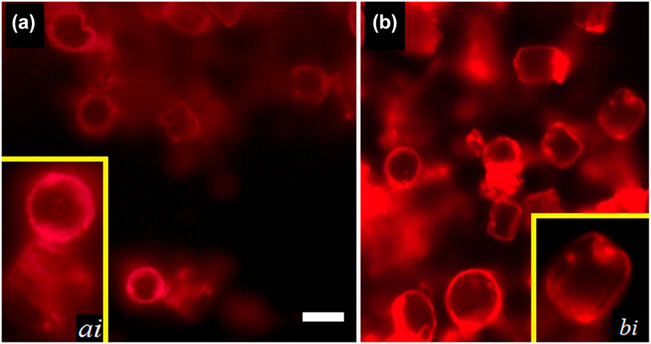
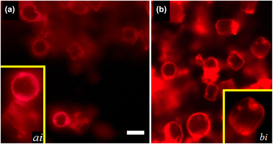
Such in vitro staining method is quite general and, according to our recent work, Reference Vona, Lo Presti, Cicco, Palumbo, Ragni and Farinola34 it can be applied to triethoxysilyl functionalized emitting molecules with π-conjugated backbones, such as the arylene-type red dye T-BTZ-T in Fig. 7. By this approach, we achieved uniformly stained nanostructured silica shells isolated from Thalassiosira weissflogii diatoms by covalent binding of the dye on the frustule surface (Fig. 9).
Two-dimensional fluorescence microscopy of two samples of dye stained frustules with different densities: (a) 0.1 mg/mL and (b) 1.0 mg/mL. Insets (ai, bi): focus on single diatom frustules. Bar, 10 μm. Reproduced with permission from Materials Research Society. Reference Vona, Lo Presti, Cicco, Palumbo, Ragni and Farinola34
Besides their suitability for the investigation of silica biomineralization mechanism in living diatoms, in vivo and in vitro chemical modification methods of diatoms shells with organic dyes may also be profitable routes to new luminescent nanostructured materials with potential application in photonics, due to the combined effect of the dye photoluminescence and the photonic crystal behavior of frustules.
This perspective was mentioned by Fuhrmann et al. after the in vivo incorporation of rhodamine laser dyes, into frustules of Coscinodiscus granii and Coscinodiscus wailesii diatoms. Reference Kucki and Fuhrmann-Lieker35 A blue-shift of the emission spectra of these dyes was, for instance, observed in the resulting luminescent biosilica, due to the influence of the biosilica nanostructure on the dye photoluminescence.
IV. DIATOM FRUSTULES IN DRUG DELIVERY AND BIOSENSING
In the last decades, mesoporous silica nanoparticles have been synthesized and applied for drug delivery applications. Although these materials can be produced with different size, morphology, and tunable properties related to the specific applications, their synthesis involves complex chemical processes, and toxic and expensive reagents. Diatoms’ silica shells possess a combination of structural, mechanical and chemical features that can offer several advantages over existing synthetic silica nanoparticle delivery systems: the frustules can be easily functionalized, protected, and engineered for controlled drug loading and successive delivery through the nanopores.
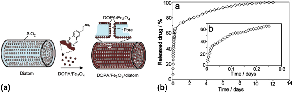
Very recently, Losic et al. demonstrated the potential of diatom silica as a promising biomaterial for the delivery of therapeutic agents to a desired site. Reference Losic, Yang, Sinn, Simovic, Thierrya and Addai-Mensaha36 In particular, for hydrophobic drugs, which are poorly soluble in blood and thus difficult to be directed to their target, a controllable drug delivery system was developed by functionalization of the frustule surface with dopamine-modified iron oxide nanoparticles; these magnetic diatoms were loaded with indomethacin, a conventional antiinflammatory drug and the drug release of indomethacine from the DOPA/Fe3O4 modified diatoms was investigated (Fig. 10).
Functionalization of diatom surface with dopamine modified iron oxide nanoparticles to obtain magnetically guided drug delivery microcarriers (a); drug release of indomethacin from DOPA/iron oxide modified diatoms over (a) 12 days and (b) 6 h. Reproduced with the permission from the Royal Society of Chemistry. Reference Losic, Yang, Sinn, Simovic, Thierrya and Addai-Mensaha36

Diatom based carrier systems for both poorly soluble and water soluble drugs have been suggested for oral and implant applications, with tunable drug delivery behavior by surface functionalization. Modification of the chemical (hydrophobic and hydrophilic) properties of the diatom surface was in fact demonstrated as a powerful tool to control the drug loading and release characteristics, thus improving the drug delivery applications. Reference Sinn, Bariana, Yu, Addai-Mensah and Losic37 Further to the need of producing biosilica-based nanomaterials (and not only micromaterials), nanoparticles for drug delivery purposes were directly obtained by processing diatomite. These nanoparticles exhibited no specific and strong cytotoxic effect in vitro on 3T3 cells (fibroblast-like cells useful for keratinocytes in vitro model). The low cytotoxicity depends on the cleaning and administration conditions, together with biosilica–cells interface. Reference Le, Bonani, Speranza, Sglavo, Ceccato, Maniglio, Motta and Migliaresi38
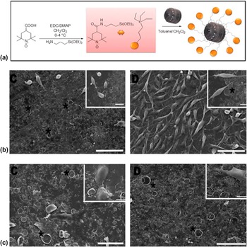
In our previous work, we have proposed the nanostructured biosilica of Thalassosiria weissflogii diatoms as a natural, easily available, multifunctional material for regenerative medicine applications. Reference Cicco, Vona, De Giglio, Cometa, Mattioli-Belmonte, Palumbo, Ragni and Farinola39,Reference Vona, Urbano, Bonifacio, De Giglio, Cometa, Mattioli-Belmonte, Palumbo, Ragni, Cicco and Farinola40 The covalent immobilization of the TEMPO radical scavenger on the diatom mesoporous silica surface has been combined with the drug delivery of the antibiotic ciprofloxacin. Further, fibroblast and osteoblast-like cells growth has been tested on TEMPO functionalized frustules showing that biosilica from diatoms can represent a smart support for cell growth and tissue regeneration (Fig. 11).
(a) Synthesis of the TEMPO-APTES derivative and subsequent immobilization on frustule biosilica; (b) SEM images of L fibroblasts seeded onto FT-coated (C, D) substrates for 7 days; several mitotic events (arrows) are evident. The FT-coated substrate surface is covered by diatom shells (*); (c) SEM images of MG63 osteoblast-like cells seeded onto FT-coated (C, D) substrates for 7 days showing an almost uniform layer of cells. Diatom shells (*). Scale bars: 50 µm; inset scale bars: 10 µm. Reproduced with permission from Wiley. Reference Cicco, Vona, De Giglio, Cometa, Mattioli-Belmonte, Palumbo, Ragni and Farinola39

In a very recent work, nanoporous silica-based materials have been genetically engineered, leading to biosilica frustules as versatile ‘backpacks’ for the immuno-targeted delivery of poorly water-soluble anticancer drugs to tumor sites. Reference Delalat, Sheppard, Ghaemi, Rao, Prestidge, McPhee, Rogers, Donoghue, Pillay, Johns, Kroger and Voelcker41
In addition to applications in drug delivery in situ, innovative biosilica-based materials have been also envisaged as fillers or as injectable substitutes to fit bone defects with irregular or complex shape and size. Reference Bonilla, Trujillo, Demirdögen, Perilla, Elcin and Ribelles42,Reference Wang, Wang, Draenert, Albert, Schroder, Mailander, Mitov and Muller43
Diatom-based biosensors were developed by the immobilization of a biological sensing element (such as an enzyme, DNA, antibody, or cell) onto the frustule surface. The large surface area combined with the photonic crystal behavior has enabled their use as optical transducers of biomolecular interactions. Antibody-functionalized diatom biosilica frustules were investigated as microscale biosensor platforms for selective and label-free photoluminescence (PL)-based detection of immunocomplex formation by Rorrer’s research group. Reference Gale, Gutu, Jiao, Chang and Rorrer44 An antibody rabbit immunoglobulin G (IgG) was covalently attached to Cyclotella sp. diatom frustules by silanization, demonstrating that functionalization with the nucleophilic IgG antibody amplifies the intrinsinc blue PL of diatoms’ biosilica by a factor of six.
The photonic properties of Coscinodiscus wailesii diatom frustules were altered by growth in the presence of nickel sulfate in sea water, altering the size of the pores of the valves and quenching the intrinsic photoluminescence of the amorphous silica. Reference Townley, Woon, Payne, White-Cooper and Parker45 By monitoring the changes in the photoluminescence intensity of diatoms frustules, it is thus possible to detect traces of metal pollutants in aquatic environment. In recent years, rationally designed optical biosensors have demonstrated unprecedented detection sensitivity, compared with conventional colloidal plasmonic (light-metal interaction-based) sensors. Reference Lee, Lee, Lee, Lim, Seong, Lee, Chang, Oh and Choo46,Reference Haes and Van Duyne47 In particular, photonic crystals can increase the local field intensities and lead to additional Raman enhancement to improve the detection limit. The unique photonic crystal features of diatoms are capable of enhancing localized surface-plasmon resonances of self-assembled metal nanoparticles on the surface of diatom frustules, with a resulting significant enhancement of the Raman signals of the molecules adsorbed to the nanoparticles surface.
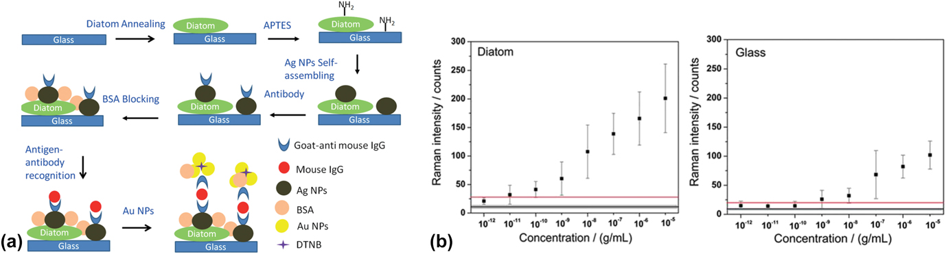
An interesting work reports on a SERS (surface-enhanced Raman scattering)-immunoassay sensor based on diatom photonic crystal platforms. Reference Yang, Zhen, Ren, Campbell, Rorrer and Wang48 In this immunoassay, silver nanoparticles were self-assembled onto diatom frustules and then functionalized with model antibody-goat anti-mouse IgG. The selectivity to detect specific antigens was tested by challenging the immunoassay sensors with complementary antigen (mouse IgG) and noncomplementary antigen (human IgG), respectively (Fig. 12). Immunoassay based on specific recognition between an antigen and a complementary antibody has become an essential and reliable tool for clinical diagnosis and disease detection.
Schematic diagram of the SERS immunoassay using diatom biosilica with self-assembled Ag NPs (a); dose–responses curves of Raman peaks (1331 cm−1) for IgG detection for diatoms’frustule surface and glass surface (b). Reproduced with permission from Wiley. Reference Yang, Zhen, Ren, Campbell, Rorrer and Wang48

The results showed that diatom frustules improve the detection limit of mouse IgG to 10 pg/mL, which is ∼100× better than conventional colloidal SERS sensors on flat glass.
V. DIATOM FRUSTULES FOR ELECTRONICS
In principle, frustules are unsuitable for electronics applications due to the insulating properties of silica. Yet, they have been used as nanostructured templates to generate a wide variety of semiconducting or conducting materials having a reproducible hierarchically ordered architecture. Various conversion protocols can be adopted to transform the biogenic silica shells into chemically different electroactive materials with preserved shell nanostructure and morphology. For instance, nanostructured materials for optoelectronics were obtained by feeding diatoms in vivo with metals such as germanium; in particular, Jeffryes et al. observed that upon the metabolic insertion of germanium into the shells of living Pinnularia sp. diatoms, frustule shape was preserved with the exception of the absence of a thin layer of biosilica containing a sub-array of 50 nm nanopores that normally underlies the layer of 200 nm pores in frustules [Figs. 13(a)–13(c)]. Reference Jeffryes, Solanki, Rangineni, Wang, Chang and Rorrer49,Reference Jeffryes, Gutu, Jiao and Rorrer50 Hence, after their isolation by removal of the organic protoplasm with hydrogen peroxide, the Ge-doped frustule valves strongly resemble bi-dimensional photonic crystal slabs. Electroluminescence in the UV-green (300–500 nm) and red-NIR regions (640–780 nm) was observed for a light emitting device fabricated by spin coating of the Ge-doped valves onto an ITO coated glass substrate and depositing a further layer of hafnium silicate dielectric and an Al cathode [Figs. 13(d) and 13(e)]. Conversely, no electroluminescence was observed for the reference diode fabricated by nondoped biosilica valves.
TEM images of (a) Pinnularia sp. valve and (b) pore array before metabolic insertion of Ge; (c) pore array after in vivo Ge insertion. (d) Scheme of light emitting diode and (e) electroluminescence spectra of diodes made with the Ge-doped and the bare valves. Reproduced with permission from Wiley. Reference Jeffryes, Solanki, Rangineni, Wang, Chang and Rorrer49

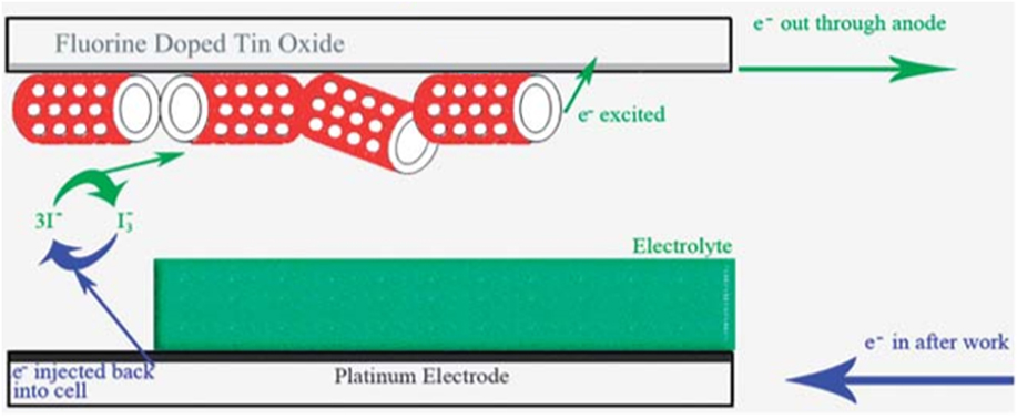
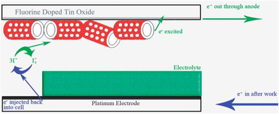
Diatom frustules have also found application in photovoltaics: titania modified biosilica shells can be used as active materials for photoanodes in dye sensitized solar cells (DSSCs). For this aim, the metabolic in vivo insertion of titania in diatoms’ biosilica was deemed unprofitable since the highest content of titania attainable by this method is quite low (3.7 wt%). A more efficient strategy recently proposed by Toster et al. is based on coating, with a titania layer, the surface of biosilica shells previously isolated from the organic cell matter by a method which preserves their nanostructure. Reference Toster, Iyer, Xiang, Rosei, Spiccia and Raston51 Such an in vitro chemical modification approach was carried out by activating via plasma treatment the silanolic groups on the surface of isolated frustules, to ensure their binding to in situ prepared titania nanoparticles. The thickness and connectivity of the titania film were improved by calcinating the coated frustules at 500 °C and repeating nanoparticles deposition three times on their surface. A DSSC photoanode was fabricated by absorbing a ruthenium dye sensitizer on the film of a blend of titania coated biosilica shells with ethyl cellulose and terpineol, which was deposited onto a fluorine doped tin oxide glass substrate. The solar cell architecture was completed by sealing this photoanode to a platinum counter electrode and back filling the cell with the iodide/tri-iodide electrolyte (Fig. 14). The presence of frustules in the photoanode induced a significant increase in energy conversion efficiency versus the more conventional titania cell (4.6 versus 3.5%, respectively), probably because light scattering and trapping ability of frustules increase the probability for photons to be absorbed by the sensitizer.
Titania coated diatoms shells in DSSC architecture. Reproduced with permission from Royal Society of Chemistry. Reference Toster, Iyer, Xiang, Rosei, Spiccia and Raston51
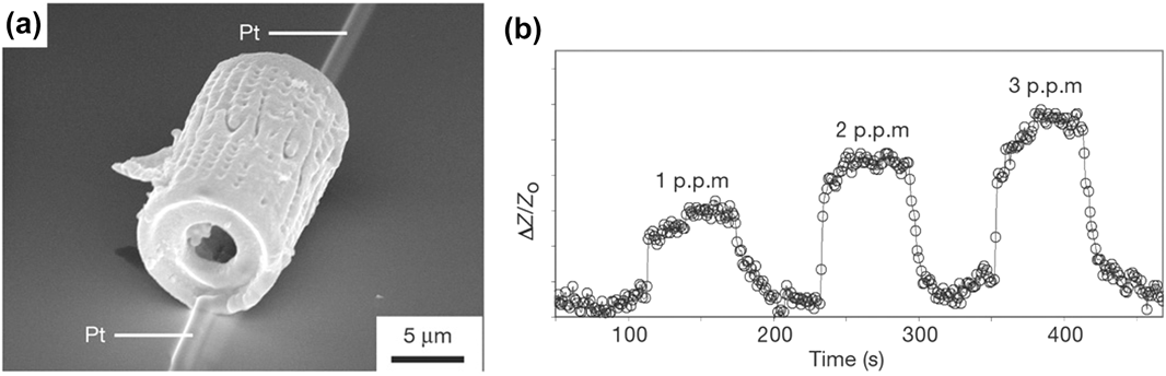
Another very intriguing approach to a wide variety of nanostructured materials with retained frustule morphology but completely different chemical composition relies on Bioclastic and Shape-preserving Inorganic Conversion (BASIC) processes. Reference Sandhage, Dickerson, Huseman, Zalar, Carroll, Rondon and Sandhage52 The BASIC reactions modify the chemical composition of the biosilica bulk of the shells converting them into nanostructured replicas with preserved morphology. For instance, the magnesiothermic reduction process is a BASIC approach useful to convert the silica diatom shells into silicon replicas, thanks to the reaction at high temperature of magnesium with the biosilica, leading to MgO/Si solid composites with retained frustule shape. Reference Sandhage, Dickerson, Huseman, Caranna, Clifton, Bull, Heibel, Overton and Schoenwaelder53 Then, these composites can be converted into pure silicon nanostructured shells by selective dissolution of magnesium oxide with HCl. This method was first applied by Sandhage et al. to Aulacoseira diatom cylindrical frustules, to generate silicon replicas exhibiting rapid changes of impedance upon exposure to NO gas, thus allowing fabrication of microscale single silicon frustule based gas sensors (Fig. 15). Reference Bao, Weatherspoon, Shian, Cai, Graham, Allan, Ahmad, Dickerson, Church, Kang, Abernathy, Summers, Liu and Sandhage54
(a) SEM image of a silicon replica connected to platinum electrodes in a simple gas sensor. (b) Electrical response of single silicon frustule sensor to NO gas. ΔZ is the impedance change upon exposure to NO gas and Z 0 is the sensor impedance in pure flowing argon. Reproduced with permission Nature Publishing group. Reference Bao, Weatherspoon, Shian, Cai, Graham, Allan, Ahmad, Dickerson, Church, Kang, Abernathy, Summers, Liu and Sandhage54

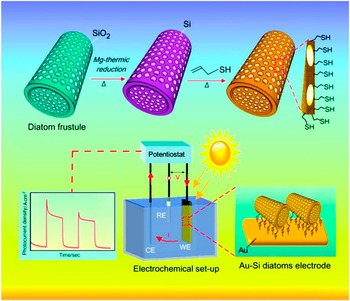
In 2014, Voelcker et al. also fabricated efficient photoanodes for photoelectrochemical energy conversion by anchoring, on gold-plated glass slides, silicon replicas obtained by magnesiothermic reduction of Aulacoseira frustules which were functionalized on the surface with thiol groups to favor their assembly on gold (Fig. 16). Reference Chandrasekaran, Sweetman, Kant, Skinner, Losic, Nann and Voelcker55
Application of thiol modified silicon replicas in photoelectrodes for solar energy conversion. Reproduced with permission from Royal Society of Chemistry. Reference Chandrasekaran, Sweetman, Kant, Skinner, Losic, Nann and Voelcker55

Efficient photoelectrodes for supercapacitors were also reported in 2015 by Losic et al. Reference Li, Xing, Huang, Li, Yu, Zhang and Losic56 In this case, the photoelectrodes are made of MnO2 replicas of frustules from various diatoms, and were obtained by a hydrothermal approach based on a preliminary heating of frustules dispersed into KMnO4 solution at 160 °C. This step allows the deposition of a nanostructured layer of MnO2 on the frustule surface. Then, the coated frustules were immersed into NaOH solution to induce selective dissolution of the underlying biosilica and recover of the MnO2 replicas. Electronically transparent graphene replicas with fine structural frustule features were also recently developed by chemical vapor deposition (CVD) of methane, and subsequent dissolution of silica with hydrofluoric acid (Fig. 17). Reference Pan, Lerch, Xu, Li, Chuang, Howe, Mahurin, Dai and Hildebrand57 These materials, besides being suitable for a in-depth diatoms’ morphogenesis investigation by deep electron microscopy of the internally visible replica morphology, represent very promising nanostructured materials for microelectronics.
SEM image of Aulacoseira sp. frustule (left). Scheme of the replication process (center) and SEM images of graphene replicas (a–c, right). Reproduced with permission from Nature Publishing group. Reference Pan, Lerch, Xu, Li, Chuang, Howe, Mahurin, Dai and Hildebrand57

Finally, diatoms frustules were used as templates for the fabrication of gold nanostructured films bearing an inverse frustule replicated nanostructured surface, according to a procedure which is much less expensive than lithography, commonly used for surface nanopatterning. In this case, gold was evaporated on a frustule coated silicon substrate and a glue coated glass was then sandwiched on the gold film. Finally the frustule-silicon substrate was removed to yield a nanopatterned gold surface. Reference Losic, Mitchell and Voelcker58 Such an approach allows fabrication of gold nanostructures with localized surface plasmon resonance (LSPR) properties enabling their applications in optical biosensors.
VI. CONCLUSIONS
Diatoms represent natural factories for large scale and low cost production of mesoporous biosilica suitable for a variety of applications. The pill-box structure with micro- and nano-scale porosity, high surface area, excellent biocompatibility, and high permeability make this biosilica a promising material for drug delivery applications. Moreover, due to the surface reactive silanolic groups, chemical modification and biofunctionalization can be used to generate a new class of bioactive silica nanostructures that can be conjugated with biomolecules such as proteins. The availability of these biomodified nano-constructs opens up entirely new and exciting research directions in the field of biosensors and biomaterials. Finally, the photonic crystal behavior of diatoms’ shells can also pave the way to their use in optics and photonics.
In conclusion of this review article, we would emphasize that, in our opinion, the impact expected from these studies goes even beyond the applications outlined here. Diatoms nanotechnology represents a groundbreaking example of a more general approach to low-impact and high-throughput manufacturing of smart materials for photonics, electronics and bio-medicine based on biotechnological production from microorganisms.