The pituitary tumor-transforming gene (Pttg1), which encodes the PTTG protein, was isolated in 1997 from rat pituitary tumour cells (Ref. Reference Pei and Melmed1). PTTG is a multifunctional protein that is involved in cell cycle regulation as the human securin, mediating sister chromatid separation during mitosis (Ref. Reference Zou2). Highly expressed in various tumours, PTTG can induce human cellular transformation (Ref. Reference Hamid, Malik and Kakar3). PTTG is associated with more aggressive tumour behaviour and has been identified as one of 17 key signature genes associated with metastatic disease (Ref. Reference Ramaswamy4).
PTTG binding factor (PBF) was identified through its interaction with PTTG and is thought to promote the nuclear localisation of PTTG (Ref. Reference Chien and Pei5). Similarly to PTTG, PBF possesses transforming ability and is overexpressed in several tumour types (Refs Reference McCabe6, Reference Stratford7, Reference Watkins8). In this review, we discuss the current data available describing PTTG and PBF and their roles in endocrine tumourigenesis.
PTTG characterisation
Rat Pttg1 was cloned and characterised after demonstrating high expression in pituitary tumour GH4 cells, with no expression in normal pituitary tissue (Ref. Reference Pei and Melmed1). The human PTTG1 homologue was subsequently cloned from the thymus (Ref. Reference Dominguez9), fetal liver (Refs Reference Zhang10, Reference Lee, Seong and Choe11) and testis (Ref. Reference Kakar and Jennes12). Human PTTG is a 202-amino-acid protein, which shares 89% homology with rat PTTG. PTTG is encoded by an open reading frame of 609 bp, which is located on chromosome 5q33 (Ref. Reference Zhang10). Both rat and human PTTG show potent transforming ability in vitro and in vivo. NIH3T3 mouse fibroblast cells overexpressing PTTG formed foci in monolayer cultures, demonstrated anchorage-independent growth in soft agar and induced tumour formation when injected subcutaneously into nude mice (Refs Reference Pei and Melmed1, Reference Zhang10, Reference Kakar and Jennes12). The ability of PTTG to induce human cellular transformation was subsequently confirmed using the human embryonic kidney (HEK293) cell line (Ref. Reference Hamid, Malik and Kakar3).
PTTG tissue expression and gene family
In normal (nonmalignant) tissues, PTTG is highly expressed in the testis and thymus, and lower levels are also present in the placenta, colon, small intestine, brain and pancreas (Refs Reference Dominguez9, Reference Zhang10, Reference Kakar and Jennes12). In fetal tissues, PTTG is highly expressed in the liver (Refs Reference Zhang10, Reference Kakar and Jennes12). As in the case for most tissues, PTTG is only expressed at low levels in normal pituitary tissue (Ref. Reference Saez13). However, PTTG was found to be overexpressed in pituitary tumours (Ref. Reference Pei and Melmed1) and several studies have replicated this observation (Refs Reference Saez13, Reference Hunter14, Reference McCabe6, Reference Zhang15). Subsequently, elevated PTTG expression has been observed in several neoplasms. Increased levels of PTTG were observed in thyroid (Refs Reference Heaney16, Reference Boelaert17, Reference Kim18), colorectal (Refs Reference Heaney19, Reference Kim20), ovarian (Ref. Reference Puri21), breast (Refs Reference Solbach22, Reference Ogbagabriel23), liver (Ref. Reference Cho-Rok24), lung (Ref. Reference Kakar and Malik25) and oesophageal (Ref. Reference Shibata26) carcinomas, in addition to haematopoietic neoplasms (Refs Reference Dominguez9, Reference Saez27) and malignant astrocytes (Ref. Reference Tfelt-Hansen28). PTTG overexpression has also been observed in numerous human cancer cell lines (Refs Reference Dominguez9, Reference Zhang10, Reference Kakar and Jennes12).
Two intronless genes homologous to PTTG1, have been identified (Refs Reference Prezant, Kadioglu and Melmed29, Reference Chen30). At the amino acid level, there exists 91% identity between PTTG1 and PTTG2 (Refs Reference Prezant, Kadioglu and Melmed29, Reference Chen30), 89% identity between PTTG1 and PTTG3, and 84% identity between PTTG2 and PTTG3 (Ref. Reference Chen30). All members of the PTTG gene family encode proteins of 202 amino acids, containing two conserved proline-rich motifs at the C-terminus. Located on chromosome 4p15.1, PTTG2 mRNA is expressed at low levels in the pituitary, liver, spleen, prostate, testis, ovary, small intestine and colon but not in the thymus or peripheral blood leukocytes. Conversely, PTTG3, which is located on chromosome 8q13.1, was not detected in any of these tissues. When expression in tumours was assessed, PTTG2 mRNA was found to be present in pituitary, testicular, liver and ovarian tumour tissues and ovarian tumour cell lines. PTTG3 mRNA was found in only ovarian tumour tissue and ovarian tumour cell lines (Refs Reference Prezant, Kadioglu and Melmed29, Reference Chen30). A tangible role for PTTG2 and PTTG3 in tumourigenesis is yet to be determined.
Regulation of PTTG expression
Oestrogen
Oestrogen was the first regulator of PTTG expression to be identified. PTTG expression is upregulated by oestrogen both in vitro in rat pituitary growth hormone/prolactin-secreting (GH3) cells and in vivo in pituitary lactotroph tumours developed by Fischer 344 rats in response to oestrogen administration (Ref. Reference Heaney31). A positive correlation between Pttg1 expression and pituitary tumour growth suggested that PTTG might confer varying oestrogen responsiveness between rat species (Ref. Reference Yin32). The activity of 4.2 kb of the mouse Pttg1 promoter, in which putative oestrogen response elements were identified, is increased by oestrogen in luciferase reporter assays (Ref. Reference Heaney31). Two putative oestrogen receptor-α (ER-α)-binding sites are present in the human PTTG1 promoter region, but are yet to be confirmed as functional oestrogen-binding elements.
Thyroid-stimulating hormone (TSH) induction of PTTG1 mRNA has been demonstrated in both primary human thyroid cells and FRTL-5 rat thyroid cells (Ref. Reference Heaney16). Because oestrogen and TSH both stimulate PTTG1 expression in the thyroid (Ref. Reference Heaney16), and oestrogen can increase serum TSH (Ref. Reference Marqusee33), it is possible that the upregulation of PTTG by oestrogen is mediated by TSH (Ref. Reference Heaney16).
Transcription factors
SP1
The SP1 transcription factor is important in basal PTTG1 transcription (Refs Reference Pei34, Reference Kakar35, Reference Wang and Melmed36, Reference Clem, Hamid and Kakar37, Reference Zhou38). Mutation of the SP1-binding site at around −520 bp upstream of the human PTTG1 translational start site reduces promoter activity by approximately 70%. Overexpression of SP1 stimulates PTTG1 promoter activity (Ref. Reference Clem, Hamid and Kakar37).
In human hepatocellular carcinoma (HCC) cell lines thyroid hormone receptor (TR)-mediated T3 repression of PTTG1 mRNA and protein expression is contingent upon an intact SP1 site (either −594 or −520 bp) (Ref. Reference Chen39). SP1 siRNA reduces PTTG expression, whereas SP1 increases PTTG1 promoter activity with or without T3. SP1, which is also downregulated by T3, binds directly to this region, whereas neither TR-α nor TR-β bind, even in the presence of retinoic acid receptor (RXR) (Ref. Reference Chen39).
PTTG is significantly raised in the heart of a model for cardiac hypertrophy that is null for the TGF-β (transforming growth factor-β)-inducible early gene (TIEG). Promoter studies suggest that PTTG expression is downregulated by TIEG, which is known to bind to SP1 sites (Ref. Reference Rajamannan40).
NF-Y
Mutation of an NF-Y-binding site located just downstream of the SP1 site causes a 25% reduction in promoter activity (Ref. Reference Clem, Hamid and Kakar37). This transcription factor has been functionally implicated in the regulation of PTTG by p53, whereby p53 is thought to interact with NF-Y and prevent it from binding to the PTTG1 promoter. By this mechanism, p53 might reduce transcription initiation from the promoter, thus decreasing PTTG expression (Ref. Reference Zhou38).
Oct-1
One further mechanism for PTTG upregulation might involve the Oct-1 transcription factor. Binding and activation of the PTTG1 promoter by Oct-1 has been demonstrated in HEK293 cells. Oct-1 overexpression induces endogenous PTTG expression and conversely, Oct-1 knockdown downregulates PTTG expression. Additionally, Oct-1 and PTTG expression are concordant in most of the pituitary, breast and colorectal tumours analysed (Ref. Reference Zhou41).
β-Catenin and γ-catenin
The PTTG1 promoter also contains a TCF-4 (T-cell factor 4)-binding element (TBE), suggesting that PTTG1 is a target gene for the β-catenin–TCF pathway. TCF-4 binds to the PTTG1 TBE and activation of this promoter region was seen by both β-catenin (Refs Reference Zhou42, Reference Hlubek43) and γ-catenin (Ref. Reference Pan44). PTTG is upregulated by β-catenin in HEK293 cells and correlates with β-catenin expression in oesophageal squamous cell carcinoma (ESCC) tissue (Ref. Reference Zhou42) and colorectal adenomas and carcinomas (Ref. Reference Hlubek43).
Growth factors
There is increasing evidence of PTTG upregulation by growth factors. PTTG was determined to be mitogenic in a human glioma cell line (U87), and its expression is induced by epidermal growth factor (EGF) and TGF-α via the EGF receptor (EGFR), and by hepatocyte growth factor through its receptor, c-Met (Ref. Reference Tfelt-Hansen28). Insulin and insulin-like growth factor enhance PTTG1 mRNA expression in the MCF-7 breast cancer cell line (Ref. Reference Thompson and Kakar45) and malignant astrocytes via the phosphotidylinositol 3-kinase (PI3K) and mitogen-activated protein kinase (MAPK) pathways (Ref. Reference Chamaon46). Levels of PTTG1 mRNA are strongly correlated with both IGF1 and FGF2 in human pituitary adenomas (Ref. Reference Chamaon47). PTTG is also upregulated by FGF-2 in NIH3T3 cells (Ref. Reference Heaney31) and in uterine leiomyomas, which are benign tumours of myometrial smooth muscle tissue (Ref. Reference Tsai48). PTTG is therefore upregulated by several tumour-promoting factors.
To assess whether PTTG upregulation is a direct effect of these regulators or secondary to increased cell proliferation, EGFR-mediated PTTG expression was studied in mouse pituitary folliculostellate TtT–GF cells. Both EGF and TGF-α upregulated Pttg1 expression and cell proliferation. The PI3K, protein kinase C (PKC) and MAPK signalling pathways mediate EGF-induced Pttg1 expression, which peaks at the S–G2 transition, declines thereafter, and is abrogated through the blockade of cells in the early S phase, suggesting that regulation of PTTG1 expression is mainly cell-cycle dependent and that high levels of PTTG in tumours might occur through increased cell proliferation (Ref. Reference Vlotides49).
Genetic and epigenetic regulation
Analysis of the main promoter regions of PTTG1 in 25 pituitary adenomas failed to identify any mutations that might have increased PTTG1 expression (Ref. Reference Kanakis50). Epigenetic studies using prostate cancer cell lines suggest that neither methylation status nor loss of heterozygosity is responsible for the altered expression of PTTG1 in tumours (Ref. Reference Hidalgo51). Histone acetylation modification has recently been implicated in PTTG1 regulation. Histone acetyltransferase (HAT) p300 upregulated PTTG expression and PTTG1 promoter activity in 293T cells. The HAT activity of p300 was essential for this regulation and ChIP assays demonstrated increased histone H3 acetylation of the PTTG1 promoter with p300 overexpression (Ref. Reference Li52).
PTTG structure
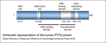
Human PTTG has an amino (N)-terminal basic domain and a carboxyl (C)-terminal acidic domain that can largely be divided in functional terms into the N-terminal regulatory domain and the C-terminal functional domain (Refs Reference Dominguez9, Reference Zhang10, Reference Kakar and Jennes12) (Fig. 1). Assessment of the overall structure of PTTG determined that it was virtually devoid of any secondary or tertiary structure, classifying it as a natively unfolded protein (Ref. Reference Sanchez-Puig, Veprintsev and Fersht54). However, more recent NMR studies suggest that although the N-terminal half of PTTG is entirely disordered, the C-terminal half demonstrates partial order, including an α-helical structure of the segment D150–F159, a region shown to interact with separase (Ref. Reference Csizmok55).
Schematic representation of the human PTTG protein. The following functional domains are displayed in white (amino acids are given in brackets): KEN box (9–11), destruction box (D-box) (61–68), PBF-binding domain (123–154) and SH3-interacting domain (163–173). The KEN box and D-box are targets for APC-mediated ubiquitylation, which signals the degradation of PTTG during mitosis (Ref. 53). The DNA-binding domain is found between amino acids 61 and 118 (Ref. 54) and a transactivation domain lies between 119 and 164 (Refs 9, 55, 56). Abbreviations: APC, anaphase-promoting complex; PBF, PTTG binding factor; PTTG, pituitary tumor-transforming gene; SH3, Src-homology-3.

PTTG phosphorylation
Cell-cycle-dependent PTTG expression has been observed. In both HeLa (human cervical carcinoma) and JEG-3 (human choriocarcinoma) cells, PTTG expression was low at the G1–S boundary, gradually increased during the S phase, peaking at the G2–M transition, and then decreased as cells entered G1 (Refs Reference Zou2, Reference Yu56). Similarly, in HeLa cell extracts, PTTG expression levels varied throughout the cell cycle, being low in G1 and S and peaking during mitosis (Ref. Reference Ramos-Morales57).
PTTG is also phosphorylated in a cell-cycle-dependent manner, peaking during mitosis (Ref. Reference Ramos-Morales57). The sole confirmed phosphorylation site of human PTTG is the serine 165 (S165) residue. S165 is phosphorylated by CDC2, a cyclin-dependent kinase that functions at the G2–M transition in association with cyclins A and B to drive the cell into mitosis (Ref. Reference Ramos-Morales57). Mutation of S165 to either abrogate phosphorylation or mimic constitutive phosphorylation did not prevent PTTG degradation during mitosis, inhibition of chromatid separation or FGF-2 induction. However, loss of phosphorylation results in increased cell transformation and proliferation, whereas increased phosphorylation has the opposite effect (Ref. Reference Boelaert58).
PTTG can also be phosphorylated by the DNA-dependent protein kinase (DNA-PK), but the site of phosphorylation and its functional significance remain unclear (Ref. Reference Romero59).
PTTG subcellular localisation
Initial studies of PTTG subcellular localisation suggested that PTTG was predominantly a cytosolic protein with partial nuclear localisation (Refs Reference Dominguez9, Reference Saez13). By contrast, both immunofluorescent detection and subcellular fractionation in interphase JEG-3 cells transfected with PTTG–EGFP demonstrated mainly nuclear localisation. This was replicated in NIH3T3, GH3 rat and AtT20 mouse pituitary tumour, SKOV-3 human ovarian cancer, MCF-7 and COS-7 monkey kidney epithelial cell lines (Ref. Reference Yu56). The predominance of nuclear or cytoplasmic PTTG might be cell-specific because PTTG was localised primarily in the nucleus in HeLa, COS-7 and DU145 cells, but distributed diffusely between the nucleus and cytoplasm in A549, DLD-1 and NIH3T3 cells (Ref. Reference Mu60).
The functional activities of PTTG described in this review all require PTTG to be localised to the nucleus. No function of PTTG in the cytoplasm has yet been identified; rather its presence here may represent a form of negative regulation. Despite lacking a nuclear localisation signal (NLS), the small molecular weight of PTTG should allow diffusion across the nuclear membrane. However, PBF was shown to promote the nuclear localisation of PTTG, with its own NLS being essential for this facilitation (Ref. Reference Chien and Pei5).
During mitosis, PTTG colocalises with mitotic spindles in JEG-3 cells (Ref. Reference Yu56) and chromosomes in H1299 non-small cell lung carcinoma cells (Ref. Reference Yu61).
PTTG may also be a secretory protein. Confocal laser-scanning microscopy analysis of human pituitary adenomas and immunoelectron microscopy of the AtT20 cell line identified PTTG expression in the Golgi complex and vesicles. Furthermore, secreted PTTG has been detected in the culture medium of mouse pituitary tumour cell lines (Ref. Reference Minematsu62).
PTTG is the human securin
The cell-cycle-dependent expression of PTTG, with increased levels at mitosis, its subcellular localisation and evidence of phosphorylation by a kinase that is active at the G2–M transition, was consistent with the identification of PTTG as a human securin (Ref. Reference Zou2). Securins are proteins that are involved in the metaphase to anaphase transition during mitosis, when sister chromatid separation occurs. Defects in this separation can cause aneuploidy; therefore, several control mechanisms are in place that prevent chromatid separation before all pairs have bioriented on the mitotic spindle (Ref. Reference Nasmyth63).
In eukaryotic cells, following chromosomal replication in the S phase, until their separation in anaphase, the two sister chromatids are held together by the cohesin complex. In vertebrates, most of the cohesin dissociates from along the arms of the chromosomes during prophase (Ref. Reference Waizenegger64). This dissociation appears to be dependent on two mitotic kinases, Aurora B and polo-like kinase 1, although the precise mechanism is unknown (Refs Reference Losada, Hirano and Hirano65, Reference Sumara66). At the metaphase to anaphase transition, the cysteine protease separase is then required for the cleavage and removal of the remaining cohesin, most of which are in the centromeric region (Ref. Reference Waizenegger64).
Control of the metaphase to anaphase transition is dependent on the anaphase-promoting complex (APC), a ubiquitin protein ligase that ubiquitinates several proteins involved in cell cycle regulation, thereby targeting them to the 26S proteasome for destruction.
Following the clearance of the spindle-assembly checkpoint, which delays APC activation until all chromosomes are correctly bioriented on the mitotic spindle, PTTG is targeted for destruction (Ref. Reference Peters67). PTTG degradation is dependent on both the destruction box (D-box) and the KEN box (Ref. Reference Zur and Brandeis53). Separase is bound and inactivated by PTTG to prevent the premature separation of sister chromatids. Hence, at the metaphase to anaphase transition, PTTG degradation by APC releases separase to cleave the RAD21 cohesin subunit (Refs Reference Zou2, Reference Jallepalli68). Separase can also be inhibited by phosphorylation at residue Ser1126 by CDC2. Interestingly, PTTG and CDC2 cannot bind simultaneously to separase. Therefore, APC mediates the activation of separase through two mutually exclusive inhibitory mechanisms, thereby allowing cell cycle progression (Refs Reference Stemmann69, Reference Gorr, Boos and Stemmann70, Reference Holland and Taylor71).
Although the APC co-activator CDC20 is essential for sister chromatid separation in other species, CDC20 knockdown in the human osteosarcoma U2OS cell line did not result in either PTTG or cyclin B stabilisation, or mitotic block (Ref. Reference Baumgarten, Felthaus and Wasch72). Alternatives to this possibly redundant mechanism of mitotic control might involve the recently discovered interaction between PTTG and the mitotic checkpoint protein BubR1, which can bind CDC20 and delay anaphase. BubR1 can compete with CDC20 for PTTG binding, thereby stabilising PTTG, and hence may inhibit anaphase in a CDC20-independent manner (Ref. Reference Kim73).
PTTG might also have a role in the G1–S phase transition. PTTG interaction with SP1 modified binding to its motif in the cyclin D3 promoter in JEG-3 and HCT116 (human colorectal) cells, thereby inducing its expression and G1–S transition, independently of p21 (Ref. Reference Tong74). A role for PTTG has also been demonstrated in the transition into the M phase, whereby it can inhibit APC activity through substrate competition allowing the stabilisation and accumulation of cyclin B (Ref. Reference Marangos and Carroll75).
PTTG and genetic instability
Genetic instability can be a temporary or a persistent condition found in many types of cancer, causing a series of mutational events that result in gross genetic alterations. The most common type of alteration is termed chromosomal instability (CIN) and includes aneuploidy (in which entire chromosomes are lost or gained) and chromosomal rearrangements (such as insertions, deletions and translocations). Defects in mitosis, such as abnormal chromosome condensation, incorrect sister chromatid cohesion and segregation, and a defective spindle checkpoint, can result in CIN. Therefore, the role of PTTG as a securin in sister chromatid segregation made it a strong candidate for inducing CIN following the dysregulation of its expression.
Overexpression of PTTG induces aneuploidy
PTTG was initially found to induce aneuploidy in MG-63 osteosarcoma cells. Transiently or stably expressed PTTG–EGFP caused an increase in the severity and frequency of aneuploidy in MG-63 cells, which are not usually prone to such aberrations (Ref. Reference Yu76). In H1299 cells, asymmetrical cytokinesis resulted from high levels of PTTG expression, the most common type being complete nonsegregation, yielding one daughter cell that contained all chromosomes and another devoid of a nucleus. Double mutation of the D-box and the KEN box generated a PTTG protein that could not be degraded and similarly resulted in asymmetrical cytokinesis. Overall, these studies demonstrated that PTTG accumulation, through either overexpression or lack of degradation, caused aneuploidy in single, live human cancer cells (Ref. Reference Yu61).
Other studies also examining the effect of PTTG overexpression on mitotic disruption have demonstrated the induction of asymmetrical cytokinesis by nondegradable PTTG mutants in HeLa cells (Ref. Reference Hagting77) and a higher number of PTTG-positive cells in aneuploid cases, versus those with a normal karyotype, in a series of malignant melanomas (Ref. Reference Winnepenninckx78).
Assessment of intrachromosomal instability as opposed to more gross chromosomal changes using a fluorescent intersimple sequence repeat PCR assay found that PTTG expression correlated with genetic instability in thyroid tumours in vivo and induced genetic instability in vitro (Ref. Reference Kim79).
PTTG deletion causes genetic instability
Following the identification of PTTG as a human securin, a PTTG1-null phenotype might be predicted to be lethal. However, Pttg1-null mice and cells are surprisingly viable (Refs Reference Wang, Yu and Melmed80, Reference Jallepalli68). Pttg1 −/− mice are both viable and fertile, although female subfertility was apparent (Ref. Reference Wang, Yu and Melmed80). These mice exhibit testicular and splenic hypoplasia, thymic hyperplasia and thrombocytopaenia. Because PTTG is abundantly expressed in the normal testis and thymus, the converse weight changes observed in these tissues indicate cell-type-specific differences in PTTG effects on cell growth (Ref. Reference Wang, Yu and Melmed80). Mouse embryonic fibroblasts derived from Pttg1 −/− mice exhibited delayed progression of the G2–M transition and aberrant chromatid separation (Refs Reference Wang, Yu and Melmed80, Reference Mei, Huang and Zhang81). PTTG-deficient cells were also derived from the HCT116 cell line by PTTG inactivation by homologous recombination (Ref. Reference Jallepalli68). These cells were defective at the metaphase to anaphase transition, but most of the cells eventually progressed to anaphase and continued the cell cycle, remaining viable (Ref. Reference Jallepalli68). These results suggest that PTTG is important for the maintenance of chromosome stability and cell cycle progression, but at least partially compensatory mechanisms are present that permit cell survival in the absence of PTTG (Refs Reference Wang, Yu and Melmed80, Reference Jallepalli68).
Despite an initially high rate of chromosomal loss in PTTG1 −/− HCT116 cells, repeated passaging caused the cells to regain chromosomal stability. However, these cells still maintained reduced separase activity (Ref. Reference Pfleghaar82). Mutation of the inhibitory phosphorylation site of separase in Pttg1-null mouse embryonic stem cells caused premature sister chromatid separation but did not affect cell viability, suggesting the existence of other mechanisms that limit the activation of separase even when PTTG and the inhibitory phosphorylation site are eliminated (Ref. Reference Huang83). Recent studies with the PTTG1-null HCT116 cell line have suggested that PTTG might be required for directing the centrosomic location of separase during mitosis (Ref. Reference Tong84).
Although these studies suggest that PTTG is potentially redundant in sister chromatid separation, prolonged study of Pttg1 −/− mice determined that loss of PTTG severely impaired glucose homeostasis, leading to diabetes during late adulthood (Ref. Reference Wang85). This was caused by a decrease in pancreatic β-cell proliferation, accompanied by atypical nuclear morphology indicative of abnormal cell division. Therefore, any mechanisms for sister chromatid separation that are compensatory for the loss of PTTG are not sufficient for maintaining β-cell proliferation and subsequent intact islet cell function (Ref. Reference Wang85). Further investigation of these Pttg1 −/− mice showed that diminished β-cell mass was caused by β-cell apoptosis in early postnatal life, followed by senescence in adulthood, possibly as a result of DNA damage (Ref. Reference Chesnokova86).
PTTG, p53 and apoptosis
The transcription factor p53 is activated by cell stress, such as DNA damage or oncogene activation, and can induce a varied response ranging from cell cycle arrest to programmed cell death or apoptosis. Important in the prevention of tumourigenesis, loss of p53 function can lead to malignant progression (Ref. Reference Vousden87).
Overexpression of PTTG in JEG-3 and MCF-7 cells, which express wild-type p53, and in p53-deficient MG-63 cells, resulted in significant cell death by apoptosis. Therefore, PTTG can induce both p53-dependent and p53-independent apoptosis (Refs Reference Yu56, Reference Yu76). PTTG–EGFP induced aneuploidy in MG-63 cells, but not in MCF-7 and JEG-3 cells, suggesting that p53 can prevent PTTG-induced aneuploidy (Ref. Reference Yu76). PTTG can upregulate p53 expression at the transcriptional level. In HEK293 and MCF-7 cells, PTTG stimulated the expression of c-Myc, which subsequently bound to the promoter of p53, thereby inducing its expression (Ref. Reference Hamid and Kakar88). Induction of the transactivation function of p53 by PTTG overexpression was demonstrated by the upregulation of the expression of BAX, a proapoptotic molecule and a known target of p53. Furthermore, overexpression of both PTTG and p53 induced apoptosis in HEK293 cells. These results suggest that PTTG can induce apoptosis, both dependently and independently of p53; however, if these mechanisms were to fail, PTTG could promote the survival of aneuploid cells and, consequently, support tumour growth (Ref. Reference Hamid and Kakar88).
Both in vitro and in vivo binding assays determined that PTTG and p53 can interact directly. This interaction blocked the specific binding of p53 to DNA and inhibited its transcriptional activity (Ref. Reference Bernal89). In contrast to previous data describing the activation of p53 and apoptosis by PTTG (Ref. Reference Yu76), PTTG binding also inhibited the ability of p53 to induce cell death (Ref. Reference Bernal89). Furthermore, depletion of PTTG was shown to induce apoptosis through the activation of p53 in SH-J1 human hepatoma cells, suggesting that the overexpression of PTTG can negatively regulate p53 (Ref. Reference Cho-Rok24). Similarly, in a study using UV irradiation to induce apoptosis in HeLa cells, RNAi-mediated knockdown of PTTG expression increased, and overexpression of PTTG decreased, UV-induced apoptosis (Ref. Reference Lai90).
Drug-induced DNA damage inhibited PTTG expression but only in the presence of functional p53. A mechanism was proposed whereby p53, activated by DNA damage, interacts with the transcription factor NF-Y and prevents it binding to the PTTG1 promoter, thereby reducing transcriptional initiation of PTTG expression. This suggests that PTTG1 is a p53 target gene, which is suppressed in response to DNA damage (Ref. Reference Zhou38). Subsequently, p53 was found to directly bind to the PTTG1 promoter, thus suppressing PTTG expression (Ref. Reference Kho91). DNA damage induced by UV irradiation caused a reduction in PTTG levels through increased proteasomal degradation and decreased protein synthesis, independently of p53. However, PTTG was required for cell cycle arrest following UV irradiation. The loss of PTTG resulted in premature entry into mitosis and increased apoptosis (Ref. Reference Romero92). Recent studies have shown that the SKP1–CUL1–βTrCP E3 ubiquitin ligase is responsible for PTTG degradation after UV irradiation (Ref. Reference Limon-Mortes93).
PTTG in DNA damage and repair
Ku-70 is part of a heterodimer that binds to the ends of DNA and recruits the catalytic component of DNA-PK, an enzyme involved in nonhomologous end-joining (NHEJ) repair of double-strand breaks in DNA. The Ku heterodimer can bind PTTG and the catalytic component of DNA-PK is capable of phosphorylating PTTG. In the presence of DNA breaks, PTTG was unable to interact with Ku-70, suggesting that PTTG and Ku form an inhibitory complex from which Ku dissociates following DNA damage, triggering DNA repair by a DNA-PK-dependent pathway (Ref. Reference Romero59). PTTG overexpression can repress both Ku-70 DNA-binding activity and double-stranded DNA (dsDNA) damage repair activity (Ref. Reference Kim20). Thus, aberrantly high levels of PTTG expression may sequester Ku proteins, adversely affecting DNA repair and contributing to genetic instability (Refs Reference Zhou38, Reference Kim20). Further studies have demonstrated reduced cell proliferation accompanied by altered patterns of end resection in NHEJ assays following DNA damage in PTTG-deficient HCT116 cells in comparison with normal HCT116 cells (Ref. Reference Bernal94).
Overall, these studies have identified an association between PTTG and proteins involved in the cellular response to DNA damage, p53 and Ku-70. The relationship between these proteins and genetic instability and apoptosis is clearly complex and appears to be dependent on the presence of DNA damage. The PTTG–p53 interaction is particularly complicated and warrants further investigation in both the presence and absence of DNA damage.
PTTG as a gene transactivator
The similarity of the human PTTG peptide sequence to DNA-binding proteins, its partial nuclear localisation and the presence of an acidic C-terminal region suggest that PTTG has a role in the transcriptional activation of other genes. A fusion protein containing PTTG and the Gal4 DNA-binding domain was able to transactivate reporter genes under the control of Gal-responsive elements in both yeast and mammalian cells (Ref. Reference Dominguez9).
The acidic C-terminal region of PTTG contains a number of glutamic acid and proline residues, which are characteristic of transactivation domains (Refs Reference Dominguez9, Reference Wang and Melmed95). This proline-rich region contains two PxxP motifs, which are potential binding sites for Src-homology-3 (SH3) domains, mediators of intracellular signal transduction pathways. Mutation of these PxxP motifs resulted in the loss of in vitro transforming activity and in vivo tumour-inducing potential of PTTG (Refs Reference Zhang10, Reference Hamid, Malik and Kakar3), and also abrogated murine PTTG transactivating ability, suggesting that the transactivation and transforming abilities of PTTG are associated (Ref. Reference Wang and Melmed95). Several genes involved in cellular growth and angiogenesis have been shown to be transactivated by PTTG.
FGF-2, VEGF and angiogenesis
The upregulation of FGF-2 expression by PTTG was first demonstrated in NIH3T3 cells (Ref. Reference Zhang10). An increased concentration of FGF-2 in the cell medium suggested that PTTG also stimulated the secretion of FGF-2. Both expression and secretion of FGF-2 were abrogated by mutation of the SH3-interacting domain (Refs Reference Zhang10, Reference Ishikawa96). Conditioned medium from PTTG-transfected NIH3T3 cells induced the proliferation, migration and tube formation of human umbilical vein endothelial cells (HUVECs) in vitro. The addition of a neutralising FGF-2 antibody abrogated these effects. Furthermore, mutation of the PxxP domain significantly reduced the proangiogenic effect of PTTG. These studies suggest that FGF-2 mediates the induction of angiogenesis by PTTG, through its transactivation by the SH3-interacting domain of PTTG (Ref. Reference Ishikawa96).
Studies within our group have shown that both PTTG and FGF-2 expression are upregulated in differentiated thyroid cancer, with their levels of expression demonstrating a significant positive correlation. Increased FGF-2 mRNA expression was independently associated with lymph node invasion and distant metastasis, whereas increased levels of PTTG were associated with tumour recurrence (Ref. Reference Boelaert17). PTTG overexpression in primary human thyroid cells increased FGF-2 expression. A mutant PTTG that could not be phosporylated (S165A) could still upregulate FGF-2, whereas abrogation of the PxxP motif reduced this effect (Ref. Reference Boelaert58). Furthermore, PTTG upregulation of FGF-2 was determined to be a direct, promoter-specific effect through the use of luciferase assays in COS-7 cells (Ref. Reference Chien and Pei5).
Further studies from our group indicated that pituitary tumours with increased PTTG expression also demonstrated increased expression of VEGF and its receptor KDR, both of which were positively correlated with PTTG expression (Ref. Reference McCabe97). PTTG was able to stimulate both FGF-2 and VEGF expression in human fetal neuronal NT-2 cells, a cell line that exhibits low background PTTG expression, as well as in MCF-7 and JEG-3 cells. Again the phosphorylation status did not alter PTTG upregulation of FGF-2 and VEGF, whereas mutation of the SH3 domain reduced this effect (Ref. Reference McCabe97). Although FGF-2 can regulate VEGF expression in endothelial cells, depletion of secreted FGF-2 in the cell media failed to influence the effect of PTTG on VEGF expression, suggesting that PTTG can upregulate VEGF expression independently of FGF-2 transactivation and secretion (Ref. Reference McCabe97).
PTTG was reported to be highly expressed in HCC, with PTTG, FGF-2 and VEGF all positively correlated and associated with intratumoural microvessel density (Ref. Reference Fujii98). Similar results were obtained in studies of PTTG and FGF-2 in endometrial carcinoma (Ref. Reference Wang99) and of PTTG and VEGF in pituitary adenomas (Ref. Reference Minematsu100).
To investigate whether other angiogenic genes might be regulated by PTTG, our group used angiogenesis-specific cDNA arrays to identify angiogenic genes that were differentially expressed in primary human thyroid cells with and without PTTG overexpression (Ref. Reference Kim18). Several genes were differentially expressed on PTTG overexpression, including the upregulation of proangiogenic genes such as VEGF and ID3, and the downregulation of antiangiogenic factors such as thrombospondin-1. The transactivation of ID3 was determined to be dependent on the putative SH3-interacting domain (Ref. Reference Kim18). Subsequently, the upregulation of ID3 by PTTG in thyroid cancer was shown to be mediated by the stimulation of VEGF and its receptor KDR (Ref. Reference Kim101).
In addition to FGF-2 and VEGF, interleukin-8 (IL-8) expression was upregulated in HEK293 cells overexpressing PTTG and in tumours developed in nude mice following injection of these cells (Ref. Reference Hamid, Malik and Kakar3). The expression and secretion of the matrix metalloproteinase MMP-2 was also increased in these models. Conditioned medium taken from PTTG-transfected HEK293 cells induced the proliferation, migration and tube formation of HUVECs in vitro and the addition of an MMP-2 antibody significantly decreased these effects (Ref. Reference Malik and Kakar102).
PTTG has also been shown to regulate pituitary hormone expression. Transfection of the C-terminal amino acids 147–202 of PTTG into GH3 cells significantly decreased prolactin (PRL) expression and secretion and conversely induced GH expression and secretion. Mutation of the SH3-binding motif abrogated these effects. The repression of PRL expression was determined to be transcriptional using promoter studies (Ref. Reference Horwitz103).
c-Myc
A cDNA expression array was used to identify target genes whose expression is regulated by PTTG (Ref. Reference Pei104). Genes encoding c-Myc, MEK1, MEK3, PKCβ-1 and the heat-shock protein HSP70 showed increased expression following rat PTTG expression. c-Myc is a transcription factor involved in the regulation of cellular proliferation, and its deregulation is associated with several tumours. PTTG was subsequently found to stimulate c-Myc transcription by directly binding to the promoter in a complex with the ubiquitously expressed upstream stimulatory factor (USF1). Furthermore, PTTG-mediated induction of p53 is an indirect event, with the induction of c-Myc expression by PTTG being a prerequisite for p53 upregulation through the binding of c-Myc to the p53 promoter in a heterodimer with its binding partner, MAX (Ref. Reference Hamid and Kakar88).
Interaction with SP1
The chromatin immunoprecipitation (ChIP)-on-chip assay was used to analyse 20 000 gene promoters to determine whether PTTG could bind as a transcription factor in JEG-3 cells (Ref. Reference Tong74). Overall, 746 promoters demonstrated PTTG binding, with the binding site for SP1 identified more frequently in promoters to which PTTG bound compared with the remaining promoters. Co-immunoprecipitation and pull-down assays confirmed that PTTG could bind SP1 both in vivo and in vitro through its N-terminal 120 amino acids. This study suggests a global role in transcriptional regulation for PTTG (Ref. Reference Tong74).
Other interacting factors
Yeast two-hybrid assays identified an interaction between the C-terminal 75 amino acids of rat PTTG and the molecular chaperone HSJ2 and ribosomal protein S10 in testicular cells. The functional significance of these interactions remains unknown (Ref. Reference Pei105).
Protein microarray screening confirmed the interaction between PTTG and ribosomal protein S10. Approximately 80 proteins were found to interact with PTTG, including transcription factors such as TBPL1 and ABT1, providing further evidence that PTTG is important in transcriptional regulation (Refs Reference Tong84, Reference Tong and Eigler106). An interaction with Aurora-A was also identified and confirmed using His-tag pull-down assays and co-immunoprecipitation assays in HCT116 cells. PTTG overexpression was subsequently found to inhibit Aurora-A kinase activity and to result in abnormally condensed chromatin (Ref. Reference Tong84). However, the interaction between PTTG and PBF remains the best characterised.
PTTG binding factor
PBF characterisation and structure
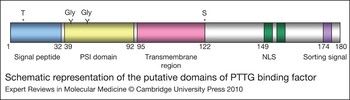
Using a yeast two-hybrid screen to identify proteins that interact with PTTG, PBF [also known as PTTG1-interacting protein (PTTG1IP)] was isolated and characterised (Ref. Reference Chien and Pei5). Located on chromosome 21q22.3, PBF encodes a protein of 180 amino acids with a predicted molecular mass of 22 kDa (Ref. Reference Chien and Pei5). PBF had previously been cloned and termed C21orf3 (also known as C21orf1). Initial protein prediction studies suggested that C21orf3 was a cell surface glycoprotein because of a potential N-terminal signal peptide, a transmembrane domain, an endocytosis motif and two putative N-glycosylation sites (Ref. Reference Yaspo107) (Fig. 2). Furthermore analysis identified potential sites for post-translational modifications such as phosphorylation sites for cAMP- and cGMP-dependent kinase, PKC and casein kinase II and five glycosylation sites for N-linked and O-linked oligosaccharides (Ref. Reference Chien and Pei5). Additionally, an extracellular N-terminal cysteine-rich putative protein-binding domain, common to plexins, semaphorins and integrins and thereby referred to as a PSI domain, is also found in PBF (Ref. Reference Bork108). By contrast, an entirely different function was purported by the presence of a bipartite NLS near the C-terminus, suggesting that PBF enters the cell nucleus (Ref. Reference Chien and Pei5).
Schematic representation of the putative domains of PTTG binding factor. A putative, cleavable signal peptide is located at the N-terminus between amino acids 1 and 32. A putative PSI domain forms the remaining extracellular region between amino acids 39 and 92, next to the putative transmembrane domain located between amino acids 95 and 122. A putative NLS and endocytosis motif are found at the C-terminus from amino acids 149 and 174, respectively. The site of interaction with PTTG, within the C-terminal 30 amino acids, is highlighted. Abbreviations: NLS, nuclear localisation signal; PBF, PTTG binding factor; PTTG, pituitary tumor-transforming gene.

Pull-down and co-immunoprecipitation assays in COS-7 cells demonstrated that PTTG and PBF specifically bind both in vitro and in vivo. Deletion analysis identified the PTTG-binding domain in PBF to be located within the C-terminal 30 amino acids (Fig. 2) and the converse domain in PTTG to be between amino acids 123 and 154 (Ref. Reference Chien and Pei5).
PBF expression and regulation
Northern blot analysis established that PBF is widely expressed in normal human tissues (Refs Reference Chien and Pei5, Reference Yaspo107). A potential involvement of PBF in tumourigenesis was proposed when PBF expression was found to be increased in pituitary tumours compared with normal pituitary tissue by studies within our group (Ref. Reference McCabe6). Subsequently, PBF expression was found to be increased in thyroid cancer and independently associated with early tumour recurrence (Ref. Reference Stratford7).
PTTG overexpression in primary human thyroid cultures and PTTG-knockout HCT116 cells resulted in a significant increase in PBF mRNA expression. However, PTTG with a mutated SH3-interacting domain failed to stimulate PBF expression. The increase in PBF expression was also replicated in NIH3T3 cells stably transfected with wild-type PTTG. Conversely, PTTG1 expression was not stimulated in PBF stable cell lines (Ref. Reference Stratford7).
Differential hybridisation screening, comparing C3H10T1/2 mouse embryonic fibroblasts constitutively overexpressing Runx2 with wild-type cells, identified Pbf as a target gene for the Runx2 transcription factor. Runx2 bound to consensus sequences present in sequence −1100 to −3000 bp 5′ to Pbf in gel-shift assays and Runx2 overexpression increased the activity of a 1.8 kb Pbf promoter fragment in reporter assays (Ref. Reference Stock109).
Unlike Pttg1, neither EGFR nor TGF-α had any effect on Pbf expression in TtT–GF cells (Ref. Reference Vlotides49). However, treatment of ER-α-positive MCF-7 cells with diethylstilbestrol and 17β-oestradiol induced significant PBF mRNA and protein expression (Ref. Reference Watkins8).
PBF localisation and function
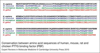
At the protein level, PBF shares no significant homology with other human proteins but is highly conserved between species (77% homology to rat, 75% mouse and 60% chicken; Fig. 3), suggesting it has unique functionality and is of evolutionary importance. However, its primary function remains unknown.
Conservation between amino acid sequences of human, mouse, rat and chicken PTTG binding factor (PBF).

The presence of an NLS at the C-terminus of PBF prompted subcellular localisation studies. PTTG and PBF were transfected into COS-7 cells and their subcellular localisation was subsequently examined by immunofluoresence staining and subcellular fractionation studies (Ref. Reference Chien and Pei5). The expression of haemagglutinin-tagged PBF (HA–PBF) was located mainly in the nucleus, with significant staining in the cytoplasm also. Mutation of the NLS shifted PBF expression predominantly to a perinuclear and cytoplasmic location, suggesting that the NLS is required for the nuclear localisation of PBF. PTTG, tagged with green fluorescent protein (GFP–PTTG), was observed predominantly in the cytoplasm, with partial nuclear localisation, whereas co-transfection of HA-PBF resulted in increased nuclear translocation of GFP–PTTG. This effect was abrogated when the mutated NLS was used, indicating that PBF with an intact NLS is required for PTTG translocation into the nucleus (Ref. Reference Chien and Pei5).
Luciferase reporter assays were used to assess the interaction of PTTG and PBF in FGF-2 transactivation. Neither PTTG nor PBF alone significantly altered FGF-2 promoter activity. However, co-expression of PTTG and PBF induced activity by more than three fold, indicating that PBF is required for PTTG to transactivate FGF-2. Overall, these data suggest that PBF binds PTTG and promotes its transactivation function by facilitating its nuclear translocation (Ref. Reference Chien and Pei5).
In addition to nuclear localisation, PBF has been found to be present within intracellular vesicles by immunofluorescent studies. PBF demonstrated strong colocalisation with the late endosomal marker CD63. Furthermore, the loss of the C-terminal 30 amino acids results in PBF accumulation at the plasma membrane, suggesting that the tyrosine-based sorting signal within this region is functional (Ref. Reference Smith110). These studies support the original prediction on cloning that PBF is a cell surface glycoprotein that is internalised upon ligand binding (Ref. Reference Yaspo107).
PBF is a proto-oncogene
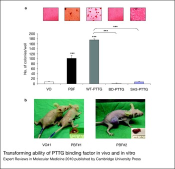
Stably transfected NIH3T3 clones with a high expression of PTTG or PBF were selected for colony formation assays to assess the transforming potential. Both PTTG and PBF overexpression led to significant colony formation (Fig. 4a). However, mutant forms of PTTG, which were unable to either stimulate PBF mRNA expression (SH3-interacting domain) or interact with PBF (amino acids 123–154), were unable to induce colony formation. This suggests that PBF is itself a transforming gene in vitro and might mediate the transforming ability of PTTG. Furthermore, injection of NIH3T3 cells stably expressing PBF into nude mice induced tumour formation, demonstrating its tumourigenic ability in vivo (Ref. Reference Stratford7) (Fig. 4b).
Transforming ability of PTTG binding factor in vivo and in vitro. (a) In vitro cell transformation by PBF and PTTG as determined by soft agar assays using NIH3T3 cells stably transfected with PBF, wild-type PTTG and mutated PTTG [mutated in the binding domain (BD) and SH3 domain]. Photomicrographs of cell colonies are shown above a graph quantifying the mean colony number per well. (b) In vivo tumourigenesis by PBF as determined by tumour growth in nude mice injected with NIH3T3 cells overexpressing PBF, compared with vector-only (VO) control. Reproduced from Ref. 7 (© 2005 The Endocrine Society). Abbreviations: PBF, PTTG binding factor; PTTG, pituitary tumor-transforming gene. ***P < 0.001.

PTTG and PBF may therefore act independently or together, or possibly both, to induce or advance tumourigenesis. The role of each in individual tumours will be discussed in the following sections.
PTTG and PBF in endocrine cancer
Pituitary tumours
Several studies have demonstrated increased PTTG expression in pituitary tumours. PTTG1 expression was more than 50% higher in pituitary tumours compared with matched normal pituitaries. In hormone-secreting tumours, PTTG1 expression was significantly higher in those that had invaded the sphenoid bone compared with those retained in the pituitary fossa, suggesting that PTTG may be a marker for invasiveness in these tumours (Ref. Reference Zhang15). In a study of 36 pituitary adenomas, positive immunostaining was detected in all but four adenomas (Ref. Reference Saez13).
PTTG1 mRNA expression was approximately fivefold higher in 111 pituitary adenomas (85 nonfunctioning, 15 GH, 4 PRL, 4 ACTH and 3 TSH) compared with normal pituitary (n = 10). Western blot analysis confirmed an increase at the protein level also. PBF mRNA expression was significantly increased and a significant positive correlation between PTTG1 and PBF expression was observed in pituitary adenomas but not in normal pituitary tissue, suggesting that an association between PTTG1 and PBF mRNA expression might be important in tumourigenesis. However, neither PTTG nor PBF expression was associated with clinical parameters (Ref. Reference McCabe6).
Immunohistochemical analysis of 45 pituitary adenomas found nuclear PTTG expression in 89% of tumours, which correlated with Ki-67 expression. Both PTTG and Ki-67 were associated with more aggressive tumour behaviour, and in those patients that were followed up for more than a year, immunopositivity for PTTG and Ki-67 predicted recurrence (Ref. Reference Filippella111). PTTG1 was one of nine genes associated with aggressive–invasive human PRL tumours. An initial case–control analysis, comparing patients in remission (nine controls) and patients with persistent or recurrent tumours (14 cases), found that PTTG1 did not correlate with clinical outcome (Ref. Reference Wierinckx112). However, a subsequent larger study of 94 patients, following surgery to remove PRL tumours, found that PTTG was highly upregulated in aggressive–invasive tumours and was associated with recurrence and progression (Ref. Reference Raverot113). Furthermore, PTTG expression was positively associated with recurrence in 35 cases of nonfunctioning pituitary adenomas, with highest expression in the early recurrent group (Ref. Reference Noh114).
Mice heterozygous for retinoblastoma (Rb) deletion develop highly penetrant pituitary tumours. Pttg1 was upregulated in the pretumourous pituitary gland of Rb +/− mice. PTTG depletion in both Pttg1 −/− and Rb +/−Pttg1 −/− mice resulted in reduced cell proliferation and increased p21 expression, and delayed and reduced tumour onset in the latter (Ref. Reference Chesnokova115).
Further analysis of the Pttg1 −/− and Rb +/−Pttg1 −/− mice identified pituitary-specific senescent features, including increased levels of p53, p19, p21 and cyclin D1, apoptosis block and elevated senescence-associated β-galactosidase (SA-β-gal) expression. Senescence in Pttg1-null mice was not due to telomere shortening but was more likely induced by aneuploidy and activation of DNA damage pathways (Ref. Reference Chesnokova116). Deletion of p21 from the Rb +/−Pttg1 −/− background restored pituitary tumour development, highlighting the contribution of p21 to the protective effect of PTTG deletion (Ref. Reference Chesnokova117). Rb +/− mice also spontaneously developed medullary thyroid (C cell) tumours. Although thyroid tumour development was abrogated in Rb +/−Pttg1 −/− mice, thyroidal p21 levels were normal and p21 ablation did not restore tumour development. Senescence was detected in 23 of 26 GH-producing pituitary adenomas with high PTTG levels. Therefore, either PTTG deletion or overexpression promotes pituitary cell aneuploidy and p53/p21-dependent senescence, particularly in GH-secreting cells. High levels of p21 in the pituitary might therefore constrain tumour growth, thus accounting for the lack of progression of benign pituitary adenomas to carcinomas (Ref. Reference Chesnokova117). Interestingly, PTTG overexpression has very recently been shown to elicit the same response causing chromosome instability, which subsequently induced p53-dependent senescence through activation of the DNA damage response pathway in normal human fibroblasts (Ref. Reference Hsu118).
Transgenic PTTG overexpression was targeted to the mouse pituitary using the glycoprotein hormone α-subunit (α-GSU) promoter. Male transgenic mice showed plurihormonal focal pituitary transgene expression with LH-, TSH- and, unexpectedly, also GH-cell focal hyperplasia and adenoma, with MRI demonstrating large and irregularly shaped pituitary glands (Ref. Reference Abbud119). In contrast to the PTTG-null mice, bitransgenic αGSU.PTTGxRb +/− mice have enlarged pituitary glands and a 3.5-fold increase in the frequency of anterior lobe tumours originating from α-subunit-expressing cells compared with singly transgenic Rb +/− mice (Ref. Reference Donangelo120).
Analysis of the main promoter regions of PTTG1 in 25 pituitary adenomas failed to identify any mutations that might have altered PTTG1 expression (Ref. Reference Kanakis50). Recent evidence suggests that Rb inactivation or phosphorylation might induce PTTG through the release of the E2F transcription factors (Ref. Reference Zhou121).
In rat pituitary lactotroph tumours, PTTG was maximally induced by oestrogen early in pituitary transformation, and this was accompanied by increased FGF2 and VEGF and angiogenesis (Ref. Reference Heaney31). Four out of eight primary human pituitary tumour cultures (three gonadotroph, five nonfunctioning) demonstrated a reduction of PTTG expression by 64% in response to the ER antagonist ICI-182780. This was associated with reduced proliferation, as determined by PCNA expression (Ref. Reference Heaney, Fernando and Melmed122).
Thyroid cancer
PTTG1 expression was initially found to be significantly upregulated in human thyroid tumours compared with normal thyroid tissue (Ref. Reference Heaney16). Furthermore, FRTL-5 cells stably transfected with PTTG exhibited increased FGF-2 expression and were transforming in vitro, in contrast to WT or SH3-mutant PTTG, confirming the role of this domain in PTTG induction of transformation in thyroid cells (Ref. Reference Heaney16). An immunohistochemical study of 60 differentiated thyroid cancers found that 65% overexpressed PTTG. High PTTG expression was significantly associated with lymph node and distant metastases, and with persistent disease (Ref. Reference Saez123).
When PBF was examined in thyroid tumours, both PTTG and PBF mRNA and protein expression levels were significantly increased in thyroid tumours (n = 27) compared with normal thyroid tissue (n = 11), and mRNA data were significantly and positively correlated (Refs Reference Stratford7, Reference Boelaert17). FGF2 and FGFR1 mRNA expression is increased in cancers compared with normal thyroid, and FGF2 expression was independently associated with lymph node involvement and distant metastases (Ref. Reference Boelaert17). PTTG1 and PBF expression were both independently associated with early tumour recurrence (Refs Reference Stratford7, Reference Boelaert17). Sequencing of the coding region of PBF in 24 thyroid tumours showed no mutations (Ref. Reference Stratford7).
The TRβPV/PV mouse expresses a dominant-negative form of thyroid hormone β receptor (TRβ), which results in decreased sensitivity to the thyroid hormone (T3) in target tissues (Ref. Reference Ying124). In a manner similar to human thyroid cancer, TRβPV/PV mice spontaneously develop follicular thyroid carcinoma, with pathological progression from hyperplasia to vascular invasion, capsular invasion, anaplasia and eventually metastasis. PTTG expression was significantly increased in both the primary thyroid tumour and the lung metastases of TRβPV/PV mice. A mechanism was proposed by which PTTG normally binds TRβ and undergoes T3-mediated proteasomal degradation. However, when PTTG was associated with PV, T3 was unable to bind and activate degradation, thus resulting in elevated PTTG levels (Ref. Reference Ying124).
Thyroid tumour progression may be driven by PTTG-induced angiogenesis, as described earlier (Refs Reference Kim18, Reference Kim101). This is supported by a study that examined the effects of PTTG deletion in TRβPV/PV mice by creating the double-mutant line TRβPV/PVPttg1 −/− (Ref. Reference Kim125). PTTG ablation resulted in reduced thyroid size because of decreased thyrocyte proliferation, possibly through induction of p21. Interestingly, although PTTG deletion did not prevent tumour initiation, there was a significant decrease in vascular invasion accompanied by decreased expression of FGF-2, FGFR1 and VEGF (Ref. Reference Kim125).
PTTG and PBF not only have a critical role in thyroid tumourigenesis, but also impede the treatment of these cancers and metastases with radioiodine. PTTG overexpression was found to decrease iodide (I−) uptake in FRTL-5 cells and human primary thyroid cells, and sodium iodide symporter (NIS) expression was reduced in human thyrocytes (Ref. Reference Heaney16). In vivo, high PTTG expression is significantly associated with decreased radioiodine uptake during follow-up (Ref. Reference Saez123). In thyroid cancers demonstrating increased PTTG and PBF expression, NIS expression is significantly reduced compared with normal thyroid and demonstrated a significant negative correlation with expression of PTTG1 and PBF (Ref. Reference Boelaert126).
In vitro PTTG and PBF overexpression significantly reduced NIS mRNA expression and 125I uptake in primary human thyroid cultures. A mutant PTTG with a disrupted SH3-interacting domain (PTTG SH3−) also repressed NIS mRNA, suggesting that the transactivation capability of PTTG is not required for the inhibition of NIS mRNA expression. However, PTTG SH3− exhibited a reduced ability to repress NIS function compared with WT PTTG, indicating that this repression is at least partially dependent on an intact SH3-interacting domain (Ref. Reference Boelaert126).
Promoter studies using both human primary thyroid cultures and FRTL-5 cells identified the human NIS upstream enhancer as the element responsible for mediating the repression of NIS by PTTG and PBF. This was further refined to a putative overlapping PAX8 and USF1-binding site where both sites were essential for PBF repression but PTTG only required an intact USF1 site (Ref. Reference Boelaert126). A post-translational mechanism of NIS repression by PBF has also recently been demonstrated. PBF was found to bind NIS in vitro and modulate its subcellular localisation away from the plasma membrane, thereby reducing I− uptake. PTTG and PBF are therefore potential therapeutic targets for improving radioiodine uptake and hence prognosis of thyroid cancer (Ref. Reference Smith110).
Ovarian cancer
PTTG cloned from several ovarian tumours was identical to that defined from nonmalignant tissue, suggesting that the primary structure of PTTG is unaltered as in other tumours. In situ hybridisation showed increased PTTG1 expression in epithelial, sex-cord, stromal cell and germ cell ovarian tumours compared with normal ovary (Ref. Reference Puri21). SVOV-3 ovarian carcinoma cells stably transfected with antisense PTTG1 demonstrated reduced endogenous PTTG and FGF-2 expression, and in vitro transforming ability (Ref. Reference Chen127).
Mice in which Pttg1 expression was targeted to ovarian surface epithelium using the Mullerian inhibitory substance type II receptor (MISIIR) gene promoter failed to develop ovarian tumours. PTTG overexpression resulted in increased ovary size, hyperplasia of the endometrium, and increased serum LH and testosterone levels. These data suggest that PTTG can initiate precancerous growth but is not sufficient to induce ovarian tumourigenesis, possibly requiring a hormonal trigger or another event to initiate cellular transformation (Ref. Reference El Naggar128). PBF function has not yet been examined in ovarian cancer.
Endocrine-related cancer
Breast cancer
PTTG is overexpressed in breast cancer (Refs Reference Saez13, Reference Puri21), and PTTG1 mRNA expression is significantly correlated with lymph node invasion and tumour recurrence within a 5-year period (Ref. Reference Solbach22).
In contrast to low levels of cytoplasmic PTTG expression in normal breast tissue (n = 18), abundant cytoplasmic and nuclear PTTG was observed in all 90 breast tumours examined. PTTG expression was highest in invasive ductal carcinoma and metastatic breast tumours and cell lines. Correlations between PTTG expression and lymph node invasion, mitotic activity and ER expression were observed (Ref. Reference Ogbagabriel23).
In a cDNA microarray that compared invasive ductal breast carcinomas with normal breast tissue, PTTG1 was one of the most significantly deregulated proliferation-associated genes. Analysis of 44 patients with invasive ductal breast cancer found that a 10-unit increment of PTTG immunopositivity was associated with a 2.3-fold overall risk of death as a result of breast cancer, suggesting that PTTG is a potential proliferation marker in invasive ductal breast cancer (Ref. Reference Talvinen129). These data were further supported in a larger immunohistochemical study of 310 patients where PTTG was defined as a prognostic factor of breast-cancer-specific survival, independent of patient age, nodal status and tumour size (Ref. Reference Talvinen130).
An intronic single-nucleotide polymorphism (SNP) in PTTG1 (C1892G) showed a significant association with breast cancer in a case–control study of 698 primary breast cancer patients and 1492 controls (Ref. Reference Lo131). However, neither genotypic nor allelic frequencies of the SNP rs1862392 found in the PTTG1 promoter differed between 749 cases of breast cancer and 1493 matched controls in a Swedish cohort (Ref. Reference Brendle132).
PTTG has been shown to have prognostic potential in breast cancer, being one of four mRNA markers on a membrane array that could detect five circulating tumour cells per millilitre of peripheral blood assayed. The sensitivity and specificity were 86% and 88%, respectively, in blood taken from 92 breast cancer patients and 100 normal controls (Ref. Reference Chen133).
PTTG1 expression was also examined in ER-positive breast tumour samples from postmenopausal women treated with primary surgery followed by adjuvant Tamoxifen (Tam) alone. PTTG1 was significantly overexpressed in patients who developed acquired resistance to endocrine therapy, because they relapsed under Tam treatment and developed metastases compared with those who did not relapse. High PTTG1 expression was correlated with shorter relapse-free survival and was identified as a prognostic marker associated with endocrine therapy resistance in breast tumour samples (Ref. Reference Ghayad134).
Recently, a central role in breast cancer aetiology has been associated with PBF. Immunohistochemical analysis of eight normal breast specimens and 146 TMA tumour samples demonstrated that PBF expression was low or absent in normal breast tissue, but was strongly expressed in epithelial cells of all types and grades of breast tumour assessed. The region of the PBF promoter −399 to −291 relative to the translational start site contains between one and six repeats of an 18-bp sequence housing a putative ERE half-site. This region was bound by ER-α and conferred most of the responsiveness of PBF to oestrogen. Subjects with greater numbers of ERE repeats showed higher PBF mRNA expression, and PBF protein expression positively correlated with ER-α status. PBF induced significant cell invasion through Matrigel, with PBF knockdown significantly abrogating oestrogen-mediated MCF-7 cell invasion. Furthermore, secretion of PBF was critical in the induction of cell invasion (Ref. Reference Watkins8). Hence, both PTTG and PBF have potentially critical roles in breast cancer, although they have only been studied independently thus far.
Colorectal carcinoma
PTTG1 overexpression was observed in all 48 colorectal carcinomas and in all but one of 20 colorectal polyps compared with normal colorectal mucosa. High PTTG levels were associated with more vascularised tumours and lymph node invasion (Ref. Reference Heaney19).
DNA microarray analysis and real-time PCR showed PTTG1 overexpression in human colorectal carcinomas compared with the corresponding normal mucosa, which correlated with aberrant nuclear β-catenin expression. β-Catenin overexpression resulted in increased PTTG1 promoter activity whereas siRNA knockdown of β-catenin reduced PTTG in HCT116, SW480 and DLD1 colon cancer cell lines (Ref. Reference Hlubek43).
Analysis of 22 colorectal tumours showed that PTTG was induced compared with matched normal colon, and was higher in Dukes’ stage C&D than in Dukes’ stage A&B colorectal tumours (Ref. Reference Kim20). Genetic instability was also higher in Dukes’ stage C&D tumours and PTTG overexpression caused a dose-dependent increase in genetic instability in HCT116 cells. By repressing Ku70 activity and inhibiting the NHEJ dsDNA repair pathway as discussed previously, PTTG might be a critical gene in the development of genetic instability in colorectal cancer (Ref. Reference Kim20). Our group is currently assessing the role of PBF in colorectal tumourigenesis.
Uterine leiomyomas
Significantly elevated PTTG expression was observed in 23 leiomyomas compared with matched normal myometria independent of menstrual cycle but associated with increased cell proliferation. In cultured leiomyoma cells, PTTG was not responsive to either oestrogen or progesterone but was significantly induced by FGF-2. In turn, both FGF2 and VEGF were upregulated by PTTG overexpression, suggesting the presence of a positive feedback loop in leiomyoma cells (Ref. Reference Tsai48).
Other cancers
PTTG1 expression was found to be higher in ESCC tissues compared with paired normal samples studied (Refs Reference Shibata26, Reference Zhou42). Regulation by β-catenin was evident through a significant correlation between PTTG expression and aberrant β-catenin localisation (Ref. Reference Zhou42). PTTG1 expression was associated with more aggressive disease (Ref. Reference Shibata26).
A large immunohistochemical study performed on 113 primary ESCC specimens showed a high prevalence of PTTG overexpression (60.2%), which was significantly associated with both regional and distant lymph node metastasis, advanced tumour stage and reduced survival (Ref. Reference Ito135).
Positive PTTG immunostaining was found in nine of ten lung adenocarcinomas (Ref. Reference Saez13). In a larger study, immunostaining of PTTG was observed in 64% of patients (n = 136) with small-cell lung cancer (SCLC) and in 97.8% of patients (n = 91) with non-small-cell lung cancer (NSCLC). Low PTTG expression was associated with shorter mean survival in SCLC patients, whereas strong PTTG expression in NSCLC was associated with reduced survival and a more aggressive phenotype (Ref. Reference Rehfeld136).
Compared with corresponding noncancerous liver tissues, PTTG1 was significantly overexpressed in 62 HCC specimens. Patients with high PTTG1 expression levels had lower five-year disease-free and overall survival rates than those with lower PTTG1 expression (Ref. Reference Fujii98). Another study found that expression of PTTG1 was increased by more than two-fold in 39 cases of 44 HCCs compared with normal liver tissues. Depletion of PTTG1 using siRNA inhibited the growth of the invasive sarcoma SH-J1 hepatoma cell line and activated p53, inducing p21 expression and apoptosis (Ref. Reference Cho-Rok24).
PTTG and PBF as therapeutic targets
The presence of high levels of PTTG expression in several forms of cancer, and its association with tumour progression, has highlighted PTTG as a potential therapeutic target. Several studies have investigated methods of inhibiting PTTG expression, some of which have been discussed in the previous sections.
PTTG1 repression through siRNA knockdown reduced the transforming ability of H1299 cells and human A2780 ovarian carcinoma cells in in vitro assays and suppressed in vivo tumour growth following the injection of these cells into nude mice (Refs Reference Kakar and Malik25, Reference El Naggar, Malik and Kakar137).
Adenoviral vector encoding siRNA against PTTG1 (Ad.PTTG1-siRNA) depleted PTTG in HCT116 colorectal cancer cells, which exhibited a cytotoxic effect in a p53-dependent manner. In vivo, Ad.PTTG1-siRNA markedly attenuated the tumour growth of Huh-7 hepatoma cells subcutaneously injected or SH-J1 tumour xenografts established in nude mice (Ref. Reference Cho-Rok24).
Transfection of the human cervical cancer cell line HELA-S3 with antisense PTTG1 mRNA successfully downregulated PTTG mRNA and protein, and both inhibited growth and increased apoptosis (Ref. Reference Solbach138).
Female Wistar-Furth rats (4–5 weeks old) subcutaneously injected with GH3 cells stably transfected with control vector, wild-type or mutant C-terminal PTTG (147–202) all developed tumours by 9 weeks. Excised tumour tissue from transfectants overexpressing the wild-type PTTG C-terminus was smaller than that from control vector or mutant PTTG1 C-terminus transfectants. Therefore, this dominant-negative peptide of PTTG might be a useful therapeutic tool (Ref. Reference Horwitz103).
Interestingly, PBF has itself been used in a potential therapeutic tool to knock down PTTG. A fusion protein (F-PBFβ-TrCP) was engineered comprising PBF attached to the first 256 amino acids of the F box protein β-TrCP, which is one subunit of a ubiquitin protein ligase complex, to specifically target PTTG for degradation. When an F-PBFβ-TrCP-expressing vector was transfected into HeLa cells, PTTG protein levels, but not mRNA levels, were significantly reduced. This was accompanied by reduced FGF-2 expression, cell growth and ability to form colonies in soft agar (Ref. Reference Mo139).
Conclusions
PTTG is a complex, multifunctional protein involved in a wide range of cellular processes such as cell cycle regulation, genetic instability and gene transactivation. Our understanding of the role of PTTG in these processes and the mechanism by which aberrant expression of PTTG contributes to tumourigenesis continues to develop. However, the dysregulation of PTTG and subsequent effects are dependent on the type of cancer and several other contributing factors, and require further investigation. PTTG represents a potential prognostic marker and shows increasing promise as a therapeutic target.
Despite being discovered shortly after PTTG, its binding partner PBF is considerably less well characterised. PBF is now also emerging as a proto-oncogene, which is upregulated in several cancers and warrants further investigation as a prospective prognostic or therapeutic tool. Currently, in pituitary, thyroid and breast cancer, PTTG and PBF show distinct overlaps in certain functional processes. However, as the function of PBF is further elucidated, it remains to be seen to what extent the roles of PTTG and PBF are dependent or independent in endocrine and other cancers.
Acknowledgements and funding
We sincerely thank the reviewers of this manuscript who provided genuinely useful insight and support.