Introduction
Dysphagia is a common complaint reported by post-laryngectomy patients. The cause may be anatomical and/or physiological. It may be influenced by the manner of reconstruction,Reference Jaber, Greenbaum, Sappington, Burgette, Kramer and Borrowdale 1 , Reference Davis, Vincent, Shapshay and Strong 2 the occurrence of complications such as pharyngo-cutaneous fistula,Reference Jaber, Greenbaum, Sappington, Burgette, Kramer and Borrowdale 1 or the administration of radiotherapy (though this has been disputed by certain reports).Reference Lippert, Folz, Rudert and Werner 3
Post-laryngectomy dysphagia may occur because of: difficulties in propulsion of the food bolus through the neopharynx, neopharyngeal or oesophageal stenosis, neuromuscular dysfunction, cricopharyngeal hypertonia, or an anterior pharyngeal diverticulum. In 1962, Kirchner and Scatliff reported an anterior vallecular pseudo-diverticulum as a cause of dysphagia in a post-laryngectomy patient.Reference Kirchner and Scatliff 4
A pseudo-epiglottis is a fold of mucous membrane and/or scar tissue extending from the lateral pharyngeal wall on either side to the base of the tongue.Reference Jaber, Greenbaum, Sappington, Burgette, Kramer and Borrowdale 1 Most often, it is an incidental finding, but it can become symptomatic when a significant anterior pouch forms. The pseudonyms for this pouch are neopharyngeal pseudo-diverticulum, anterior pharyngeal diverticulum and pseudo-vallecula.
Methods and results
Two patients reported to the ENT out-patient department of a tertiary care hospital. In case one, the patient was a 57-year-old gentleman, who had undergone laryngectomy and post-operative radiotherapy for laryngeal carcinoma 8 months previously. At presentation, he complained of dysphagia, regurgitation and an inability to use the voice prosthesis effectively. In case two, the patient was a 52-year-old gentleman, who had undergone laryngectomy for laryngeal carcinoma. He experienced regurgitation of feeds and halitosis six weeks after surgery. He was undergoing radiotherapy.
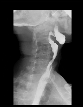
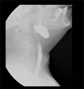
Both patients underwent a barium swallow test. This showed an anterior pharyngeal diverticulum between the base of the tongue and the neopharynx, with stasis of barium in the pouch (Figures 1 and 2).
Barium swallow image of case one showing the diverticula.

Barium swallow image of case two showing the diverticula.

Both patients underwent transoral micro-endoscopic potassium titanyl phosphate (KTP) 532 nm laser assisted excision of the pseudo-epiglottis under general anaesthesia. Weerda's laryngoscope was used for the exposure. One ribbon gauze piece was kept in the diverticulum. Another ribbon gauze piece soaked in methylene blue was placed behind the pseudo-epiglottis. The intervening band was excised using a KTP 532 nm laser at 7 W. Rigid hypopharyngoscopy and oesophagoscopy were performed to assess the neopharynx and cervical oesophagus.
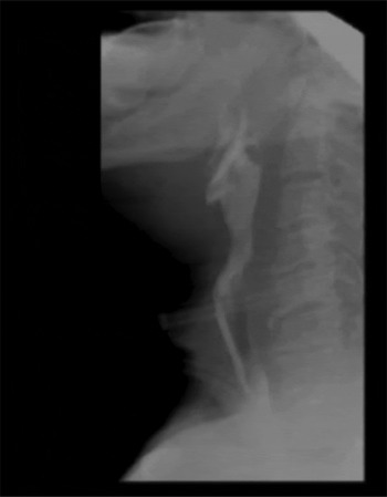
Post-operatively, the patients’ swallowing improved. A contrast-enhanced radiograph showed an absence of pooling in the previously demonstrable diverticulum (Figure 3). The first patient died one and a half years after the procedure because of secondary lung cancer. The second patient died after three years as a result of regional recurrence. The patients were followed up until their demise. The patients had no difficulty in swallowing semi-solids. Rigid telescopic examination showed no evidence of pharyngeal diverticulum recurrence.
Post-resection barium swallow image demonstrating free passage of contrast.

Discussion
In 1962, Kirchner and Scatliff reported an anterior vallecular pseudo-diverticulum as a cause of dysphagia in a post-laryngectomy patient.Reference Kirchner and Scatliff 4 The posterior tissue band has been described as a pseudo-epiglottis. Approximately 35–75 per cent of post-laryngectomy patients may have pseudo-diverticula.Reference Nayar, Sharma and Arora 5 , Reference Muller-Miny, Eisele and Jones 6 It forms during the healing phase, along with scar development. It has been previously proposed that the formation may be related to: the type of pharyngeal closure (muscular or non-muscular), the shape of neopharyngeal closure (with longitudinal closures being more prone as compared to the ‘T’ shaped onesReference Lippert, Folz, Rudert and Werner 3 ), the presence of prior pharyngo-cutaneous fistulaReference Jaber, Greenbaum, Sappington, Burgette, Kramer and Borrowdale 1 and the administration of radiotherapy. Radiotherapy as a cause has been disputed.Reference Lippert, Folz, Rudert and Werner 3
The lack of muscle, in addition to the posterior movement of a pseudo-epiglottis, impedes the passage of the food bolus. This leads to collection in the diverticulum, which further occludes the upper end of the neopharynx. The clinical presentation includes the regurgitation of undigested food, the sensation of food for a prolonged period after eating, and halitosis. It may mimic velopharyngeal insufficiency.Reference Jaber, Greenbaum, Sappington, Burgette, Kramer and Borrowdale 1 In patients with a voice prosthesis, the sound may be distorted because of inadvertent vibration during tracheoesophageal speech through the collected material. The pseudo-epiglottis with diverticulum may be missed during rigid telescopic examination as it is folded on the base of tongue. It can be identified better by a transnasal flexible endoscopic evaluation, especially during swallowing. The diverticulum can be documented by a contrast-enhanced radiograph,Reference Lippert, Folz, Gottschlich and Werner 7 which will also rule out a stricture lower down in the neopharynx.
The treatment initially is conservative, and involves washing down food with liquids, changing head posture and increasing the effort of swallowing. For persistent severe symptoms, surgery is required. A transoral micro-endoscopic approach is preferred over an external approach. The pseudo-epiglottis can be excised using laser,Reference Hartey, Saeed and Farrington 8 monopolar cautery,Reference Lippert, Folz, Rudert and Werner 3 coblatorReference Jaber, Greenbaum, Sappington, Burgette, Kramer and Borrowdale 1 or harmonic scalpel.Reference Jaber, Greenbaum, Sappington, Burgette, Kramer and Borrowdale 1
In the few reported cases in which the endoscopic approach was used for excising the pseudo-epiglottis, no complications were reported.Reference Hartey, Saeed and Farrington 8 The post-operative development of a fistula after an external approach for pseudo-epiglottis excision has been reported.Reference Gacek 9 As experience with such a procedure is limited, a true estimate of long-term success without recurrence is not available. Laser-assisted micro-endoscopic resection is a relatively safe, quick and effective procedure for the management of pseudo-epiglottis.
-
• A wide possibility of lesions should be considered in post-laryngectomy dysphagia patients, including anterior pharyngeal diverticulum
-
• Transnasal flexible endoscopy and contrast radiography can help identify an anterior pharyngeal diverticulum
-
• An anterior pharyngeal diverticulum is a common incidental finding
-
• Transoral micro-endoscopic laser-assisted resection of the pseudo-epiglottis is a relatively safe, quick and effective treatment