Introduction
There is increasing evidence that challenging cognitive stress tests that tap proactive semantic interference (PSI) in which old semantic learning interferes with new semantic learning and the failure to recover from proactive semantic interference (frPSI), despite repeated learning trials are early markers of amnestic Mild Cognitive Impairment (aMCI) and prodromal Alzheimer’s disease (AD) (Loewenstein et al., Reference Loewenstein, Curiel, Duara and Buschke2018a; Reference Loewenstein, Curiel, Duara and Buschke2018b; Matías-Guiu et al., Reference Matías-Guiu2017). Measures of PSI and frPSI on the Loewenstein-Acevedo Scales of Semantic Interference and Learning (LASSI-L) have consistently been more predictive of preclinical and prodromal AD than traditional list-learning tests such as the Free and Cued Selective Reminding Test or Hopkins Verbal Learning Test – Revised (HVLT-R) (Loewenstein et al., Reference Loewenstein2017b; Matias-Guiu et al., Reference Matias-Guiu2018). PSI and frPSI deficits have also been associated with loss of brain volume as visualized by magnetic resonance imaging (MRI) (Loewenstein et al., Reference Loewenstein2017a; Reference Loewenstein2017b), cerebral metabolism using fluorodeoxyglucose (FDG) positron emission tomography/computed tomography (PET/CT) imaging (Matias-Guiu et al., Reference Matias-Guiu2018) and amyloid burden (Loewenstein et al., Reference Loewenstein2016; Loewenstein et al., Reference Loewenstein, Curiel, Duara and Buschke2018a). Most of these studies and earlier work have primarily focused on the number of correct responses elicited by measures of PSI and frPSI (Crocco et al., Reference Crocco, Curiel, Acevedo, Czaja and Loewenstein2014; Curiel et al., Reference Curiel2013); however, recent evidence suggests that semantic intrusion errors on PSI and frPSI subscales are particularly sensitive to early AD (Torres et al., Reference Torres2019) and can distinguish between patients clinically diagnosed with aMCI who are amyloid positive from non-AD patients who are amyloid negative, suggesting that semantic intrusion errors may be an early cognitive marker of AD, with high specificity (Loewenstein et al., Reference Loewenstein, Curiel, Duara and Buschke2018a). Indeed, Sanchez et al. (Sánchez et al., Reference Sánchez2017) found that middle-aged AD probands had more frPSI intrusion errors than controls and that the number of errors was related to widespread disconnectivity in cortico-limbic circuits.
Amyloid load has been associated with semantic intrusion errors, even after controlling for volume in medial temporal lobe areas (e.g. hippocampus or entorhinal cortex) (Loewenstein et al., Reference Loewenstein, Curiel, Duara and Buschke2018a). However, Dickerson (Dickerson et al., Reference Dickerson2011) has argued that “AD signature regions” of neurodegeneration are not only limited to medial temporal lobe structures, but occurs within a number of brain regions within the neocortex. In fact, subtypes of AD are known in which the medial temporal regions are spared and atrophy in the neocortex occurs especially in the precuneus, posterior cingulate, and superior parietal brain regions (Ferreira et al., Reference Ferreira, Pereira, Volpe and Westman2019; Murray et al., Reference Murray2011). Thus, a singular focus on the hippocampus and the entorhinal cortex may not capture the neurodegeneration present in preclinical and clinical AD. There are also important factors such as age and APOE ϵ4genotype that may be related to early cognitive change; however, it is likely that associations may be indirect and work through mechanisms of increased amyloid burden. In addition, important covariates such as sex and language/culture that may be related to both neuroimaging and semantic intrusions have not yet been evaluated.
In the current study, we examined the relationships of amyloid burden, volumetric loss in AD-prone regions, age, and APOE ϵ4 genotype with regards to semantic intrusion errors in a structural equation modeling (SEM) framework. SEM allows for the specification of theoretical models, and multiple relationships may be estimated simultaneously. Although previous research suggests associations between amyloid burden and semantic intrusions (Cid et al., Reference Curiel Cid2020; Loewenstein et al., Reference Loewenstein, Curiel, Duara and Buschke2018a), there are lingering questions as to whether semantic intrusions are related to volumetric loss in AD-prone regions and if the presence of brain amyloid is related to volumetric loss in AD-prone regions once factors such as age are accounted for. We further examined the effects of age and APOE ϵ4 on semantic intrusion errors, and whether these two factors have a direct versus indirect effect on this cognitive deficit. A model estimating these relationships simultaneously, while controlling for important covariates, represents the most comprehensive evaluation of factors that may contribute to PSI or frPSI errors. To our knowledge, this study is the first to examine the direct and indirect effects of age, APOE ϵ4 genotype, amyloid positivity, and volumetric reductions in AD-prone brain regions as it relates to semantic intrusion errors (a novel and sensitive measure of early AD. These predictors have been examined in isolation, but not examined in a way that we can determine the direct and indirect effects of these relationships on the outcome using causal modeling approaches. Utilizing confirmatory path analysis, we examined the causal modeling among the variables and expand the understanding of the underpinnings of sensitive semantic interference effects. Such analyses will provide insights into the causal relationships of these important factors.
Materials and methods
In this cross-sectional study, we examined 212 participants enrolled in the baseline of 1Florida Alzheimer’s Disease Research Center from October 2015 to December 2017. All participants consented to participate in this Institutional Review Board-approved study. The 1Florida ADRC baseline evaluation includes extensive clinical and neuropsychological evaluations, MRI and amyloid PET/CT images to assess fibrillar amyloid plaques, and analyses of APOE genotype. Participants with an ϵ4 allele in their APOE genotype (ϵ4/ϵ4, ϵ3/ϵ4, or ϵ2/ϵ4) were categorized as ϵ4 positive; those without an ϵ4 allele were considered as APOE ϵ4 negative.
This sample included a broad array of cognitive diagnoses in those individuals with MMSE scores ranging from 16 to 30. Diagnostic categories are described below:
Cognitively Normal (CN; n = 29 [14%])
Participants were classified as CN if there were: (a) no subjective cognitive complaints made by the participant and/or a collateral informant; (b) no evidence by clinical evaluation and no history of memory or other cognitive declines, after an extensive interview with the participant and an informant; (c) all memory (e.g. HVLT-R) (Benedict et al., Reference Benedict, Schretlen, Groninger and Brandt1998) or delayed paragraph recall from the National Alzheimer’s Coordinating Center Uniform Data Set (NACC UDS) (Beekly et al., Reference Beekly2007)) and non-memory measures (e.g. Category Fluency (Lucas et al., Reference Lucas1998), Trails A and B (Reitan, Reference Reitan1958), WAIS-IV Block Design subtest (Wechsler, Reference Wechsler2008)) were less than 1.0 SD below normal limits for age, education, and language group; (d) Global Clinical Dementia Rating (CDR) (Morris, Reference Morris1993) scale score of 0; (e) had a negative amyloid scan as read by an experienced rater (RD).
Mild Cognitive Impairment (MCI; n = 99 [47%])
Participants were diagnosed with MCI if they: (a) fulfilled Petersen’s criteria (Petersen et al., Reference Petersen2014) for MCI; (b) subjective cognitive complaints reported by the participant and/or collateral informant; (c) impaired delayed recall (i.e. 1.5 SD or greater, below the mean, accounting for age, education, and language of testing) for either the HVLT-R or delayed paragraph recall from the NACC UDS and/or 1.5 SD below expected levels on non-memory measures as described for the CN group; (d) Global CDR scale score of 0.5; (e) no evidence of clinical dementia.
Mild Dementia (DEM; n = 42 [20%])
Participants were diagnosed with mild dementia if they: (a) fulfilled criteria a, b, and c as specified for the aforementioned MCI group (b) Global CDR scale score of 1.0; (c) clinical course and history-consistent dementia with loss of independence in everyday activities of daily living.
Cognitive Impairment not MCI (PreMCI; n = 36 [17%])
Participants in this group did not meet formal criteria for MCI or CN as described above. In general, these participants either had a clinical history consistent with MCI and a global CDR of 0.5, but lacked cognitive deficits upon neuropsychological testing (see CN neuropsychological group criteria) or had no clinically relevant memory concerns (patient and informant), a Global CDR of 0, and impaired neuropsychological testing (see MCI neuropsychological group criteria). A longitudinal study of individuals diagnosed with PreMCI found that they are at greater risk for transition to MCI or dementia (Loewenstein et al., Reference Loewenstein2012).
Loewenstein-Acevedo Scales for Semantic Interference and Learning (LASSI-L)
The LASSI-L was not used for diagnostic determination in this study to avoid potential issues of circularity. This cognitive stress test represents a novel paradigm that employs controlled learning and cued recall in an effort to maximize the storage of a list of to-be-remembered target words belonging to three distinct semantic categories (fruits, clothing, and musical instruments) (Loewenstein et al., Reference Loewenstein2016). Participants were tested in their preferred language (English vs. Spanish) and the LASSI-L has been previously shown to be culturally fair and valid in either language (Curiel Cid et al., Reference Curiel Cid2019; Matías-Guiu et al., Reference Matías-Guiu2017).
During the administration of the LASSI-L, the examinee is instructed to remember a list of 15 common words representing three semantically distinct categories. A unique aspect of the LASSI-L paradigm is the presentation of a second competing list of to-be-remembered words that is presented in the same manner as the first list. The second list introduces different words, but shares the same previously presented semantic categories, in order to elicit a considerable amount of PSI. Unlike other traditional memory assessment paradigms, the readministration and subsequent recall of this second list of words measure the individual’s ability to recover from the effects of PSI (frPSI).
For the current study, we focused on subscales of the LASSI-L that have been sensitive to elevated amyloid load, presumably underlying AD (e.g. number of intrusion errors on Cued B1 and Cued B2). These intrusion errors primarily involve words from the first list of semantically similar target items or other nontarget words sharing a similar semantic category. Intrusion errors produced on the Cued B1 and Cued B2 trials are extremely sensitive to PSI and frPSI deficits thought to reflect deficits in source memory and inhibitory control (Cid et al., Reference Curiel Cid2020; Loewenstein et al., Reference Loewenstein, Curiel, Duara and Buschke2018b; Torres et al., Reference Torres2019).
Assessment of neurodegeneration using MRI
All participants described above underwent structural MR imaging using a Siemens Skyra 3T scanner at Mount Sinai Medical Center, Miami Beach, Florida, USA. Brain parcellation was obtained using a 3D T1-weighted sequence (MPRAGE) with 1.0 mm isotropic resolution using FreeSurfer version 6.0 software (http://surfer.nmr.mgh.harvard.edu). We calculated the volumes of bilateral AD-prone regions specified by Dickerson (Dickerson et al., Reference Dickerson2011) and from our previous work (Loewenstein et al., Reference Loewenstein2017a; Reference Loewenstein2017b;), which included a composite score of regions within the hippocampus, entorhinal cortex, amygdala, parahippocampal gyrus, inferior temporal lobule, temporal pole, supramarginal, superior parietal, precuneus, rostral middle frontal, and superior frontal areas. All volumes of the brain regions that went into the composite were normalized by dividing by the total intracranial volume.
Amyloid imaging
PET/CT imaging was obtained using a 3D Hoffmann brain phantom to establish a standardized acquisition and reconstruction method. Participants were infused with (18F) florbetaben 300 MBQ over a 3-minute period. Scanning commenced 70–90 min after the infusion for a duration of 20 min. We scanned all participants on a Siemens Biograph 16 PET/CT scanner operating in 3D mode (55 slices/frame, 3mm slice thickness 128 × 128 matrix). The PET data was reconstructed into a 128 × 128 × 63 (axial) matrix with voxel dimensions of 0.21 × 0.21 × 0.24 cm. Thirty-three participants hadflorbetapir as their amyloid tracer and 134 participants had florbetaben. Reconstruction was performed using manufacturer-supplied software and included corrections for attenuation, scatter, random coincidences, and dead time. Images for regional analyses were processed using Fourier analysis followed by direct Fourier reconstruction. Images were smoothed with a 3 mm Hann filter. Following reconstruction, image sets were inspected and, if necessary, corrected for inter-frame motion. Images were obtained from the top of the head to the top of the neck and computed tomography (CT) data was employed for initial attenuation correction and image reconstruction in the sagittal, axial, and coronal planes.
The PET/CT scans, including the outline of the skull, co-registered linearly (i.e. trilinear interpolation) with 12 degrees of freedom, onto the volumetric MRI scan using a T1-weighted (MP-RAGE) (Lizarraga et al., Reference Lizarraga2016; Smith et al., Reference Smith2004) region-of-interest (ROI) boundaries were defined manually using the structural MRI for anatomical reference, and criteria that have been proven to provide highly reproducible outcomes (Desikan et al., Reference Desikan2006). This registration process ensured that the florbetaben PET/CT image had the same accurate segmentation and parcellation as in the MRI scan. Average activity was calculated in the ROIs corresponding to cerebellar gray matter and cerebral cortical regions. A composite Standardized Uptake Value Ratio (SUVR) was calculated by the ratio of the mean volume-weighted SUVR of five bilateral cortical regions (frontal, temporal, parietal, anterior, and posterior cingulate cortex), to the cerebellar gray matter (Rowe et al., Reference Rowe2008). The Centiloid method has been widely used to create a common metric by which total amyloid uptake can be placed on the same scale for different amyloid tracers (Jack et al., Reference Jack2017; Rowe et al., Reference Rowe2017). Using normalization to the whole-brain cerebellum, for florbetaben, the Centiloid formula is ([SUVR × 153.4] −154.9) and for flobetapir, the Centiloid formula is ([SUVR × 183] −177). This created a centiloid score for each participant.
Visual ratings of amyloid PET scans
All Aß-PET scans were interpreted, using a methodology similar to that described by Seibly (Seibyl et al., Reference Seibyl2016), by an experienced reader (RD) who was blind to the cognitive and clinical diagnosis. Using the same methodology, Loewenstein and colleagues (Reference Loewenstein2018a) reported an interrater reliability of 98% for amyloid visual reads between RD and an independent rater. Tracer uptake was assessed in six cortical regions (orbitofrontal, frontal, parietal, lateral temporal, occipital, and precuneus/posterior cingulate cortex, combining values from the left and right hemispheres. A final dichotomous (A+ vs. A−) diagnosis was rendered. Visual amyloid reads are considered as the gold standard in the field.
Modeling framework
As part of the testing protocol, all participants were administered the LASSI-L, which was not used for diagnostic classification. We focused on semantic intrusion errors that occurred on Cued B1 and Cued B2 since it has been found that performance deficits on these subscales that measure PSI and frPSI are more common among MCI participants with presumptive AD (amyloid positive) relative to MCI due to non-AD conditions (amyloid negative) (Loewenstein et al., Reference Loewenstein2018a). What is unknown is whether an amyloid load is directly related to semantic intrusions or whether it exerts its effect through neurodegeneration as measured by reduction in MRI volumes. The effects of age and APOE ϵ4 would also need to be considered, either as a primary risk factor for elevated amyloid load, neurodegeneration, and/or as directly affecting cognitive performance (semantic intrusion errors) rate.
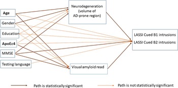
Adopting a causal modeling approach through confirmatory path analyses, we propose a model that examines several relevant relationships simultaneously as depicted in the path diagram (Figure 1). In the path analysis, a mediating variable is a dependent variable in one relationship and an independent variable in another. Our predictors of interest, age, and APOE ϵ4 genotype were hypothesized to influence the two mediating variables, amyloid positivity and neurodegeneration, which in turn influence cognitive performance. We also hypothesized that amyloid positivity has a direct effect on neurodegeneration in AD-prone regions. Additionally, age and APOE ϵ4 genotypes may exert a direct effect on cognitive performance. These hypothesized relationships fit the temporal and biological sequence that was related to the cognitive outcomes (intrusion errors on LASSI-L Cued B1 and Cued B2 subscales). APOE genotype conferred at birth and age are not modifiable. In contrast, amyloid load and neurodegeneration occur as part of the aging process, thus are mediators in the model.
Path diagram of the final model.

Covariates such as sex and educational attainment may exert effects on neurodegeneration, amyloid positivity, and cognitive performance on the LASSI-L. We also account for factors that might be related to LASSI-L performance in our model such as language of testing (English vs. Spanish) and global cognitive impairment assessed by the Mini-Mental State Examination (MMSE) (Folstein et al., Reference Folstein, Folstein and Mchugh1975). Global cognitive status such as MMSE affects the performance of LASSI-L test and the intrusion errors made and is also related to biological markers of neurodegeneration and amyloid load, thus, is controlled in the model.
Statistical analyses
Three regression models were evaluated simultaneously in the path analysis: (1) A probit model of amyloid positivity regressed on age and APOE ϵ4, while controlling for sex, education, and MMSE. The probit model was estimated because the dependent variable, namely, visual amyloid read was binary; (2) a multiple linear regression model with brain volume in AD-prone regions regressed on age, APOE ϵ4, and amyloid positivity, while controlling for sex, years of education, and MMSE; (3) a multiple linear regression model with LASSI-L Cued B1 or Cued B2 intrusions regressed on the two mediators (amyloid positivity and neurodegeneration), in addition to age and APOE ϵ4, while controlling for sex, education, MMSE and testing language.
If the two paths involved in the mediation relationship were both statistically significant, it indicated an indirect mediation effect, which was calculated by multiplying the two parameters involved in the mediation relationship (MacKinnon et al., Reference Mackinnon, Fairchild and Fritz2007). For example, the effect of age on neurodegeneration was multiplied by the effect of neurodegeneration on Cued B1 intrusions to obtain the indirect effect of age on Cued B1 intrusions. The Bootstrap method was used to calculate the 95% confidence interval (CI) of the indirect and total effect (MacKinnon et al., Reference Mackinnon, Fairchild and Fritz2007).
All models were evaluated using model fit statistics including the Comparative Fit Index (CFI) and Tucker–Lewis Index (TLI) for which values above 0.90 indicate good fit and values above 0.95 indicate excellent fit. The Root-Mean-Square-Error of Approximation (RMSEA) was also used for which values less than 0.05 indicate excellent fit. Maximum Likelihood Robust for missing data (full-information MLR) estimation was used to obtain estimates in the presence of missing data (Arbuckle et al., Reference Arbuckle, Marcoulides and Schumacker1996). We used a sandwich estimator for the standard errors that is robust to non-normality (Yuan and Bentler, Reference Yuan and Bentler2000). Analyses were conducted using the SAS 9.4 (SAS, 2019) and Mplus 8.4 software packages (Muthen and Muthen, Reference Muthen and Muthen1998–Reference Muthen and Muthen2012).
Results
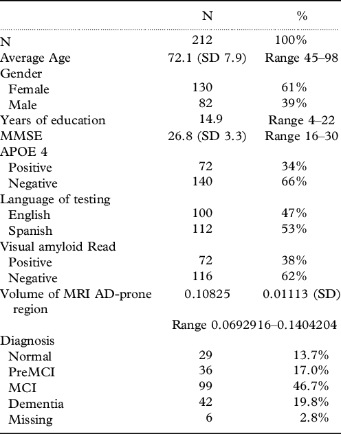
This investigation included 212 participants who underwent both MRI and amyloid PET/CT imaging with information on age, sex, education, language of testing, and MMSE. The average age of the sample was 72.1 years (range 45–98), 130 participants were female (61%), the average years of education was 14.9 years (range 4–22). Close to half of the participants (47%) were tested in English. The average MMSE score was 26.8 (range 16–30) (Table 1).
Characteristics of study participants

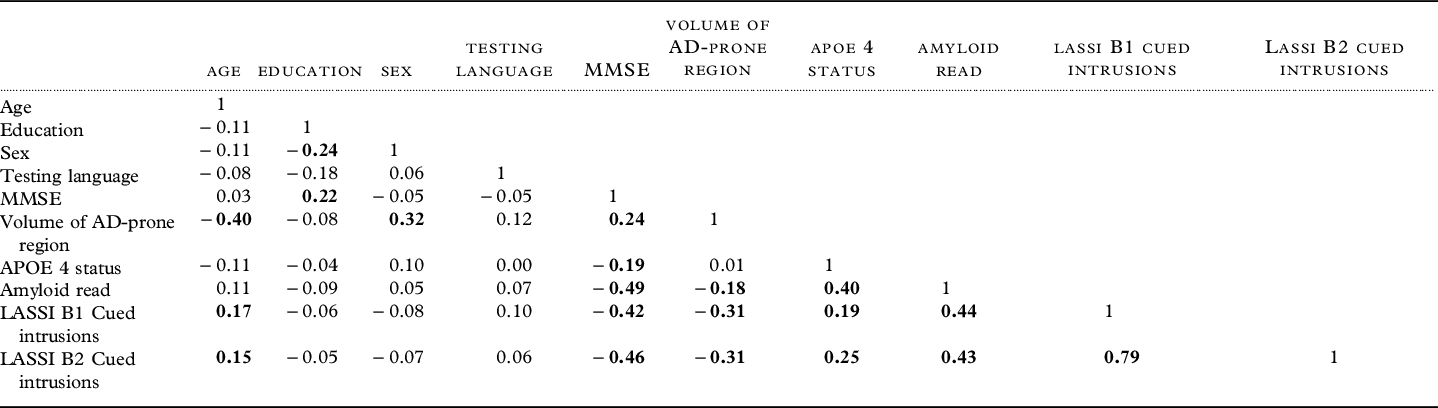
To give readers a general idea of the relationships among variables involve in the path analysis, we present in Table 2, the Pearson’s correlation coefficients among all variables, with correlation coefficients that are statistically significant at the 0.05 level bolded.
The Pearson’s correlation coefficients among variables involved in the path analyses*

* Numbers in bold indicating statistical significance at the 0.05 level.
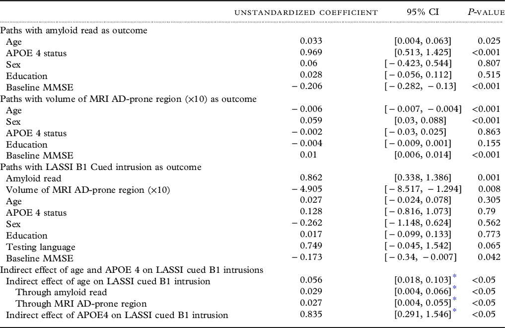
Table 3 presented the path analysis results of intrusion errors on Cued B1(measuring PSI). In the probit regression model, with amyloid positivity as the outcome, age, APOE ϵ4, and MMSE were all statistically significantly associated with amyloid positivity. For a 1 year increase in age, amyloid positivity increased by a z-score of 0.033, which means with each additional year in age, the probability of amyloid positivity slightly increases. Positive APOE ϵ4 was associated with an increased probability of amyloid positivity. Higher MMSE score was associated with a lower likelihood of being classified as amyloid positive.
Path analysis of LASSI-L Cued B1 intrusions

* Bootstrap method was used to calculate the CI of the indirect effect due to the non-normality of the product term.
In the regression model with brain volume in AD-prone regions as the outcome, we initially set up the model to include a path from amyloid positivity to brain volume in AD-prone regions; however, this path was not statistically significant, and was removed for the purpose of model parsimony. The same occurred when an amyloid load was expressed as a centiloid score instead of a binary visual read. As shown in Table 3, younger age, female sex, and higher MMSE score were statistically significantly associated with a greater brain volume of AD-prone region.
In the regression model with LASSI-L Cued B1 intrusion errors as the outcome, amyloid positivity, brain volume in AD-prone regions, and MMSE were statistically significantly associated with cognitive performance deficits (LASSI Cued B1 intrusions). Positive amyloid load was associated with more intrusion errors on this subscale. Greater brain volume of AD-prone regions was associated with a lower number of intrusion errors made on LASSI-L Cued B1. Higher MMSE score was associated with lower Cued B1 intrusions. Age, sex, education, testing language, and APOE ϵ4 were not found to have a direct effect on Cued B1 intrusions (Table 3).
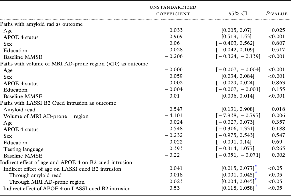
A similar pattern of results was observed for LASSI-L Cued B2 intrusions, as displayed in Table 4. Amyloid positivity, and brain volumes in AD-prone regions exerted effects on Cued B2 intrusions. Greater brain volumes of AD-prone regions were associated with less Cued B2 intrusions. Higher MMSE score was associated with less Cued B2 intrusions. Age and APOE e4 did not have a direct effect on the number of Cued B2 intrusions.
Path analysis of LASSI-L Cued B2 intrusion

* Bootstrap method was used to calculate the CI of the indirect effect due to the non-normality of the product term.
Indirect effects
Neurodegeneration and amyloid load were the two hypothesized mediators in our model. The path from age to brain volumes in AD-prone regions, and the path from brain volumes to cognitive performance (LASSI Cued B1 intrusion errors) were both statistically significant; therefore, neurodegeneration served as a mediator between the relationship of age and cognitive performance as measured by semantic intrusion errors. The indirect effect of age on Cued B1 intrusions through neurodegeneration was 0.027, (95% CI [0.004, 0.055]). The two paths involved (age to amyloid and amyloid to Cued B1 intrusions) were both statistically significant; therefore, amyloid also served as a mediator between the relationship of age and LASSI-L Cued B1 intrusions. The indirect effect of age on Cued B1 intrusions through amyloid was 0.029, 95% CI [0.004, 0.066]. The total indirect effect of age on LASSI B1 intrusions through both mediators was 0.056, 95% CI [0.018, 0.103]. Amyloid load also served as a mediator between the relationship of APOE ϵ4 and cognitive performance on LASSI-L Cued B1 intrusions. The indirect effect of APOE ϵ4 on Cued B1 intrusions through amyloid load was 0.835, 95% CI [0.291, 1.546].
Similar mediating relationships were found for the path analysis of Cued B2 intrusions, with age having an indirect effect on Cued B2 intrusions via MRI neurodegeneration and amyloid load. The total indirect effect of age through both mediators was 0.041, 95% CI [0.015, 0.077]. The indirect effect of APOE ϵ4 on Cued B2 intrusions through amyloid load was 0.53, 95% CI [0.118, 1.058].
Discussion
The present study is the first to examine the direct and indirect effects of age, APOE ϵ4 genotype, amyloid positivity, and volumetric reductions in AD-prone brain regions as it relates to semantic intrusion errors reflecting PSI and the frPSI. A unique feature of our confirmatory path analyses was that other important factors such as sex, education, language of evaluation, and global cognitive impairment were taken into account in the model.
For both LASSI-L Cued B1 and Cued B2 intrusions (sensitive to PSI and frPSI effects), both amyloid positivity and decreased brain volumes in AD-prone regions were directly related to these outcomes. APOE4 status did not evidence direct effects on these LASSI-L cognitive outcomes, but rather exerted their effects on amyloid positivity which in turn related to PSI and frPSI. Based on our models, the indirect effect of APOE ϵ4 genotype on Cued B1 and Cued B2 intrusions was 0.84 and 0.53 more intrusion errors on average. Similarly, age did not have a direct relationship with LASSI-L scores, but exerted its effects indirectly through its effects on amyloid positivity and AD-prone brain volumes. The effect of 8 years older in age (or 1 SD) on PSI and frPSI was on an average 0.44 and 0.32 more intrusion error. Although relatively modest, these effect sizes are both of clinical and theoretical interest, particularly given the overall results of the model.
Amyloid positivity was directly related to LASSI-L intrusions even after controlling for age, sex, education, language of testing, and MMSE. However, when substituting centiloid score for dichotomous visual amyloid read, centiloid score was not associated with LASSI-L intrusions when MMSE was included in the model as a control. It is likely that there is a certain threshold at which increasing centiloid values do not have linear effects on outcomes and that is why we prefer visual reads, which remain the gold standard in the field and for which we have obtained excellent inter-rater reliability (Loewenstein et al., Reference Loewenstein, Curiel, Duara and Buschke2018b).
Our results suggested there is no direct relationship between amyloid load and volumetric loss of AD-prone regions. While amyloid load was correlated with the volume of AD-prone regions, this association disappeared after controlling for age or MMSE score. Although amyloid load has been reported to be associated with regional brain volumes (Storandt et al., Reference Storandt, Mintun, Head and Morris2009), the potential confounding effect of age and global cognitive performance was not accounted for on the relationship between amyloid load and volumetric loss.
Our previous work has not indicated that hippocampal or entorhinal cortex volumes have independent effects when controlling for amyloid positivity. This may reflect difficulties in the reliance of single medial temporal lobe structures in assessing cognitive function across a broad array of disease severity. Consequently, for this analysis, we chose to include additional neocortical areas in the frontal, temporal and parietal regions that have been shown to be signature regions demonstrating atrophy among AD patients, even in the preclinical stage (Dickerson et al., Reference Dickerson2011).
Our findings that the language of testing did not have an association with LASSI-L cognitive measure is consistent with data obtained by Curiel et al., (Curiel Cid et al., Reference Curiel Cid2019) for English versus Spanish-speaking subjects, and emphasizes the lack of language and cultural bias in the LASSI-L paradigm (Capp et al., Reference Capp2019). Age is known to be associated with lower cognitive scores, brain atrophy, and is also associated with increased risk for AD. In the current study, we found that the effect of age on PSI and frPSI, as reflected by LASSI-L intrusions, was indirect, through its relationship with amyloid load and reductions in brain volume. APOE4 status was found to have only an indirect effect on PSI and frPSI intrusions through its effect on amyloid positivity.
The strengths of this study are (1) a large, well worked up sample of participants, (2) comprehensive clinical and neuropsychological evaluations and diagnosis (diagnostic determination was made independently of the LASSI-L), and (3) biomarker evaluation using amyloid PET and MRI scans. While participants in this study did have a broad range of global cognitive impairment, as reflected in their MMSE scores, from CN to dementia, we addressed this potential confounder by controlling for overall cognitive impairment, by entering MMSE into our path analytic models. As indicated in Figure 1, even after accounting for the level of global cognitive status, age continued to have an association with amyloid positivity and MRI volumes, ApoE ϵ4 status continued to be related only to amyloid status, and LASSI-L PSI and frPSI intrusions continued to be directly associated with amyloid positivity and decreased regional brain volumes on MRI scans.
The current results expand findings from samples confined to amnestic MCI (Loewenstein et al., Reference Loewenstein2018a) and extend these findings across the entire range of cognitive function in a large cohort. We believe that calculating an AD-prone volumetric score which includes the medial temporal region, but also includes neocortical regions, as described by Dickerson (Dickerson et al., Reference Dickerson2011), more fully captures the extent of volumetric loss across the full cognitive spectrum. A strength of confirmatory path analyses is that this represents causal modeling in what otherwise would be seen as correlational analyses. In addition to controlling for an extensive list of covariates in the model, the temporal and directionality of these relationships support the causal modeling.
We have shown that PSI and frPSI intrusions have a higher frequency among MCI patients who are amyloid positive versus amyloid negative (Curiel Cid, Reference Curiel Cid2020; Loewenstein et al., Reference Loewenstein2018a). The current investigation demonstrates that volumetric loss in AD-prone regions also has an independent influence on these LASSI-L subscales. Intrusion errors on PSI and frPSI appear to reflect the effects of deficits in source memory and inhibition and possibly a disconnection between medial temporal and frontal lobe structures. For example, Sanchez et al. (Sánchez et al., Reference Sánchez2017) found that among clinically normal middle-age offspring of a parent with late-onset AD, there were limbic-neocortical disconnections (including frontal regions) on fMRI that were related to frPSI intrusions on the LASSI-L. This most likely captures different aspects of AD such as synaptic disconnection (even in areas in which there is not yet discernable volumetric loss on structural MRI), neuroinflammation, or microglial activation. This may also reflect the underlying influences of abnormal tau proteins which may be a part of the AD cascade. Clearly, further research should be conducted that includes measures of both tau and synaptic disconnectivity by fMRI.
The current investigation clearly expands our understanding of the underpinnings of PSI and frPSI intrusions in a large cohort. It should be noted that although we obtained excellent model fit in our confirmatory path analyses, there are other variables that may have functioned as mediators and resulted in equal or better model fit. Candidate variables for future research include functional MRI (fMRI) scans and tau PET scans. This should provide an even greater understanding of the biological determinants of PSI and frPSI on the LASSI-L. We currently focused on specific LASSI-L variables that have previously been shown to be most highly related to AD biology and diagnoses (Loewenstein et al., Reference Loewenstein2018a). Future research should focus on other potentially important measures of the LASSI-L, including retroactive semantic interference (RSI), maximum learning achieved through semantic cues at acquisition and retrieval, as well as delayed recall.
Conflict of interest
None of the authors have conflicts of interest involving this manuscript, although Drs. Loewenstein and Curiel hold intellectual property rights to the LASSI-L at the University of Miami.
Acknowledgements
We wish to acknowlege the contributions of all of our research participants. and study team at Mount Sinai Medical Center, Miami, Beach, Florida.
Source of funding
This research was supported by the National Institute of Aging Grants number 5 P50 AG047726602 1Florida Alzheimer’s Disease Research Center, 1 R01 5R01AG055638-02 and R01 AG061106-02 University of Miami.
Description of authors’ roles
D. Loewenstein designed and carried out the study, supervised the data collection, formulated the research question, and assisted with writing the paper. D. Zheng formulated the research question, carried out the statistical analysis, and wrote the article. R. Curiel-Cid and R. Duara designed the study, carried out the study, supervised the data collection, and edited the paper. M. Kitaigorodsky and E. Crocco carried out the study and reviewed the paper.