INTRODUCTION
The phylum Bryozoa Ehrenberg, 1831 comprises three extant classes, Gymnolaemata, Stenolaemata and Phylactolaemata, but the morphology and ontogeny of the latter is distinctively different from that of the other two; it is represented exclusively by freshwater species and its relationship to the other classes has been questioned by some recent authors (Mundy et al., Reference Mundy, Taylor, Thorpe, Larwood and Nielsen1981; Mackey et al., Reference Mackey, Winnepenninckx, de Wachter, Backeljau, Emschermann and Garey1996; Giribet et al., Reference Giribet, Distel, Polz, Sterrer and Wheeler2000; Glenner et al., Reference Glenner, Hansen, Sørensen, Ronquist, Huelsenbeck and Willerslev2004).
Some bryozoans release numerous small eggs that undergo development in the plankton, but the majority brood their embryos and release mature larvae with a short free-swimming phase. Larval release is induced by light stimulus in most shallow water species. Larval morphology reflects adaptations in relation to locomotion and sensory perceptions and is not related to the morphology of the adult form (Reed, Reference Reed, Giese, Pearse and Pearse1991). Two basic larval types occur among the Gymnolaemata: planktotrophic larvae, commonly referred to as cyphonautes, and lecithotrophic coronate larvae (Zimmer & Woollacott, Reference Zimmer, Woollacott, Woolacott and Zimmer1977). The little that is known about larval morphology in the Stenolaemata is limited to a very few species (Nielsen, Reference Nielsen1971; Santagata, Reference Santagata2008a).
With the exception of the Membraniporoidea all known species that produce a distinctive planktotrophic cyphonautes larva, larval morphology has not been used for taxonomic or systematic purposes in the Bryozoa. Yet, the evolution of larval traits and morphologies might be expected to reflect, for example, the hypothesized switch from planktotrophy to non-planktotrophy (possibly in the Ordovician in stenolaemates, and in the Cretaceous in gymnolaemates; see Taylor & Larwood, Reference Taylor, Larwood, Taylor and Larwood1990), and larval traits might therefore be employed in the classification of extant taxa (Santagata & Zimmer, Reference Santagata and Zimmer2000).
Zimmer & Woollacott (Reference Zimmer, Woollacott, Woolacott and Zimmer1977) presented a classification scheme for bryozoan larvae based on published accounts of 45 species of Gymnolaemata. The classification uses gross morphological features, revealed by histological and scanning electron microscopy (SEM) techniques and recognizes seven main larval types, which do not reflect distinctions at lower taxonomic levels, such as between species. The scheme is important, however, in providing a framework for the classification of further larval types. Recently, Santagata (Reference Santagata2008a) employed SEM, light and confocal laser scanning microscopy (CLSM) to describe four new larval types to be added to Zimmer & Woollacott's classification scheme.
Previously, larval imaging employed either light microscopy (Ryland, Reference Ryland1970) or a combination of light microscopy (LM) and SEM (Reed, Reference Reed, Chia and Rice1977, Reference Reed1988; Reed & Cloney, Reference Reed and Cloney1982; Reed et al., Reference Reed, Ninos and Woollacott1988; Reed & Woollacott, Reference Reed and Woollacott1982, Reference Reed and Woollacott1983). Both SEM and LM methods allow for very detailed morphological examination, but are not very suitable for large-scale surveys of larval morphological types because material preparation may be very time consuming or, as in LM, does not show the surface elements of the larvae adequately.
Epi-fluorescence microscopy, with use of stains which target DNA and mitochondria, has been used to study the surface cells of bryozoan larvae and their superficial morphology (Santagata & Zimmer, Reference Santagata and Zimmer2000). While this method allows a relatively simple and rapid examination of larval surface morphology, it has limitations. One, which is a common feature of epi-fluorescence microscopy, is that a clearly noticeable uniform glow occurs during the imaging of those parts of the specimen that are out of focus. This obscures the fine details of the specimen. Also contamination of the image by sample autofluorescence occurs which is very hard to filter out. Therefore, a simple method of larval imaging is required, suitable for large-scale studies, which would allow identification of larval type based on the already established system or to facilitate recognition of novel larval types.
To this end, a technique for identifying larval morphological characters using CLSM is proposed here. This method is sufficiently rapid, requires little preparation, uses live larvae and has all the advantages of confocal microscopy. Using the proposed method, several larvae were examined and identified using the Zimmer & Wollacott (1977) larval system.
MATERIALS AND METHODS
Bryozoan colonies were collected from locations along the South Wales coast. For an optimization of the method, species were chosen based on those known to produce many larvae and for which the onset of larval release is relatively easy to monitor. Representatives of the following species were studied: Flustrellidra hispida (Fabricius), Alcyonidium spp., Phaeostachys spinifera (Johnston) and Bowerbankia spp. Unfortunately, some taxa (such as Bowerbankia spp.) have a short reproductive period and thus offer a supply of live material for only a rather limited part of the year. The brooding colonies were placed in a light-insulated tank and kept at ambient sea temperature with constant seawater and air supply. Live extraction of larvae was facilitated by the known positive phototaxis of the larvae just after their release. The brooding colonies were transferred into small transparent containers which had a fibre optic light source (to lower the heat shock) directed at one side. On release, larvae would swim towards, and congregate by, the spotlight source where they could be easily collected using a micropipette. Larvae were usually released within 20 minutes of exposure to the light.
Once collected, larvae were anaesthetized using magnesium seawater (MSW). This was prepared using 7.5% w/v MgCl2 6H2O diluted with an equal volume of seawater (Ross & Ross, Reference Ross and Ross1999). It was found that some larvae (in particular the larger ones), when exposed to the laser beam, became active regardless of the time for which they were sedated. Therefore, modification of the concentrations of MgCl2 6H2O in the MSW (7.5%, 10%, 15% and 20%) were evaluated; although higher concentrations of MgCl2 6H2O had a faster immobilizing effect, they also reduced efficacy of staining and at the higher concentrations tended to result in high larval mortality. Therefore, the concentration recommended originally (Ross & Ross, Reference Ross and Ross1999) was used for all types of larvae.
When fully immobilized (a period of approximately 30 minutes) larvae were transferred to a watch glass for staining with Mitotracker orange (MO) CMTMRos (Molecular Probes M7510), a cell-permeant derivative of tetramethylrosamine which binds specifically to mitochondria. Its excitation and emission spectral maxima are 554 nm and 576 nm respectively. This fluorochrome stain was selected as it had been previously tested successfully for imaging bryozoan larvae with epi-fluorescence microscopy (Santagata & Zimmer, Reference Santagata and Zimmer2000). The protocols for stain preparation and working solution concentrations were based on the recommendations of Molecular Probes (Invitrogen) and Santagata & Zimmer (Reference Santagata and Zimmer2000).
The MO stain was diluted and prepared as required immediately before use, as a diluted stain has a relatively short shelf life compared to its lyophilized form. The working solution of the fluorochrome was prepared as follows: 50 µg of MO diluted in 585 µl of 100% DMSO (spectrophotometric grade, Sigma)—to make 200 µM; 150 µl of this solution was mixed with 100 ml of MSW (see above), producing a working solution of approximately 300 nM. All dilutions were performed in containers shielded from light as the fluorochrome is light sensitive.
Once anaesthetized, larvae were stained for approximately 20–30 minutes in MO solution, washed twice in the MSW to remove the fluorochrome, then placed on the slide for imaging.
Although it is possible to image actively moving larvae using a confocal microscope, the resulting images cannot be used for the three-dimensional reconstruction. One of the biggest advantages of using a confocal microscope is the possibility to electronically rebuild a three-dimensional image (3-D) of the entire organism using a collection of optical slices, which can be assembled in z-stacks (vertical axis parallel to the sample plane). For successful 3-D reconstruction, however, the series of images must be taken with the x and y planes fixed (i.e. perpendicular lateral dimensions of the specimen must not change during imaging). Then, once the specimen is fully immobilized, several images representing different slices in the z-axis can be assembled together. Further, once a 3-D image is reconstructed it can be digitally flattened to include all important features observed in an individual slice of the z-stack, thus giving a much fuller and sharper two-dimensional image of the organism than could be achieved with an epi-fluorescence microscope, for example.
Larvae were imaged under an inverted microscope (Carl Zeiss LSM 510 META). A green helium neon 543 nm laser was used for the imaging with the main dichroic beam splitter for 488/543 nm, with an optional secondary dichroic beam splitter set to 545 nm. Manufacturer's instructions were followed for the operation of the microscope. Channel settings (such as pinhole size, detector gain and amplifier offset) were adjusted on a sample-by-sample basis, as was the laser transmission power. The latter was determined in many cases by the condition of the stained material (such as time since larval staining). The stained larvae were placed into an 8-well chambered cover glass (LabTech® II no. 155409) designed specifically for live-cell imaging on an inverted microscope. This cover glass system significantly simplifies preparation of the slides by essentially inverting the positions of the cover glass and slide glass and making the latter redundant, thus potential damage to the larvae by the cover glass was avoided. Additionally, larvae could easily be moved and positioned once under the microscope with a preparation needle or a very fine paintbrush (size 000). An automatic subject-seek function (implemented in the microscope) was not used as it operates by short exposures to the laser and may bleach the sample and weaken the fluorochrome stain; manual stage adjustment was used instead.
Photo-bleaching is a common problem in confocal microscopy (Amos et al., Reference Amos, White and Fordham1987; Longin et al., Reference Longin, Souchier, Ffrench and Bryon1993) and is one of the major drawbacks of using live material, because anti-fading agents (such as buffered glycerol) are usually toxic or require special fixing media and can only be applied to fixed non-live material (Login et al., 1993). Because of the photo-bleaching effect, a balance between period of exposure and image size had to be achieved—higher image resolution (in pixels) required longer exposure to the laser beam and thus bleached the sample more quickly. On average, approximately 20 minutes of working time per sample was enough to bleach the fluorochrome, at which point no further imaging was possible. Thus the resolution chosen (1024 × 1045 pixels, 8 bit depth) was optimal to acquire maximum information without bleaching the specimen. The images are reproduced here as they were taken using the microscope, without any manipulation. The only adjustments were made to the contrast of some images (in order to show less stained organs), and the channel colouring of the images (so called pseudocolour) was changed (e.g. red) to vary the relative perception of contrast of some organs. For each species, larval type was identified according to the system of Zimmer & Woollacott (Reference Zimmer, Woollacott, Woolacott and Zimmer1977). The improvements of the CLSM method over epi-fluorescence microscopy are clear from the images both in electronic format and in print.
RESULTS AND DISCUSSION
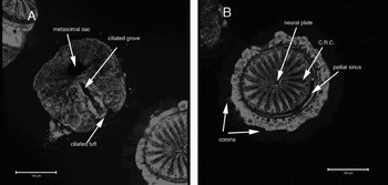
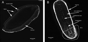
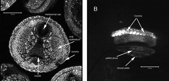
Some representative images are reproduced here. The larvae of Phaeostachys spinifera imaged using the CLSM method are shown in Figure 1A, B. These show many details very clearly: the metasomal sac, ciliated grove with a ciliated tuft, neural plate, ciliated ray cells, pallial sinus and corona are visible. This larva belongs to Type E of Zimmer & Woollacott (Reference Zimmer, Woollacott, Woolacott and Zimmer1977). Three-dimensional image reconstruction of Flustrellidra hispida (Figure 2A) shows many surface characters. Once a 3-D image is processed it can be rotated to a required angle to show particular characters, and then electronically flattened for reproduction in print. For example the images in Figure 2A, B were flattened from the lateral–aboral and oral sides, respectively. The larva belongs to a shelled lecithotrophic type, and the valves, metasomal sac, neural pore and corona are clearly visible. Figure 2B shows an image of a Flustrellidra hispida larva viewed from its oral pole. This image, although giving a good resolution at the posterior end of the larva is indistinct at the anterior part (indicated as M.A. on Figure 2B). This was caused by a sudden movement of the larva during the imaging and resulted in the distortion of the x–y plane and as a result, misalignment of the z-stack images during the three-dimensional assembly. It is an example of a potential problem associated with sudden movement of larvae during z-stack image acquisition. Figure 3A, B shows the larva of Alcyonidium hirsutum (Fleming) in oral and lateral views respectively. Here the metasomal sac, oral epithelium and cells of the corona are clearly seen, as well as the opening of the mouth. This larva fits best Type E in the classification of Zimmer & Woollacott (Reference Zimmer, Woollacott, Woolacott and Zimmer1977).
(A) Confocal laser scanning microscopy (CLSM) image of the oral pole of a Phaeostachys spinifera Type E larva, stained with Mitotracker® orange. The image is pseudocoloured; (B) CLSM image view of the aboral pole of a Phaeostachys spinifera Type E larva, stained with Mitotracker® orange.

(A) Confocal laser scanning microscopy (CLSM) lateral–aboral view image of a Flustrellidra hispida shelled lecithotrophic larva, stained with Mitotracker® orange; (B) CLSM image of a lateral pole view of a Flustrellidra hispida shelled lecithotrophic larva, stained with Mitotracker® orange. Misaligned z-stack planes (M.A.) are marked by a yellow line. These were caused by sudden movement of the larva during imaging; see text for discussion.

(A) Confocal laser scanning microscopy (CLSM) image of the oral pole view of an Alcyonidium hirsutum Type E larva stained with Mitotracker® orange. The image is pseudocoloured. Parts of several other larvae can be seen in the periphery; (B) CLSM lateral view image of an Alcyonidium hirsutum Type E larva stained with Mitotracker® orange.

The three-dimensional reconstruction technique is somewhat limited by the size of larvae. The larger the object under the microscope, the more difficult it is to perform a complete scan of all focal planes, therefore requiring several independent images to be taken from different larvae positioned at different angles. Thus, for identification, a larva should be imaged from the oral and aboral poles as a minimum, and preferably a lateral image should be taken as well. This enables sufficiently precise collection of external morphological characters required for the identification of larval type based on the Zimmer & Woollacott (1977) system. This is especially true for larvae with somewhat similar morphology, for example those larvae belonging to Type E or Type AE. In some species, such as Flustrellidra hispida, the larval type (i.e. shelled lecithotrophic) can be clearly identified because of its particularly distinct characters.
The CLSM method in combination with a mitochondrial stain showed good results for the surface imaging of bryozoan larvae and demonstrated that it could be used for quick assessment of major larval morphological characters and identification of larval types based on the system of Zimmer & Woollacott (Reference Zimmer, Woollacott, Woolacott and Zimmer1977). Using this method, several larvae can be imaged in an hour for later classification. This method complements recent studies that employed CLSM to investigate more complex details of larval morphology, such as musculature, although requiring fixation of larvae (Gruhl, Reference Gruhl2008; Santagata, Reference Santagata2008a, Reference Santagatab; Gruhl et al., Reference Gruhl, Wegener and Bartolomaeus2009). Additionally, the method described here could also be employed to survey other marine invertebrate larvae of comparable size.
New fluorochrome and other staining techniques have been developed recently and could be used to extend the methods used here. In particular some attention should be given to the BODIPY® 505/515 stain (Molecular Probes). This stain belongs to a group of membrane fluorochromes, which are fluorescent analogues of phospholipids capable of incorporating themselves into cell membranes. These are relatively new stains that have shown good results for cytoplasmic staining with some model organisms such as zebrafish embryos (Cooper et al., Reference Cooper, Szeto, Sommers-Herivel, Topczewski, Solnica-Krezel, Kang, Johnson and Kimelman2005). Their other advantage is a very low photobleaching rate, which means that the stain can be used for a prolonged time, and thus offers better imaging of bryozoan larvae. In addition, fluorescent semiconductor nanocrystals ‘quantum dots’ (artificially created semiconductors which can be adjusted to emit required spectra) promise to solve the problem of photo-bleaching (Dubertret et al., Reference Dubertret, Skourides, Norris, Noireaux, Brivanlou and Libchaber2002).
ACKNOWLEDGEMENTS
We thank NERC (Grant no. NER/S/J/2002/12005) and University of Wales, Swansea for financial support. We are also grateful to Dr Peter J. Hayward for assistance during the project and for valuable suggestions and comments on the manuscript.