Introduction
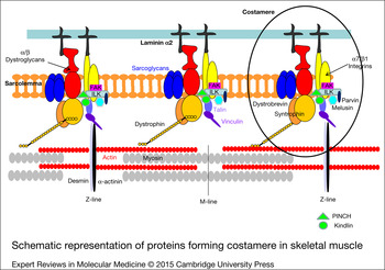
The concept of the costamere as a morphological structure in striated muscle was first introduced by Pardo et al. in 1983 (Ref. Reference Pardo, Siliciano and Craig1). The observation under an electron microscope of protein bands perpendicular to the longitudinal axis of the muscle fibre, reminiscent of ribs (‘costa’ in Latin), led them to coin the name costamere (Ref. Reference Pardo, Siliciano and Craig1). It was found that these structures are located at the subsarcolemmal level, aligned with the myofibril Z-discs (Refs Reference Pardo, Siliciano and Craig1, Reference Nelson, Colaco and Lazarides2, Reference Nelson and Lazarides3). Similar structures are oriented transversely over the M lines of the contractile apparatus and oriented parallel to the long axis of the myofibre (Ref. Reference Bloch4) (Fig. 1).
Schematic representation of proteins forming costamere in skeletal muscle. FAK, focal adhesion kinase; ILK, integrin-linked kinase; PINCH, particularly interesting cysteine- and histidine-rich protein.

Functions of costameres
Possible functions of costameres may include the assembly and stabilisation of sarcomeres (Refs Reference Pardo, Siliciano and Craig1, Reference Danowski5, Reference Trimarchi6). Specifically, these protein complexes, associated with the sarcolemma, enable muscle adhesion to the extracellular matrix (Ref. Reference Ervasti7) and provide mechanical linkage. This linkage both distributes contractile forces from the sarcomere to the basal lamina (‘inside-out’) (Refs Reference Danowski5, Reference Trimarchi6) and transmits externally applied forces to the extracellular matrix inside the myocytes (‘outside-in’) (Ref. Reference Mansour8). Given this, it could be expected that defects in costameres would compromise muscle strength directly. These defects would reduce the efficiency of the transmission of lateral forces, and/or indirectly, increase the likelihood of damage to the sarcolemma, resulting in degeneration or death of the myofibre (Ref. Reference Bloch4).
The process of sarcomere assembly is complex and remains poorly understood (Ref. Reference Rui, Bai and Perrimon9). In the initial stages of assembly, there are small aggregates associated with the membrane, known as Z-bodies, which will mature into Z-discs (Ref. Reference Heuson-Stiennon10). Integrins, α-actinin and the constituents of binding sites for integrin, vinculin and talin are the first proteins that can be observed with a periodic pattern in the plasma membrane (Refs Reference Tokuyasu11, Reference Fujita, Nedachi and Kanzaki12). There is abundant evidence suggesting that integrin-binding sites are where myofibril assembly starts in vivo. Sparrow and Schock (Ref. Reference Sparrow and Schock13) suggested that, within the myofibril maturation process, protocostameres are the site of early integrin binding and, hence, of the initiation of myofibril assembly, although other authors proposed an alternative model based on the assembly of multiple latent protein complexes (Ref. Reference Rui, Bai and Perrimon9).
The correct alignment of costameres with the Z- and M-lines is dependent on other structures in the network of filamentous proteins of the cytoskeleton, namely, the family of the intermediate filaments (IFs) (Refs Reference Ku14, Reference Omary, Ku and Toivola15). These are responsible for binding between the sarcolemma and the myofibrils adjacent to the Z-lines (Refs Reference Pierobon-Bormioli16, Reference Shear and Bloch17, Reference Street18) and presumably also the M-lines (Refs Reference Pierobon-Bormioli16, Reference Li19). Desmin, together with other associated proteins, synemin and paranemin, form the IFs of the Z-lines (Refs Reference Lazarides and Hubbard20, Reference Capetanaki and Milner21) (Fig. 1). The complex process of assembly is genetically regulated by a specific process that involves transcription factors: myocyte enhancer factor-2 (MEF2), serum response factor (SRF) and histone deacetylase (Ref. Reference Estrella and Naya22).
First, MEF2 is a transcription factor (Refs Reference Black and Olson23, Reference Naya and Olson24, Reference Potthoff and Olson25) required for the terminal differentiation of cardiac, skeletal and smooth muscle (Refs Reference Bour26, Reference Lilly27, Reference Ewen28). MEF2A, in particular, plays an essential role in muscle regeneration in adult mice through direct regulation of the microRNA cluster, the Gtl2–Dio3 locus. A subset of the Gtl2–Dio3 miRNAs represses secreted Frizzled-related proteins, inhibitors of Wnt signalling (Ref. Reference Snyder29). Second, SRF is a broadly expressed transcription factor with an essential role in the differentiation of mesoderm-derived tissues, such as muscle (Ref. Reference Miano30). In floxed SRF mice, there is dysregulation of genes encoding costamere, sarcomere and numerous other cytoskeletal proteins (Ref. Reference Balza and Misra31), such as dystrophin, α-dystrobrevin, integrin β1, melusin and β-sarcoglycan (Ref. Reference Estrella and Naya22).
As well as gene regulation through transcription factors, mechanical stimuli play an important role in regulating the expression of costamere components (Refs Reference Fluck32, Reference Gordon, Fluck and Booth33, Reference Chopard, Pons and Marini34, Reference Chopard35, Reference Anastasi36), their level of expression also varying between fibre types (Refs Reference Shear and Bloch17, Reference Bozyczko37, Reference Schroder38, Reference Schroder38, Reference Williams, Resneck and Bloch39, Reference Fluck40, Reference Li41).
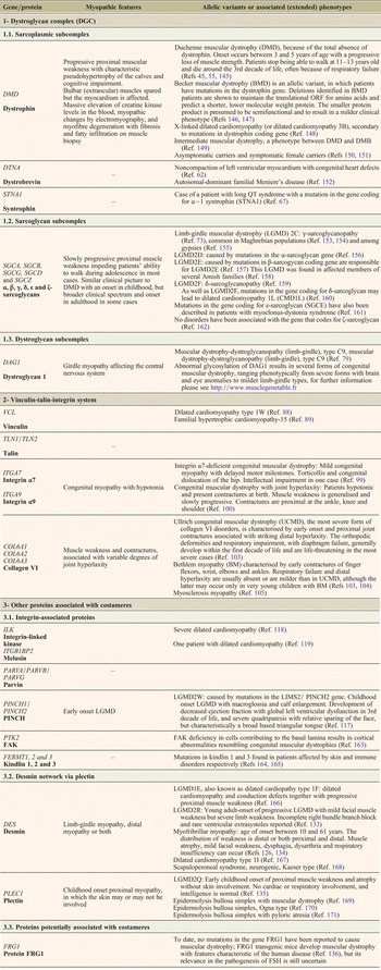
To summarise, the assembly of sarcomere proteins is a highly regulated, complex and delicate process, depending on many factors, and abnormalities in these factors, for example, as a result of mutations of genes coding for some of their components, produce a range of different myopathic processes (listed in Table 1). Below, we describe the main constituent proteins of costameres, their location within the cell and their involvement in these myopathic processes.
Muscle disorders because of mutations of genes coding costamere components

Main components of costameres
Costameres are principally composed of two complexes, the dystrophin–glycoprotein complex and the vinculin–talin–integrin system. Both regulate the interaction between the cytoskeleton and the extracellular matrix in skeletal muscle in adults (Ref. Reference Anastasi42).
The intracellular actin fibres, which are part of the contractile apparatus, are bound to laminin of the extracellular matrix by dystrophin (Refs Reference Campbell43, Reference Matsumura and Campbell44, Reference Monaco45, Reference Suzuki46). Integrin α7β1, a transmembrane laminin receptor, also helps to link the extracellular matrix and the cytoskeleton (Refs Reference Song47, Reference Song48, Reference Hodges and Kaufman49) (Fig. 1).
In addition to the aforementioned two complexes, other proteins, including desmin, plectin and melusin, are involved and these are described in more detail below.
The dystrophin–glycoprotein complex (DGC)
This structural unit is composed of dystrophin and a series of proteins (dystroglycans α and β, sarcoglycans α−ε, sarcospan, syntrophins α1, β1 and β2, and two isoforms of a subsarcolemmal protein, α-dystrobrevin-1 and -2) that form a complex for binding cytoplasmic myofibrillar contractile elements and proteins of the extracellular matrix, providing structural support to the sarcolemma (Refs Reference Yoshida50, Reference Ohlendieck51). Other more peripheral components of the complex include neuronal nitric oxide synthase (nNOS), caveolin-3 and laminin α−2 (Refs Reference Anastasi42, Reference Watkins52).
In the relation to the DGC complex, three sub-complexes can be identified, on the basis of different biochemical characteristics and localisation: the sarcoplasmic sub-complex, the sarcoglycan (transmembrane) complex and the dystroglycan sub-complex (Ref. Reference Anastasi53).
The sarcoplasmic subcomplex
This is composed of proteins present in the sarcoplasm of muscle fibres, mainly dystrophins, dystrobrevins and syntrophins.
Dystrophin
Dystrophin is a cytoskeletal protein found on the inner surface of the sarcolemma of skeletal muscle (Ref. Reference Kunkel54, Reference Hoffman55, Reference Beggs and Kunkel56). Its amino-terminal residue is a cytoplasmic actin-binding domain, whereas the C-terminal domain is associated with a transmembrane complex of glycoproteins called dystrophin-associated glycoproteins (DAGs), some of which interact directly with elements of the extracellular matrix (Ref. Reference Campbell43). Absence or dysfunction of dystrophin leads to an increase in the fragility of the membrane, in turn, weakening the transmission of mechanical forces through the sarcolemma. The result is myofibrillar necrosis, leading to a succession of cycles of degeneration and regeneration, exhausting the regenerative capacity of the muscle. This is accompanied by a loss of myofibrils, muscle being replaced by fibrous and fatty tissue, as observed in patients with Duchenne and Becker muscular dystrophies.
Dystrobrevins
Dystrobrevins are expressed in various types of tissue, including the brain and muscle. The α-dystrobrevin isoform is expressed in muscle and is homologous to the cysteine-rich C-terminal domain of dystrophin (Refs Reference Wagner, Cohen and Huganir57, Reference Blake58). This isoform is directly associated with dystrophin and the sarcoglycan complex (Refs Reference Sadoulet-Puccio, Rajala and Kunkel59, Reference Yoshida60). Nevertheless, to date, no myopathies have been described secondary to mutations in the α-dystrobrevin gene (Ref. Reference Nakamori and Takahashi61), although some cases of left ventricular noncompaction-1 have been reported (Ref. Reference Ichida62) (Table 1).
Syntrophin
Syntrophins are a family of cytoplasmic membrane-associated adaptor proteins directly associated with dystrophins and related proteins (Ref. Reference Piluso63). They serve as a link between the extracellular matrix and the intracellular downstream targets and cell cytoskeleton by interacting with F-actin. They play an important role in regulating the postsynaptic signal transduction, sarcolemmal localisation of nNOS, EphA4 signalling at the neuromuscular junction, and G-protein-mediated signalling (Ref. Reference Bhat, Adams and Khanday64). There are three isoforms encoded by different genes expressed in muscle: α−1, β−1 and β−2 (Refs Reference Adams65, Reference Ahn66). To date, only one type of heart disorder (long QT syndrome) related to mutations in these genes has been described, this being attributable to an α−1 syntrophin mutation (Ref. Reference Ueda67).
Sarcoglycan subcomplex
Sarcoglycans are glycosylated proteins with a transmembrane domain (Ref. Reference Ozawa68) that needs to be correctly assembled in a complex together with dystrophin (Ref. Reference Crosbie69) to maintain the sarcolemma (Refs Reference Holt and Campbell70, Reference Hack71). The sarcoglycan subcomplex is composed of the α, β, γ, δ, ε and ζ-sarcoglycan transmembrane proteins (Refs Reference Yoshida50, Reference Ettinger, Feng and Sanes72), of which α and δ-sarcoglycans are only expressed in muscle tissue, whereas the rest are more widely distributed (Ref. Reference Noguchi73). This complex has two functions: on the one hand, it has mechanical and non-mechanical roles in the interaction between the extracellular matrix, sarcolemma and cytoskeleton (Ref. Reference Ozawa74); and, on the other hand, it mediates membrane targeting and the stabilisation of the sarcospan protein. When mutations result in partial or total loss of sarcoglycans α, β or γ, sarcospan is also affected (Ref. Reference Crosbie75). Four types of autosomal recessive limb-girdle muscular dystrophies have been described secondary to mutations in genes that code for components of this complex (Ref. Reference Bonnemann, McNally and Kunkel76) (Table 1).
Dystroglycan subcomplex
Dystroglycans include two proteins that are part of a complex of glycoproteins associated with dystrophin. They are both coded by the same gene, DAG1 (Ref. Reference Ibraghimov-Beskrovnaya77), which, after post-translational processing, produces a transmembrane protein, β-dystroglycan and an extracellular protein, α-dystroglycan.
The dystroglycan complex strengthens the physical connection between the cytoplasmic proteins that bind to the actin cytoskeleton and components of the basement membrane, proteins of the extracellular matrix that contain globular domains of laminin (for example, laminin, agrin and neurexin) (Refs Reference Ibraghimov-Beskrovnaya77, Reference Barresi and Campbell78).
Although genetic dystroglycan deficiency leads to muscular dystrophy with cognitive impairment (Ref. Reference Hara79), several forms of congenital muscular dystrophy are the result of abnormalities in the glycosylation of mucin-like domains of α-dystroglycan, essential for its correct functioning as a receptor of the extracellular matrix in various types of tissue, including skeletal muscle and brain (Refs Reference Barresi and Campbell78, Reference Michele80) (Table 1).
The vinculin–talin–integrin system
Vinculin and talin are two cytoskeletal proteins that are essential for the linkage of actin filaments to the plasma membrane (Ref. Reference Schwartz81), whereas integrins are heterodimers composed of two subunits, α and β (Ref. Reference Hynes82), involved in the cascade of intracellular signalling (Ref. Reference Schwartz, Schaller and Ginsberg83).
Vinculin
Vinculin is associated with the cytoplasmic side of the plasma membrane, interacting with talin and α/β-catenins (together with cadherin) (Ref. Reference Lu84, Reference Burridge and Mangeat85, Reference Weiss86). In addition, there is evidence to suggest that vinculin plays a critical role in the regulation of integrin clustering, force generation and strengthening of adhesion (Ref. Reference Peng87). Mutations in the gene that codes for vinculin lead to dilated cardiomyopathy type 1W and to familial hypertrophic cardiomyopathy-15 (Refs Reference Olson88, Reference Vasile89), but have not yet been found to cause disorders affecting skeletal muscle (Table 1).
Talin
Talin is a dimeric protein that contains different domains for binding to the β-integrin subunits, F-actin, Wech, H-Ras, layilin, PIPK, focal adhesion kinase (FAK), actin, vinculin and muscle-specific α-synemin (Ref. Reference Critchley90). Hence, talin links integrins with cytoskeletal actin (Ref. Reference Zemljic-Harpf, Manso and Ross91), being essential for the structural integrity of the attachment of cells to the extracellular matrix (Ref. Reference Zhang92). As well as this structural function, talin modulates ligand binding to integrins and is involved in signal transduction, recruiting signalling proteins, such as FAK and phosphatidylinositol-(4)-phosphate 5-kinase type Iγ (PIPKIγ) to focal adhesions. Talin is coded by two genes that code for two isoforms (Tln1 and Tln2), with 74% sequence identity. Tln 1 is ubiquitous while Tln 2 is expressed in fewer tissues, mainly the heart, brain and skeletal muscle (Ref. Reference Zemljic-Harpf, Manso and Ross91). No disorders are known to be associated with mutations in either of these isoforms.
Integrin
The vinculin–talin–integrin system includes various transmembrane heterodimeric receptors that play a very important role in cell adhesion (Ref. Reference Hynes82). They link the extracellular matrix to the actin cytoskeleton and facilitate the bidirectional transmission of signals between the extracellular matrix and the cytoplasm (Refs Reference Schwartz, Schaller and Ginsberg83, Reference Yamada and Miyamoto93).
Integrins are connected to actin filaments through talin, vinculin and other related proteins, such as α-actinin, integrin-linked kinase (ILK), filamin and tensin (Ref. Reference Legate, Wickstrom and Fassler94). As well as their structural function, integrins recruit signalling proteins for the transduction of mechanical stimuli through FAK, melusin, paxillin, src, cas and PIPKI (Ref. Reference Wiesner, Legate and Fassler95).
Integrins play an important role in the process of muscular differentiation and, in particular, are mediators of cell adhesion and migration (Ref. Reference Trimarchi6). Various different heterodimers have been identified, composed of α and β chains. The α/β combination determines the binding specificity of the ligand of the integrin heterodimer for various different proteins of the extracellular matrix, including fibronectin, laminin and collagens. The most common isoforms in skeletal muscle of adults are α7B and β1D, the latter being found with vinculin in skeletal and cardiac muscle (Refs Reference Belkin96, Reference Belkin97). The affinity of integrin for its ligands depends on its activation, this usually occurring through the cytoplasmic tail of β-integrins that can be regulated by various biochemical signalling pathways (Ref. Reference Tadokoro98).
During myogenesis, two isoforms of integrin 1β are expressed. The isoform 1A is expressed in myoblasts and is under-expressed during tissue formation, whereas isoform 1D is expressed during fusion and its expression increases during the maturation of myotubes, completely displacing the isoform 1A in mature myotubes (Ref. Reference Belkin96).
Two types of congenital muscular dystrophy have been found to be because of mutations in genes coding for integrins, one to mutations in ITGA7 (Ref. Reference Hayashi99), and the other, to mutations in ITGA9 (Ref. Reference Tetreault100) (Table 1).
On the other hand, as previously mentioned, integrins interact with collagens and it is worthwhile to underline that collagen VI plays a fundamental role in costameric function maintenance, given that proteins essential for mechanotrasduction are altered in collagen VI null mice and collagen VI myopathy (Refs Reference De Palma101, Reference De Palma102, Reference Demir103, Reference Bethlem and Wijngaarden104, Reference Merlini105) (Table 1).
Other proteins associated with costameres
As well as the aforementioned complexes, other proteins are indirectly involved in the structure of the costamere, either by being associated with integrins, or by being part of or associated with IFs.
Integrin-associated proteins
These include a complex formed by ILK, particularly interesting cysteine- and histidine-rich protein (PINCH) and parvin, the IPP complex (Ref. Reference Legate106).
Regarding the proteins in the IPP complex, ILK regulates integrin-mediated signalling, and PINCH and parvins are adaptor proteins. ILK was identified in 1996 among other proteins that bound to the cytoplasmic region of integrin β1 (Ref. Reference Hannigan107). It is a serine–threonine protein kinase that, as well as binding to PINCH and parvins, also binds to the cytoplasmic regions of integrins β1 and β3, regulating integrin-mediated transduction signal (Refs Reference Hannigan107, Reference Tu108). PINCH has two isoforms, PINCH1 and 2 (Refs Reference Rearden109, Reference Zhang110) and together with ILK, is essential for controlling the change of shape, mobility and survival of cells (Ref. Reference Fukuda111). Parvins are a family of proteins composed of α-parvin (also known as actopaxin and calponin homology-containing ILK-binding protein), β-parvin (affixin) and γ-parvin that bind to ILK through a calponin-homology domain (Refs Reference Nikolopoulos and Turner112, Reference Yamaji113, Reference Olski, Noegel and Korenbaum114).
Another integrin-associated protein is melusin, a protein coded by the ITGB1BP2 gene. Melusin binds to the cytoplasmic domain of the integrin β1 subunit in costameres, acting as a specific biomechanical sensor of signal transduction (Ref. Reference Brancaccio115). In the cardiac muscle, the effect of melusin is to prevent heart failure under biomechanical stress (Ref. Reference Brancaccio116).
Some mutations in the genes coding for proteins of this complex have been associated with muscular dystrophies (Ref. Reference Nigro and Savarese117) and with heart disease (Refs Reference Knoll118, Reference Ruppert119), (Table 1).
Another protein closely associated with integrins is FAK. The phosphorylation of this protein activates hypertrophic signalling through Akt, extracellular signal-regulated kinase 1/2 and Jun N-terminal kinase/c-Jun pathways. The FAK signalling pathway regulates the expression of MEF2 transcription factors, which in turn regulate the expression of sarcomeric proteins (Ref. Reference Nadruz120) and it has been confirmed that FAK inhibition results in an abnormal maturation of costameres and myofibrils (Ref. Reference Quach and Rando121).
Finally, kindlin is a protein that binds directly to β-integrins and ILK. It has three isoforms (kindlin 1, 2 and 3) in vertebrates, although the most widely expressed isoform in cardiac and skeletal muscle is kindlin 2 (Ref. Reference Ussar122). Kindlin is found at costameres and plays an important role in myoblast differentiation in vitro (Ref. Reference Dowling123).
Desmin and Plectin
Desmin is the most important protein in IFs for the organisation of the costamere in the muscle (Ref. Reference O'Neill124). It surrounds Z-discs and links the contractile apparatus with the sarcolemma, cytoplasmic organelles and the nucleus (Ref. Reference Kouloumenta, Mavroidis and Capetanaki125) providing structural solidity and integrity to the cell during force transmission and mechanochemical signalling (Ref. Reference Goldfarb and Dalakas126).
Plectin, a protein associated with the cytoskeleton, is abundantly expressed in various types of tissue and cell types (Refs Reference Wiche127, Reference Wiche128). It has been suggested that it may interact directly with actin filaments, microtubules, microtubule-associated proteins, α-spectrin, and integrin β4, as well as most IFs (Refs Reference Herrmann and Wiche129, Reference Svitkina, Verkhovsky and Borisy130, Reference Rezniczek131).
Myofibre integrity depends on the desmin network targeting Z-discs and costameres via plectin isoforms. Among the different isoforms of plectin, 1 and 1f are localised at costameres. Plectin deficiency results in desmin detaching from Z-discs, costameres, mytochondria and nuclei; depending on which plectin isoform is lacking, desmin aggregates have a distinct morphology and form in distinct cytoplasmic compartments (Ref. Reference Konieczny and Wiche132).
Desmin deficiency leads to limb-girdle muscular dystrophy, LGMD2R (Ref. Reference Cetin133) and an allelic variant of myofibrillar myopathy (Ref. Reference Goldfarb134), whereas plectin deficiency produces LGMD 2Q (Ref. Reference Gundesli135) (Table 1).
Protein potentially associated with costameres
Facioscapulohumeral muscular dystrophy region gene 1 (FRG1) protein is one of a group of proteins whose localisation and function are still to be clearly defined. There is evidence that it participates in the processing of RNA (Ref. Reference Gabellini136) and it may be involved in F-actin bundling in Caenorhabditis elegans (Ref. Reference Liu137). It has been suggested that FRG1 in C. elegans is a multi-function protein present in various subcellular niches, but primarily in nucleoli and dense bodies. In this organism, the most important function of dense bodies is transduction of mechanical forces, essentially by anchoring sarcomeric actin to the extracellular matrix; that is, they are structures with a similar function to that of the combination of Z-discs and costameres in vertebrates (Refs Reference Liu137, Reference Lecroisey, Segalat and Gieseler138). Therefore, as well as in C. elegans, FRG1 could be involved in costameres in vertebrates.
Summary
The structure and role of costameres and their involvement in the pathophysiology of skeletal muscular disorders are still not well understood. Given that this system includes many molecules closely associated spatially and with two types of function (structural support and signal transduction), it is likely that abnormalities in any of the components have an impact on the stability of the whole system, with consequences for the structure of the membrane, or the functionality of the contractile apparatus, or both at the same time. This has been confirmed in dystrophinopathies, in which the absence of dystrophin leads to the disappearance of dystoglycans/sarcoglycans, and in sarcoglycanopathies, in which the lack of the protein as a result of mutation leads to a secondary reduction or disappearance of the remaining sarcoglycans. Moreover, a similar phenomenon has also been reported when contactin-1 gene is mutated, as this results in reduced expression of β2-syntrophin and α-dystrobrevin (Ref. Reference Compton139).
Regarding potential therapies for myopathic processes because of defects in costamere proteins, different approaches have been widely tested. However, clinical trials have only been performed for a few of them. In the case of Duchenne muscular dystrophy (DMD), for example, molecular-based pharmacologic therapies to correct gene products have been applied (Refs Reference Malik, Rodino-Klapac and Mendell140, Reference Li141). In sarcolgycanopathies, an adeno-associated virus has been used to transfect the exogenous gene (Ref. Reference Herson142) and in collagen VI myopathies, compounds that modulate the activity of the mitochondrial permeability transition pore have been assessed (Refs Reference Merlini and Bernardi143, Reference Zulian144). Unfortunately, just a few of these approaches showed the sought outcome so far.
Further research is required on selective silencing and at different stages in the processes of myogenesis and differentiation of skeletal muscle in physiological and pathological conditions to clearly establish the role of these structures in skeletal muscle. Current knowledge suggests that these proteins could be therapeutic targets for preventing the ‘de-structuring’ of the sarcolemma that leads to the degeneration of myofibres in patients with muscular dystrophy.
Acknowledgements and funding
This work was supported by grants from the Health Research Fund (FIS: PI13/00722 and PI14-00436) of the Spanish Ministry of Economy and Competitiveness and the European Union (ERDF). The work was also supported in part by the Center for Networked Biomedical Research on Neurodegenerative Diseases (CIBERNED), Carlos III Health Institute, Spanish Ministry of Economy and Competitiveness.