Newcastle disease (ND)
Newcastle disease (ND) is an economically important and highly infectious disease of both wild and captive birds (Saif et al., Reference SAIF, FADLY, GLISSON and MCDOUGALD2008). ND results from infections with virulent Newcastle disease viruses (NDV), having intracerebral pathogenicity indices (ICPI) of ≥0.7 in day-old chickens (Gallus gallus) and/or having multiple basic amino acids (at least three arginine (R) or lysine (K) residues) at the C-terminus of the fusion protein cleavage site, starting at position 113, along with a phenylalanine at position 117 (OIE, 2009). This disease is one of the most important infectious diseases of poultry because of its worldwide distribution and the potential for devastating loses. It occurs on at least six of the seven continents of the world and is enzootic in many countries. Worldwide, outbreaks of vNDV are a constant threat to poultry with clinical infection. Clinical manifestation of ND in chickens varies significantly among isolates. Infection with virulent viruses causes three well defined clinical forms: (1) viscerotropic velogenic, which is characterised by acute lethal infections, usually with haemorrhagic lesions in the intestines of dead birds, (2) neurotropic velogenic, which is characterised by high mortality following respiratory and neurological disease, but where gut lesions are usually absent, and (3) mesogenic, which causes respiratory and neurological signs, but low mortality. The presence of virulent strains (velogenic and mesogenic strains) in poultry requires monitoring and control measures even in countries where they are endemic because the existence of the virus severely impacts commercial productivity and the international trade in poultry and poultry products. Low virulence NDV (loNDV) is often referred to as lentogenic and are endemic in many countries. In addition to being classified as being of low virulence, causing mild infections of the respiratory tract, lentogens can be classified as asymptomatic enteric, in which replication appears to be primarily in the gut without respiratory signs.
Newcastle disease is considered as a major infection of commercial poultry in Pakistan among all the viral, bacterial and parasitic diseases (Rehman et al., Reference REHMAN, FAWAD, ABBAS, NAHEED, SIDDIQUE, AFZAL, MUNAWAR, AURANGZEB, ATIF and HAQ2013). Although the prevalence of ND in broiler and layer birds remains higher throughout the year, it reaches its plateau during seasonal stress periods (January-February, June-July). During September 2011-January 2012, ND killed 45 million chickens resulting in a loss of approximately US$65 million to the poultry industry in Punjab (Farooq et al., Reference FAROOQ, SALIHA, MUNIR and KHAN2014; Ali et al., Reference ALI, MUNEER, HUSSAIN, REHMANI, YAQUB and NAEEM2014). An outbreak of ND in seven southern districts of the Sindh province of Pakistan resulting in the death of approximately 167 wild peacocks in just three weeks (Abbas et al., Reference ABBAS, SHABBIR, KHALIQ, ALI and MUNIR2014; Siddique et al., Reference SIDDIQUE, NAEEM, ABBAS, ALI MALIK, RASHID, RAFIQUE, GHAFAR and REHMAN2013). The disease has affected Rock Pigeons (Columba livia) and Indian Peafowl (Pavo cristatus) in the province of Sindh. This review aims to elucidate crucial issues regarding the emergence and evolution of vNDV in the poultry industry of Pakistan. In addition, recent advances and challenges of vaccination against NDV are discussed.
Newcastle disease virus (NDV)
Newcastle disease virus (NDV), synonymous with avian paramyxovirus- 1 (APMV-1), is a member of the Avulavirus genus in the Paramyxoviridae family. Encompassing a diverse group of single-stranded, negative sense, non-segmented, enveloped RNA viruses of approximately15.2kb, NDV has a broad host range and are known to infect over 200 bird species (Alexander, Reference ALEXANDER2001). The NDV genome encodes for six major structural proteins: nucleocapsid (NC), phosphoprotein (P), matrix (M), fusion (F), hemagglutinin-neuraminidase (HN), the RNA dependent RNA polymerase (L), and for a seventh protein (V) that is produced by a frame shift within the P coding region. While the virulence of NDV is dependent on multiple genes, the fusion protein cleavage site is the critical site responsible for major changes in virulence (de Leeuw et al., Reference DE LEEUW, KOCH, HARTOG, RAVENSHORST and PEETERS2005). The less virulent viruses (loNDV) have fewer basic amino acids at this site, with leucine replacing phenylalanine at position 117 (Alexander et al., Reference ALEXANDER, ALDOUS and FULLER2012). Using reverse genetic techniques, it has been shown that the phenylalanine at position 117 and basic amino acids surrounding Q 114 are necessary for virulence (de Leeuw et al., Reference DE LEEUW, KOCH, HARTOG, RAVENSHORST and PEETERS2005). Newcastle disease virus (NDV) was first reported in 1926, in Java, Indonesia and Newcastle-on-Tyne, England (Doyle, Reference DOYLE1927), and since then various genotypes have been responsible for different ND panzootics. NDV is distributed worldwide and its continual presence in multiple avian species presents a constant threat to all poultry industries and other activities that involve the raising or keeping of birds (Anonymous, 2011).
NDV isolates may be classified into genotypes based on either the complete genome sequences or the full fusion protein sequences (Diel et al., Reference DIEL, DA SILVA, LIU, WANG, MILLER and AFONSO2012). Two different systems of classifying NDV are currently utilised worldwide with no consensus as to which is more appropriate. A system suggested by Aldous grouped NDV into six lineages and 13 sublineages and later, three additional sublineages were added (Snoeck et al., Reference SNOECK, OWOADE, COUACY-HYMANN, ALKALI, OKWEN, ADEYANJU, KOMOYO, NAKOUNE, LE FAOU and MULLER2013; Cao et al., Reference CAO, GU, ZHANG, LIU and LIU2013). A second system classified NDV into two major divisions represented by class I and class II. Both systems of classification were based on similar genomic information, therefore discrepancies between the two classification systems are nominal. At this time, ND viruses are grouped into one genotype for class I NDV isolates, and eighteen (I-XVIII) genotypes for class II NDV isolates, some with sub-genotypes (Courtney et al., Reference COURTNEY, SUSTA, GOMEZ, HINES, PEDERSEN, BROWN, MILLER and AFONSO2013; Diel et al., Reference DIEL, DA SILVA, LIU, WANG, MILLER and AFONSO2012; Snoeck et al., Reference SNOECK, OWOADE, COUACY-HYMANN, ALKALI, OKWEN, ADEYANJU, KOMOYO, NAKOUNE, LE FAOU and MULLER2013). The group VI and VII can further be subdivided into sub-genotypes VIa to VI-h and VIIa to VII-i (Miller et al., Reference MILLER, HADDAS, SIMANOV, LUBLIN, REHMANI, WAJID, BIBI, KHAN, YAQUB, CLAUDIO and AFONSO2015). A classification system of NDV was proposed based on the utilisation of the complete sequence of the F protein gene in 2012 (Diel et al., Reference DIEL, DA SILVA, LIU, WANG, MILLER and AFONSO2012). The system was based on the mean inter-population evolutionary distance between previous existing NDV genetic groups, and differences of 10% (at the nucleotide level) were proposed as the cut off value to assign new genotypes. This system grouped NDV isolates of class I into a single genotype comprised of mainly viruses that have been isolated from waterfowl and shorebirds, and occasionally from samples collected in live bird markets worldwide and captured wild birds (Kim et al., Reference KIM, KING, SUAREZ, WONG and AFONSO2007; Miller et al., Reference MILLER, DECANINI and AFONSO2010). Class II viruses were initially grouped into 15 genotypes; however, three additional genotypes have been added since 2012 (Courtney et al., Reference COURTNEY, SUSTA, GOMEZ, HINES, PEDERSEN, BROWN, MILLER and AFONSO2013; Diel et al., Reference DIEL, DA SILVA, LIU, WANG, MILLER and AFONSO2012; Snoeck et al., Reference SNOECK, OWOADE, COUACY-HYMANN, ALKALI, OKWEN, ADEYANJU, KOMOYO, NAKOUNE, LE FAOU and MULLER2013).Viruses from class II are present in both wild bird and poultry species; however, most virulent NDV (vNDV) isolates are obtained from poultry and are responsible for significant economic losses to the poultry industry worldwide (Miller et al., Reference MILLER, HADDAS, SIMANOV, LUBLIN, REHMANI, WAJID, BIBI, KHAN, YAQUB, CLAUDIO and AFONSO2015). Viruses of genotypes II, III and IV of class II were responsible for the first panzootic during 1920s to 1960s (Alexander et al., Reference ALEXANDER, ALDOUS and FULLER2012), whereas the second panzootic in Europe during the late 1960s was resulted from isolates of genotype V. Viruses from genotypes III, IV, IX and X are related to those of genotypes of I and II, but only circulate in limited areas of the world. Viruses of subgenotype VIb originated in the Middle East and were responsible for the third panzootic in pigeons during the 1980s (Alexander et al., Reference ALEXANDER, ALDOUS and FULLER2012). Genotypes VII and VIII were responsible for ND outbreaks in Asia, including Pakistan, and in Europe since 1984 or earlier (Diel et al., Reference DIEL, DA SILVA, LIU, WANG, MILLER and AFONSO2012; Shabbir et al., Reference SHABBIR, ABBAS, YAQUB, MUKHTAR, SUBHANI, HABIB, SOHAIL and MUNIR2012). Viruses from genotype VII are responsible for the fourth panzootic, which continues today, having spread from Asia, Africa, Europe and has even been isolated in South America (Miller and Koch, Reference MILLER and KOCH2013; Perozo et al., Reference PEROZO, MARCANO and AFONSO2012). The fourth panzootic of ND began around 1985 in Southeast Asia and spread to most countries of Africa and in Venezuela, South America (Perozo et al., Reference PEROZO, MARCANO and AFONSO2012). Genotypes V, VI, VII, VIII and XI emerged after 1960's and are considered ‘late’ genotypes (Miller et al., Reference MILLER, KIM, IP and AFONSO2009) and only contain vNDV strains. Currently, viruses from genotype VII are most frequently associated with outbreaks of ND in the Middle East (Radwan et al., Reference RADWAN, DARWISH, EL-SABAGH, EL-SANOUSI and SHALABY2013), and Asia (Alexander et al., Reference ALEXANDER, ALDOUS and FULLER2012). These viruses are of particular concern as some have demonstrated higher mortality in vaccinated poultry (Alexander et al., Reference ALEXANDER, ALDOUS and FULLER2012), while others may have expanded their host range and are now able to cause disease in geese (Wang et al., Reference WANG, DUAN, HU, KAI, WANG, SONG, ZHONG, SUN, WANG, WU and LIU2012). Genotype XIV contains vNDV isolates obtained in West and Central Africa between 2006 and 2008 (Snoeck et al., Reference SNOECK, OWOADE, COUACY-HYMANN, ALKALI, OKWEN, ADEYANJU, KOMOYO, NAKOUNE, LE FAOU and MULLER2013), which are divided into three sub-genotypes XIVa, b and c (Susta et al., Reference SUSTA, JONES, CATTOLI, CARDENAS-GARCIA, MILLER, BROWN and AFONSO2014).
The emergence and spread of new genotypes across the world represent a significant threat to poultry and suggest that vNDV is continuously evolving, leading to more diversity (Miller et al., Reference MILLER, KIM, IP and AFONSO2009; Miller et al., Reference MILLER, HADDAS, SIMANOV, LUBLIN, REHMANI, WAJID, BIBI, KHAN, YAQUB, CLAUDIO and AFONSO2015). However, little has been done to understand the mechanisms of maintenance and evolution of new genotypes (Alexander et al., Reference ALEXANDER, ALDOUS and FULLER2012). The majority of alterations in genome of non-segmented RNA viruses are due to either recombination or as a result of the intrinsic error rate of the polymerase. Polymerase error gives rise to a large number of genetic variants (or quasispecies), upon which natural forces select genomic changes in the NDV (Alexander et al., Reference ALEXANDER, ALDOUS and FULLER2012; Cao et al., Reference CAO, GU, ZHANG, LIU and LIU2013). The emergence of highly virulent viruses from low virulence strains is a big concern for poultry producers worldwide. Low virulence viruses, mutated to high virulence and impacted poultry sector in Ireland (1990) and Australia (1998-2000). However, studies in the past have suggested that exceptional circumstances may be required to allow the mutation from a low to the highly virulent cleavage site as F protein cleavage site of the ancestor viruses tends to be conserved (Miller et al., Reference MILLER, KIM, IP and AFONSO2009; Miller and Koch, Reference MILLER and KOCH2013). Newcastle disease viruses encode their own RNA polymerases that induce cell fusion in wild birds, favouring recombination and rapid mutation rates. A recent study has confirmed that RNA recombination can occur in all genes, except polymerase (Miller et al., Reference MILLER, KIM, IP and AFONSO2009). One possibility for enhanced evolution of virulent NDV may be due to the fact that viruses are capable of infecting a broader range of host tissues, contributing to genome diversity and can therefore improve the capacity of the vNDV for adaptation and evolution. Recently, vNDV isolates from Indonesia, Israel and Pakistan have been characterised that suggested the emergence of a fifth panzootic constituted by highly related vNDV. These virus strains belonged to a new vNDV sub-genotype (VIIi), and together with the existence of additional sub-genotypes (VIIh and XIIIa and XIIIb) related to older strains from wild birds suggested that unknown reservoirs harbour new vend isolates capable of additional panzootics (Miller et al., Reference MILLER, HADDAS, SIMANOV, LUBLIN, REHMANI, WAJID, BIBI, KHAN, YAQUB, CLAUDIO and AFONSO2015).
Molecular basis of pathogenicity
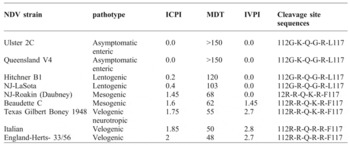
The genome of NDV encodes for six major structural proteins. Viral replication, transcription and translation occur in the cytoplasm of the host cell, while virus particles are assembled in the plasma membrane by budding (Zanetti et al., Reference ZANETTI, RODRIGUEZ, KING, CAPUA, CARRILLO, SEAL and BERINSTEIN2003). An important pathogenic marker of NDV exists in F protein (Madadgar et al., Reference MADADGAR, KARIMI, NAZAKTABAR, KAZEMIMANESH, GHAFARI, DEZFOULI and HOJJATI2013). A disulphide linkage is present between F1 and F2, and these proteins enable the virus to attach to the host cell membrane (Wen et al., Reference WEN, CHEN, ZHANG, YANG and ZHOU2007). At the cleavage site, F0 protein has two pairs of basic amino acids that can be cleaved by the host proteases (Courtney et al., Reference COURTNEY, SUSTA, GOMEZ, HINES, PEDERSEN, BROWN, MILLER and AFONSO2013). Highly virulent NDV has three or more basic amino acids, which are lysine (K) or arginine (R) present at residues 113-116 and phenylalanine (F) at position 117 (OIE, 2012). Cleavage of F0 protein is due to the presence of these basic amino acids in virulent NDV (Boostani et al., Reference BOOSTANI, POURBAKHSH, MOMAYEZ and CHARKHKAR2013). It has been found that avirulent viruses have 112G/E-K/R-Q-G/E-R-L117 and virulent viruses have 112R/K-R-Q-K/R-R-F117 amino acid sequences at the cleavage site (Courtney et al., Reference COURTNEY, SUSTA, GOMEZ, HINES, PEDERSEN, BROWN, MILLER and AFONSO2013). Most of the pathogenic APMV-1 viruses in chicken have sequence 112R/K-R-Q/K/R-K/R-R116 (OIE, 2012). The Office of International Epizootics (OIE) has accepted the F cleavage sequence as the determinant of primary virulence (Wise et al., Reference WISE, SUAREZ, SEAL, PEDERSEN, SENNE, KING, KAPCZYNSKI and SPACKMAN2004). However, if this cleavage sequence is not found, then an ICPI is required for determination of the virulence. Examples of vNDV, mNDV and LoNDV cleavage site sequences are shown in Table 1 .
Examples of ICPI, MDT, IVPI, and cleavage site for some strains of NDV.

MDT=mean death time, ICPI= intracereberal pathogenicity index,IVPI= intravenous pathogenicity index
The incubation period from the time of infection to development of disease varies from two to 15 days depending on several factors (Saif et al., Reference SAIF, FADLY, GLISSON and MCDOUGALD2008). The pathogenicity of the virus, host species and age, host immune status, secondary infections, stress, environmental conditions, the amount of virus transmitted, and the route of transmission can all play a role in determining the severity of disease and the length of incubation. Disease severity has led to the classification of NDV isolates under three distinct pathotypes (Alexander, Reference ALEXANDER2001). Infection of lentogenic NDV isolates can range from non-apparent to mild respiratory or gastrointestinal disease in adult chickens. When replication is limited to the gastrointestinal tract, the infection is often classified as asymptomatic enteric due to lack of respiratory symptoms. Young susceptible birds may develop a more serious respiratory disease that can lead to death due to increased susceptibility to secondary infection. LoNDV are categorised as lentogenic NDV and are commonly used as sources for vaccine production. Mesogenic (mNDV) isolates are considered to have intermediate virulence. Infection is typically systemic and can lead to development of a non-fatal respiratory disease. A drop in egg production can be seen in layers infected with mNDV. Rarely, symptoms of the nervous system can develop, but mortality is usually low following infection. Pigeon paramyxovirus isolates usually fall within the mNDV classification due to their intermediate virulence and neurologic symptoms.
Highly virulent velogenic (vND) viruses are also systemic and can cause high morbidity and mortality. Factors such as species of the infected bird, age, coinfection with other organisms, route of exposure, viral dose, stress, and the immune status of the individual determine disease severity (Alexander et al., Reference ALEXANDER, ALDOUS and FULLER2012). Velogenic viscerotropic ND (vvND) causes an acute infection of the gastrointestinal mucosa resulting in haemorrhagic lesions and death (Alexander, Reference ALEXANDER2001). Clinical signs may begin with weakness, increased rate of breathing, listlessness, and prostration. During the course of infection, green diarrhoea, muscular tremors, and paralysis of the extremities may be apparent. Oedema may be seen on the head, especially around the eyes. In highly susceptible flocks, mortality can be as high as 100%. Velogenic neurotropic NDV (vnNDV) isolates do not replicate in the gastrointestinal mucosa, like vvNDV (Alexander, Reference ALEXANDER2001; Saif et al., Reference SAIF, FADLY, GLISSON and MCDOUGALD2008). Infection primarily leads to respiratory distress followed by neurologic disease, and a decrease in egg production. Morbidity is similar to vvND, around 100%, but the mortality rate is lower. Mortality in adult birds is usually only 50%, but in young chickens it can be as high as 90%. Cormorant vNDV falls within the vnNDV classification due to the severe neurologic symptoms and high mortality rate in juvenile cormorants (Anonymous, 2011).
In acute and severe cases (like neurotropic velogenic strain), death is sudden and birds die without showing any clinical signs. Dead birds have haemorrhagic or necrotic lesions in the mucosa of the intestine, caecal tonsils, proventriculus and gizzard. Swollen kidneys and deposition of urates are common lesions. Clinical symptoms in turkeys may be less severe than those seen in chickens. Game birds are susceptible, and outbreaks occasionally occur, whereas ratites are less susceptible to disease development, and waterfowl are usually resistant (Alexander et al., Reference ALEXANDER, ALDOUS and FULLER2012).
Emergence and evolution of Newcastle disease virus
Virulent NDV isolates have been endemic in all areas of Pakistan since 1963 and have been reported in commercial poultry, domestic chickens, turkeys, guinea fowls, parrots, partridges, and wild crows (Khan and Huq, Reference KHAN and HUQ1963). However, new control measures were implemented in 1990 through 2005 as a result of outbreaks of avian influenza (H9N2, H7N3 and H5N1). However, the collection of surveillance samples for AI has helped elucidate the epidemiological situation of ND. NDV isolates in Pakistan belong to the original genotype XIII (now called sub-genotype XIIIa). Since 1984, genotypes VII and VIII are causing huge economic losses to the poultry industry of southeast (Diel et al., Reference DIEL, DA SILVA, LIU, WANG, MILLER and AFONSO2012; Khan et al., Reference KHAN, RUE, REHMANI, AHMED, WASILENKO, MILLER and AFONSO2010; Munir et al., Reference MUNIR, SHABBIR, YAQUB, SHABBIR, MUKHTAR, KHAN and BERG2012b; Abbas et al., Reference ABBAS, SHABBIR, KHALIQ, ALI and MUNIR2014; Farooq et al., Reference FAROOQ, SALIHA, MUNIR and KHAN2014). In Pakistan, outbreaks of genotype VII NDV have been reported recently (Siddique et al., Reference SIDDIQUE, NAEEM, ABBAS, ALI MALIK, RASHID, RAFIQUE, GHAFAR and REHMAN2013; Munir et al., Reference MUNIR, CORTEY, ABBAS, QURESHI, AFZAL, SHABBIR, KHAN, AHMED, AHMAD and BAULE2012a; Abbas et al., Reference ABBAS, SHABBIR, KHALIQ, ALI and MUNIR2014; Farooq et al., Reference FAROOQ, SALIHA, MUNIR and KHAN2014). In recent studies hemagglutinin neuraminidase (HN) gene from different outbreaks of NDV in commercial poultry farms revealed a consistent pattern of nucleotide, and crucial amino acid substitutions in prevailing field viruses of Pakistan (Munir et al., Reference MUNIR, SHABBIR, YAQUB, SHABBIR, MUKHTAR, KHAN and BERG2012b; Abbas et al., Reference ABBAS, SHABBIR, KHALIQ, ALI and MUNIR2014).
Vaccine failure can be correlated with substantial changes at the nucleotide level, which results into differences in amino acids. Furthermore, the prevailing strain of NDV had shown huge phylogenetic gaps with LaSota and Maktaswar. It has been reported that high genetic differences and phylogenic gaps among the vaccine strains and field strains enhance evolution (Miller et al., Reference MILLER, KIM, IP and AFONSO2009). Live bird market system and improper use of vaccines may contribute to the evolution of viruses. Molecular epidemiology of recent outbreaks in Pakistan have been reported and compared with three older ancestral strains as described in Table 1 and 2 (Miller et al., Reference MILLER, HADDAS, SIMANOV, LUBLIN, REHMANI, WAJID, BIBI, KHAN, YAQUB, CLAUDIO and AFONSO2015).
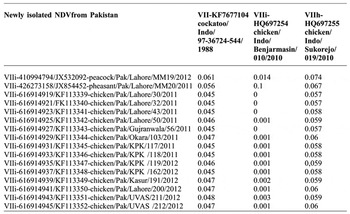
The partial and full F gene sequence of NDV isolates were compared with reference strains belonging to genotypes I-XVIII of class II viruses. The cleavage sites of the fusion protein ( Table 2 ) demonstrate that the most common configurations of the fusion cleavage site were 112RRQKR*F117, suggesting the virulent nature of these strains. ND viruses isolated worldwide, was compared to the Pakistani strains. Phylogenetic analysis of the complete F coding regions suggests that the new sub-genotype VIIi circulating in Pakistan is 99% similar to strains present in Israel and Indonesia ( Table 2 ). It has been documented that previously predominant genotypes XIIIa in Pakistan have been replaced by the new sub-genotypes VIIi during 2012-2013. Previously, genotype VII caused major outbreaks in Pakistan (Abbas et al., Reference ABBAS, SHABBIR, KHALIQ, ALI and MUNIR2014; Siddique et al., Reference SIDDIQUE, NAEEM, ABBAS, ALI MALIK, RASHID, RAFIQUE, GHAFAR and REHMAN2013; Farooq et al., Reference FAROOQ, SALIHA, MUNIR and KHAN2014). Present isolates from genotype VIIi sampled throughout Pakistan are highly related to current Indonesian isolates. The phylogenetic analysis for the current NDV isolates ( Table 2 ) demonstrated that NDV strains currently circulating in Pakistan are nearly identical to isolates of Israel, however, they have separate genetic origins from NDV isolates previously reported in the country.
Estimated pair wise evolutionary divergences among viruses of recent panzootic in Pakistan.

The numbers of base substitutions per site from between sequences are shown. The rate variation among sites was modelled with a gamma distribution (shape parameter= 1). The differences in the composition bias among sequences were considered in evolutionary comparisons. The analysis involved 35 nucleotide sequences compared to three historical strains from Indonesia. Codon positions included were 1st + 2nd + 3rd + Noncoding. All positions containing gaps and missing data were eliminated. Evolutionary analyses were conducted in MEGA5 (Tamura et al., Reference TAMURA, PETERSON, PETERSON, STECHER, NEI and KUMAR2011)
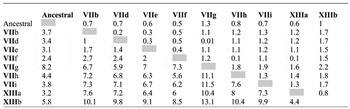
Table 2 shows the nucleotide distances of the F protein among individual viruses of highly related strains of sub genotypes VIIi. Distances of approximately 5% between the ancestral Cockatoo/Indonesia/1987 strain and viruses of new sub-genotypes VIIi have been observed ( Table 3 ). However, distances within sub-genotypes are lower than 1%, indicating that VIIi is a separate sub-genotypes. Comparison of the nucleotide sequences of NDV strains revealed that all of the new Pakistani strains, except chicken/UDL/Pak/12/2010, have >99% homology with other Indonesian strains, such as chicken/Banjarmasin/010/2010, chicken/Kudus/018/2010, chicken/Sragen/014/2010 and chicken/Gianyar/013/2010 (Miller et al., Reference MILLER, HADDAS, SIMANOV, LUBLIN, REHMANI, WAJID, BIBI, KHAN, YAQUB, CLAUDIO and AFONSO2015; Abbas et al., Reference ABBAS, SHABBIR, KHALIQ, ALI and MUNIR2014). Recently isolated NDV strains have lower identity (87-93%) with other previously characterised Pakistani strains; chicken/Pak/Lahore1/2010, chicken/ Pak/Islamabad3/2010, chicken/Pak/Rawalpindi/2010, chicken/Pak/Karachi/1/1974, chicken/Pak/Karachi/43/2008, chicken/Pak/Karachi/26/2005, chicken/Pak/Karachi/33/2007, chicken/Pak/Karachi/2010, chicken/Pak/Lahore/2010 and chicken/Pak/Islamabad1/2010 (Farooq et al., Reference FAROOQ, SALIHA, MUNIR and KHAN2014; Miller et al., Reference MILLER, HADDAS, SIMANOV, LUBLIN, REHMANI, WAJID, BIBI, KHAN, YAQUB, CLAUDIO and AFONSO2015).
Comparative evolutionary distances (%) among new and previously defined sub-genotypes based complete full fusion coding sequences.

The numbers of base substitutions per site from averaging over all sequence pairs between groups are shown. Standard error estimate(s) are shown above the diagonal. Analyses were conducted using the Maximum Composite Likelihood mode. The rate variation among sites was modelled with a gamma distribution (shape parameter = 1). The differences in the composition bias among sequences were considered in evolutionary comparisons. The analysis involved 96 nucleotide sequences. Codon positions included were 1st + 2nd + 3rd + Noncoding. All positions containing gaps and missing data were eliminated. Evolutionary analyses were conducted in MEGA 5 (Tamura et al., Reference TAMURA, PETERSON, PETERSON, STECHER, NEI and KUMAR2011).
NDV from genotype VIIi were predominantly responsible for the ND outbreaks during October 2011 through March 2012, and previous studies reported a predominance of genotype XIII in Pakistan (Khan et al., Reference KHAN, RUE, REHMANI, AHMED, WASILENKO, MILLER and AFONSO2010). All of the newer NDV strains from Pakistan, except chicken/Pak/UDL/12/2010, clustered together with Indonesian and Israeli strains, suggesting rapid spread and epizootic characteristics for this sub-genotype. Outbreaks in Pakistan have occurred mainly in poultry, however, the role of wild bird populations cannot be ignored as NDV infected wild birds have been found in unvaccinated wild birds in Pakistan, Israel and in a public zoo in Mexico (Shabbir et al., Reference SHABBIR, ABBAS, YAQUB, MUKHTAR, SUBHANI, HABIB, SOHAIL and MUNIR2012; Munir et al., Reference MUNIR, CORTEY, ABBAS, QURESHI, AFZAL, SHABBIR, KHAN, AHMED, AHMAD and BAULE2012a). This new sub-genotype (VIIi) is different from other recent strains circulating in West Africa (genotypes XIV, XVII and XVIII), Madagascar (XI), China (VII, XII and XV), North America (genotypes V), the Caribbean (XVI), or rock pigeon viruses (VI). These newer isolates appear to be more distantly related to old genotype viruses (I, II, III, IV, X), suggesting that they have an independent origin from these viruses. Although some of the strain of this new panzootic is still related to the viruses from genotype XIII, phylogenetic evidence and distance analysis suggest that the viruses cause of this pandemic did not originate directly from of genetic shift from previously described strains of genotypes XIII, but rather are the result of independent evolution from older viruses circulating in Indonesia in parrots, cockatoos and wild birds.
All available viruses of sub-genotype VIIi isolated recently in Indonesia, Pakistan and Israel (between 2010 and 2012) appear to be most closely related to the ancestral strains Lory/Indonesia/1988 (KF67105) and cockatoo/Indonesia/1990 (KF67104) than currently circulating viruses from genotype VII. Strains from Indonesia from the late 1980s and early 1990s, such as Lory/Indonesia/1988 and cockatoo/Indonesia/ 1990, appear to represent the most closely known ancestors of vNDV of sub-genotype VIIi. Viruses of genotype XIII are closely related to the ancestor strain, culex pool/Indonesia/ JKT/1979 (JX393313) (Forrester et al., Reference FORRESTER, WIDEN, WOOD, TRAVASSOS DA ROSA, KSIAZEK, VASILAKIS and TESH2013). Similarly, the ancestor strains most closely related to the viruses of genotype XIIIa from Iran and Pakistan appear to be a cockatoo/India/7847/1982 (JN942041). Therefore, viruses circulating and causing ND outbreaks in Pakistan, belong to a new sub-genotype (VIIi). These strains readily infect chickens and other bird species, and have been able to rapidly spread and persist in multiple countries.
These above-mentioned characteristics and the devastating effects from ND outbreaks due to these new sub-genotypes in Pakistan, Israel and Indonesia suggest that viruses of genotype VIIi may have the potential to start a new panzootic. The factors that allow a virus to become capable of creating a panzootic are unknown. Often there is not enough epidemiological data available to understand how a panzootic originated, or the data with this type of information are produced retrospectively (Alexander et al., Reference ALEXANDER, ALDOUS and FULLER2012). However, based on past experiences, human factors such as the movement of live birds without quarantining (Alexander et al., Reference ALEXANDER, ALDOUS and FULLER2012), a live bird marketing system for sale of birds, lack of testing to ensure a disease-free status, or the inability to contain and eradicate a disease after an outbreak is identified, are likely to increase the risk of creating a panzootic. Other human factors that may play a role are the lack of efficient veterinary and diagnostics services or cultural and socioeconomic factors (e.g. backyard poultry and unvaccinated fighting gamecocks). Moreover, lack of government incentives to conduct culling and or reporting may also contribute in virus emergence and evolution. Biological factors are likely also important, but often these are not well understood. An increased capacity of the virus to replicate in a host without producing clinical signs of disease, or the existence of highly mobile hosts are factors that should be considered as they have the potential to create natural reservoirs in wild birds (Miller et al., Reference MILLER, KIM, IP and AFONSO2009). The improved replication rate of virulent NDV in the unusual new host may also play a vital role in evolution (Wang et al., Reference WANG, DUAN, HU, KAI, WANG, SONG, ZHONG, SUN, WANG, WU and LIU2012; Dortmans et al., Reference DORTMANS, KOCH, ROTTIER and PEETERS2011). Other viral characteristics may also give viruses, additional advantages such as thermostability, UV resistance, or the ability of the viruses to move into environments that favour survival in the environment for longer periods of time (such a cold water bodies). As seen with the genotype I strains, longer survival time may increase the ability of NDV to infect more hosts before becoming inactivated (Alexander et al., Reference ALEXANDER, ALDOUS and FULLER2012). In this instance, the identification of new sub-genotype VIIi in three different countries that are not sharing borders suggested that the VIIi strains could potentially become panzootic if not contained. Viruses from the new sub-genotype VIIi may be rapidly replacing vNDV isolates from predominant sub-genotypes in certain locations as is happening with Pakistani isolates of genotype XIIIa Southeast Asia represents one of the largest concentrations of commercial and backyard poultry in the world and their poultry production system displays some common characteristics that may favour the emergence of vNDV isolates diverse enough to be considered new sub-genotypes. Indonesia and Pakistan, as well as other Asian countries, have mixed poultry production systems with a large portion of the birds raised in backyard farms, and an abundance of live birds’ markets selling birds of multiple species and ages. Historically backyard poultry and live bird markets have been considered as possible culprits in the maintenance and evolution of vNDV strains because of the difficulties in vaccinating newly hatched backyard birds. It is suspected that poorly vaccinated commercial poultry, non-vaccinated domestic poultry, and wild birds may all act as reservoirs of NDV strains, transmitting vNDV isolates to susceptible birds.
Live bird markets are a prime concern because different species or breeds of birds originating from various commercial poultry producers, as well as from backyard farms, from different areas are brought together and housed in small areas, allowing intermingling of the birds and possible transmission through contact with contaminated cages, food, water and faeces. Regardless of the existence of these high-risk production and marketing practices in Asia, other factors may add epidemiological risk to the emergence of new epizootics in Asia (Miller et al., Reference MILLER, HADDAS, SIMANOV, LUBLIN, REHMANI, WAJID, BIBI, KHAN, YAQUB, CLAUDIO and AFONSO2015; Farooq et al., Reference FAROOQ, SALIHA, MUNIR and KHAN2014). The emergence of AI has facilitated increased surveillance and improved bio-security; surprisingly, the focus on AI has not been enough to prevent the spread of vNDV (Shabbir et al., Reference SHABBIR, ABBAS, YAQUB, MUKHTAR, SUBHANI, HABIB, SOHAIL and MUNIR2012). The incidence of ND has continued at all levels of production facilities despite the use of full time veterinary services developed to control avian influenza in the most advanced commercial production facilities.
Recent advances and challenges in Newcastle disease vaccination
In addition to biosecurity measures, vaccination is an important tool for prevention and control of NDV. The primary goal of vaccination is to prevent or reduce the clinical disease from an infectious agent. Because there is no compensation policy from the government of Pakistan, it is not practical to demand that the affected farmers destroy their flocks. Before stocking the premises, basic biosecurity measures have been introduced at most farms by the Pakistan Poultry Association (PPA). A vaccine policy against NDV has been developed in coordination with the poultry industry, vaccine manufactures and officials of Food, Agriculture and Livestock (MINFAL). Under this plan, all the commercial poultry flocks in the affected poultry states are vaccinated with oil based killed vaccines or live attenuated vaccine. The two most common vaccine strains used in Pakistan are LaSota and Mukteswar (Munir et al., Reference MUNIR, LINDE, ZOHARI, STAHL, BAULE, HOLM, ENGSTROM and BERG2011; Shabbir et al., Reference SHABBIR, ABBAS, YAQUB, MUKHTAR, SUBHANI, HABIB, SOHAIL and MUNIR2012; Rehman et al., Reference REHMAN, FAWAD, ABBAS, NAHEED, SIDDIQUE, AFZAL, MUNAWAR, AURANGZEB, ATIF and HAQ2013; Umar et al.,2015; 2016a; 2016b). The LaSota vaccine is formulated from a lentogenic (low virulence) clone of the LaSota strain and is manufactured in different countries of the world and imported and administered to commercial poultry in Pakistan. However, the Mukteswar strain (a virulent strain by OIE definition and a strain of moderate virulence from genotype III) continues to be manufactured by the local vaccine companies and administered to backyard poultry (Munir et al., Reference MUNIR, LINDE, ZOHARI, STAHL, BAULE, HOLM, ENGSTROM and BERG2010; Khan et al., Reference KHAN, RUE, REHMANI, AHMED, WASILENKO, MILLER and AFONSO2010; Umar et al., Reference UMAR, SUBHAN, AZAM, MUNIR and SHAH2017a; Reference UMAR, MUNIR, AHSAN, RAZA, CHOWDHURY, AHMED and SHAH2017b). To provide the best protection, vaccine programs have adopted the method of progressive vaccinations which involves successive booster vaccines with increasingly virulent strains (Iram et al., Reference IRAM, SHAH, ISMAT, HABIB, IQBAL, HASNAIN and RAHMAN2014; OIE, 2009). Different methods for administering ND vaccines include via drinking water, eye dropper, sprays, mixing in feeds (thermostable vaccine) and injection. The drinking water method is easier and popular, but it elicits lower immunity levels. Vaccine application through sprays and aerosols is also a popular method, but controlling the size of aerosol droplets is usually a concern (Flint et al., Reference FLINT, ENQUIST, RACANIELLO and SKALKA2007; Mahmood et al., Reference MAHMOOD, SIDDIQUE, HUSSAIN, AHMAD and RAFIQUE2014; Miller and Koch, 2013). Live viruses are able to transmit between birds and present a disadvantage due to the potential for live virus vaccines to produce clinical symptoms in the flock (OIE, 2009; Senne et al., Reference SENNE, KING and KAPCZYNSKI2004). Live virus vaccines usually fail to achieve desirable immunity in the presence of maternal antibodies in young. The killed vaccines result in measurable antibody titre to all the structural proteins including the HN and F protein. One or both of these proteins are used as antigen for antibody tests. Therefore, vaccinated birds cannot be distinguished from naturally infected birds using this approach. This creates a great hindrance in doing surveillance and vaccination for NDV control. For vaccination against NDV, little effort typically has been used to match the vaccine and the field strain. The comparison of the genetic sequence of the F gene can provide a good estimate of the variability between vaccine and field strain. However, when using vaccine as a control tool, both clinical protection and virus shedding are of concern. Experimental evidence demonstrates that the closer the vaccine is to field strain, the lower the amount of virus will be shed in infected birds (Mahmood et al., Reference MAHMOOD, SIDDIQUE, HUSSAIN, AHMAD and RAFIQUE2014). Emergence of new sub genotypes poses a major problem for NDV vaccines because it results in decreased protection from vaccination (Perozo et al., Reference PEROZO, MARCANO and AFONSO2012; Ali et al., Reference ALI, MUNEER, HUSSAIN, REHMANI, YAQUB and NAEEM2014; Farooq et al., Reference FAROOQ, SALIHA, MUNIR and KHAN2014; Abbas et al., Reference ABBAS, SHABBIR, KHALIQ, ALI and MUNIR2014; Umar et al.,2015).
By virtue of greatly advanced biotechnology and the availability of a great deal of genomic information on NDV, there are new opportunities to create novel concept vaccines. Virus like particles (VLP) have been suggested as a new generation of non-egg based vaccine platform against various viral infections with solid safety profiles (Roldao et al., Reference ROLDAO, MELLADO, CASTILHO, CARRONDO and ALVES2010; Iram et al., Reference IRAM, SHAH, ISMAT, HABIB, IQBAL, HASNAIN and RAHMAN2014). VLP resembles infectious virus particles in the structure and morphology with multiple antigenic epitopes which were shown to be highly immunogenic probably due to its ability to stimulate a diverse set of host immune responses. DNA vaccines have great potential as an alternative conventional vaccine capable of inducing protective immune responses against a variety of infectious diseases (Zhao et al., Reference ZHAO, ZHANG, ZHANG, LI, SHI, GUO, DAI, CHEN, JIN, ZHAO, CUI and WANG2014; Ullah et al.,2013; Mahmood et al., Reference MAHMOOD, SIDDIQUE, HUSSAIN, AHMAD and RAFIQUE2014). These DNA vaccines can encode recombinant DNA molecule of multiple antigen gene, which reduces manufacturing cost and time as compared to the conventional vaccine that we are currently using, without carrying infections that are associated with live attenuated vaccines. These plasmid-based DNA vaccines are able to express high levels of proteins of interest in cells and can induce both humoral and cellular immune responses. They can override maternal antibodies and facilitate disease surveillance by differentiating vaccinated from non-vaccinated infected birds (Zhao et al., Reference ZHAO, ZHANG, ZHANG, LI, SHI, GUO, DAI, CHEN, JIN, ZHAO, CUI and WANG2014; Senne et al., Reference SENNE, KING and KAPCZYNSKI2004).
The current scenario of NDV outbreaks in Pakistan demands the evaluation of vaccines against field challenges. Failure of live vaccines to elicit protective immune response might be due to several reasons such as poor refrigeration, inappropriate route of vaccination, or uneven vaccination schedules. There is considerable controversy regarding the issue of vaccine failure on NDV control. Some argue that inadequate application is the main reason of vaccine failure. However, others have suggested that increasing the homology of the vaccine to the challenge virus can play a critical role in disease control (Alexander et al., Reference ALEXANDER, ALDOUS and FULLER2012; Susta et al., Reference SUSTA, JONES, CATTOLI, CARDENAS-GARCIA, MILLER, BROWN and AFONSO2014). Live vaccine applications followed by inactivated ND vaccine could be practiced to protect the birds from virulent NDVs. In the Past, mass vaccination against NDV in Pakistan has reduced virus isolation, indicating that NDV is under the control of vaccination. However, recent reports indicated the emergence of new sub genotypes of NDV thus could escape from vaccine protection. Moreover, it has been reported that the currently practiced NDV vaccines give better protection against the velogenic NDVs isolated in 1930 to 70s (Herts33/56, California 71) than the ones, which have been isolated in the past few years (Mahmood et al., Reference MAHMOOD, SIDDIQUE, HUSSAIN, AHMAD and RAFIQUE2014). Continued active surveillance of poultry farms and live bird markets to reveal new variant NDV viruses and analysing appropriate vaccine seed viruses are warranted to prevent new outbreaks in Pakistan.
Conclusions
The emergence and spread of new genotypes of NDV in Pakistan represent a significant threat to poultry and suggest that vNDV is continuously evolving; leading to novel genotypes. Wider use of NDV vaccine in animal population could enhance the immune pressure and drive the mutation resulting in genetic diversity. Several studies reported periodic changes in the genome sequence of NDV and could escape from vaccine protection. The Pakistani ND outbreaks suggest that the use of live and inactivated vaccines that typically are able to induce sufficient antibody levels may not be sufficient to control ND outbreaks under the current production practices in coming days. Therefore, improved vaccine technology and periodic updates of vaccine seed strains are required to increase immunogenicity and cross protective efficacy. These strategies could include: Maintaining up-to-date diagnostic assays, the selection of highly immunogenic vaccine seed strains, the use of effective adjuvants, and the use of new technology vaccines. Considering these new sub-genotypes, continuous monitoring of viral genetic changes and vaccine updating throughout the year is warranted to prevent new outbreaks.