Introduction
Accidental foreign body ingestion is not uncommon, and fish bones are the most common presentation in the upper digestive tract in adults.Reference Loh, Tan, Smith, Yeoh and Dong1 Fish bone ingestion is a common emergency encountered by otorhinolaryngologists, especially in the Arabian Gulf region. Most cases are straightforward and uncomplicated, and the bone can be easily removed. However, some cases require more effort, and a range of procedures.Reference Yang, Lee, Huang and Yeh2 Delayed, undiagnosed and migratory fish bones may cause catastrophic consequences, such as retropharyngeal abscess, mediastinitis, deep neck abscess,Reference Yang, Chen and Chen3 fatal arterial-oesophageal fistula, tracheoesophageal fistulaReference Loh, Tan, Smith, Yeoh and Dong1 and cardiac tamponade.Reference Osinubi, Osiname, Pal, Lonsdale and Butcher4
Surgical exploration for a migrated foreign body has been described by some otolaryngologists as ‘fishing for a needle in the deep ocean’. A pre-operative computed tomography (CT) scan of the neck is helpful in locating the site of penetration and guiding the direction of exploration. Intra-operative fluoroscopic guidance is a helpful but under-used tool, despite its great effectiveness.
We present a very rare case of an impacted fish bone which was not evident on examination, being located only on CT. During surgery, intra-operative fluoroscopy was vital in locating and aiding bimanual removal of the fish bone from the carotid space.
Case report
A 67-year-old man presented to the accident and emergency department of Sultan Qaboos University Hospital with a history of fish bone ingestion one day prior to presentation.
The patient had previously visited a local hospital, where a soft tissue neck X-ray had not shown any foreign body. However, the patient had insisted that he felt a pricking sensation due to the sharp fish bone, and his pain had increased in severity and he had become unable to swallow solids. He had tried to remove the fish bone himself, causing much trauma to the mucosa of the tonsillar pillar. The patient had visited the local hospital again, where repeated examination again failed to locate a foreign body.
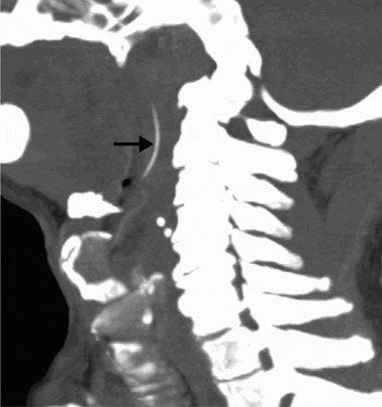
At our institution, a second neck X-ray showed a small, suspicious shadow. Computed tomography scanning showed an approximately 1.5 cm fish bone lying in a vertical position in the retropharyngeal space, about 5 mm above the level of the epiglottis (Figure 1).
Reformatted sagittal computed tomography scan of the neck, showing the fish bone in its entire length extending along the left postero-lateral pharyngeal soft tissues (arrow) of the neck.

The patient was re-examined by the ENT surgeons. Flexible endoscopy was performed, which revealed the trauma caused by the patient's own manipulations and by previous attempts at peripheral hospital, but did not show any part of the foreign body. The epiglottis and pyriform fossa were normal, with no increase in salivation.
The patient was taken to the operating theatre for surgical exploration. Under general anaesthesia, the patient was positioned and a tonsil gag fixed. A 2 cm tear along the posterior pharyngeal wall was observed. The area was probed but no fish bone was seen or felt.
A tonsillectomy was performed: the superior tonsillar pole was incised and dissected, the tonsil was excised, and the superior and inferior poles were ligated. We were unable to see or feel the fish bone on palpation or probing of the tonsillar bed.
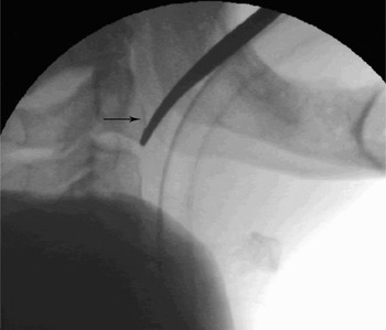
Image-guided fluoroscopy was performed, with placement of a marker to assist orientation. The fish bone was identified superior to the marker, in the superior part of the tonsillar bed and the nasopharyngeal part of the parapharyngeal space (Figure 2). The wound was explored and palpated but, again, no fish bone was found. Fluoroscopy was repeated, this time with two markers. The fish bone was again identified, this time embedded deep in the parapharyngeal space and carotid space. The bed was incised and marker was moved under fluoroscopic guidance. The tonsillar incision was probed with an upturned finger and with forceps. The latter located the fish bone, but as it was awkwardly deep-seated we were unable to remove it.
Image-guided fluoroscopic image showing the fish bone in the neck (arrow), grasped by the tip of the localising forceps.

The fish bone (Figure 3) was finally removed bimanually, by pressing behind the angle of the jaw (left side) externally while simultaneously palpating the fish bone in the nasopharyngeal bed. The external pressure over the submandibular region helped displace the bone to a more superficial plane and then out into the nasopharyngeal lumen. Fluoroscopic scanning was again performed, and showed no obvious foreign body remnants. The wound was sutured with 3/0 Vicryl and haemostasis confirmed.
The patient's fish bone, after removal from the carotid space.

Post-operatively, the patient had immediate and dramatic relief from the previous pricking sensation, despite general throat soreness due to surgery.
The patient was followed up one week later in the out-patient department, and was found to be cured, with no pain and a healed wound.
Discussion
People who swallow fish bones usually seek medical attention in the accident and emergency department. However, sometimes the fish bone cannot be found even after a series of examinations, as seen in the present case.
A common site of foreign body impaction is the tonsils, generally the inferior tonsillar pole or within the tonsillar tissue itself.
Patients who have swallowed a fish bone tend to try to remove it themselves by manipulation and induced vomiting, which causes trauma to the oropharynx. In our patient, this probably contributed to migration of the fish bone to its unusual and potentially lethal final position.
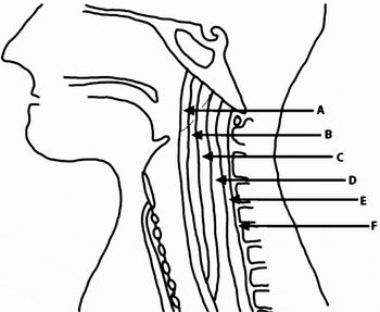
The presented case is rare due to the fish bone's migration: it pierced the buccopharyngeal mucosa of the posterior pharyngeal wall and migrated to the tonsillar bed, it subsequently entered the anterior compartment of the retropharyngeal space, and it then partly entered the carotid space (i.e. the post-styloid parapharyngeal space), from where it was retrieved (Figure 4).
Sagittal view of the fascial layers and spaces posterior to the nasopharynx, showing the fish bone with its migratory path. A = buccopharyngeal fascia; B = anterior compartment of retropharyngeal fascia; C = alar layers of prevertebral fascia; D = posterior compartment of retropharyngeal space; E = prevertebral fascia; F = prevertebral space

Fish bones can be overlooked on plain X-rays as they are often radiolucent. Therefore, CT scanning is preferred to exclude the presence of a foreign body in symptomatic patients. Our patient's fish bone was only diagnosed with the aid of CT scanning of the oropharynx and neck. During surgery, the fish bone was located away from the site suggested by the CT, being higher up behind the tonsils in the carotid space. Removal of the tonsils was necessary in this case in order to gain access to the foreign body. Intra-operative fluoroscopy guided us to the correct position of the foreign body, which was positioned vertically and could not be palpated or seen by the operating surgeon.
When attending a patient with symptomatic fish bone ingestion, the ingested fish bone should be removed as soon as possible, as the linear, sharp, pointed contours of fish bones result in a higher risk of gastrointestinal tract perforation. The first imaging investigation for impacted fish bone should be a lateral plain neck radiograph. However, interpretation of this view is occasionally difficult, as ingested foreign bodies are often mistaken for calcified normal structures in the neck.Reference Chawla, Eng and Peh5
Although most ingested foreign bodies can be located by detailed oral examination and laryngoscopic inspection, CT scanning of the neck using extra-fine slices provides the most accurate imaging investigation for confirming and localising foreign body impaction.Reference Lue, Fang and Manolidis6, Reference Braverman, Gomori, Polv and Saah7 In the presented case, CT scanning was diagnostic. If the CT scan of the neck is difficult to interpret whether the fish bone is partially or completely extra-luminal.Reference Lim, Quah and Loh8
Flexible endoscopy is a good investigation in cases of fish bone ingestion, but it may give false negative results, as in our patient.
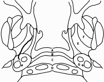
Migrating fish bones are potentially dangerous, especially in the nasopharyngeal area. Furthermore, it is important to note that while foreign bodies in this area may be asymptomatic, their disimpaction can have potentially fatal consequences.Reference Singh, Jha, Dhawan, Dwivedi and Yadav9 The presented patient's fish bone was potentially lethal as it lay in the carotid space (Figure 5). Migratory foreign bodies have been previously reported; a case of a migrating fish bone leading to cardiac tamponade has been described.Reference Osinubi, Osiname, Pal, Lonsdale and Butcher4 In the presented patient, the fish bone migration was partly due to his own manipulation of the bone, in a blind attempt to remove it. The fish bone could also have migrated due to vomiting and retching movements of the pharyngeal muscles. Neck movements, pharyngeal peristalsis and tissue reaction to the foreign body are also possible contributing factors.
Axial view of the nasopharynx showing the impacted fish bone and its relation to the fascial spaces. A = pharyngeal mucosal space; B = anterior compartment of retropharyngeal space; C = posterior compartment of retropharyngeal space; D = prevertebral space; E = parapharyngeal space (pre-styloid parapharyngeal space); F = masticator space; G = carotid space (post-styloid parapharyngeal space)

It is important to be aware that lack of evidence of a fish bone on oral examination and plain neck radiography does not exclude the presence of such a bone. If the patient has persistent symptoms of sore throat and foreign body sensation, an early CT neck scan or flexible endoscopic examination should be promptly conducted to facilitate appropriate management and to avoid life-threatening consequences.
Intra-operative fluoroscopy was vital in the presented case, as it prevented over-manipulation during surgery and avoided potential complication, in view of the critical location of the fish bone in the carotid space. Intra-operative fluoroscopy also helped to shorten the procedure time, as it corrected our initial, false impression of the foreign body's location, gained from CT scanning. There are reports of the use of fluoroscopy to locate lost surgical needles in the operating theatre, and to remove a metallic foreign body in the foot. However, we could find only one previous report of the use of fluoroscopy to aid removal of a pharyngeal foreign body in an adult.Reference Park and Carr10
• In patients with a history of fish bone impaction in the upper aerodigestive tract but no visible foreign body, plain neck X-ray and flexible endoscopy can give false negative results
• If such patients report a pricking sensation in the throat, prompt computed tomography scanning is indicated
• Intra-operative fluoroscopy can be effective in locating such foreign bodies, avoiding blind over-manipulation
• In deep-seated nasopharyngeal foreign bodies, bimanual, fluoroscopy-guided removal may be a minimally invasive alternative to major skull base surgery
• Patients should be educated to refrain from blindly attempting to remove impacted foreign bodies in the upper aerodigestive tract
In our patient, the use of digital pressure over the angle of the jaw, in the submandibular space, assisted bimanual removal of the fish bone by pushing it superficially into the lumen of the nasopharynx. This was vital to safe extraction.
In the reported patient, retrieval of the impacted fish bone was facilitated by intra-operative, fluoroscopy-guided, bimanual manipulation. Failure could have been catastrophic for the patient, as he would have had to undergo major skull base surgery, with a strong possibility of cardiac tamponade should the bone have entered the carotid artery.