Relevance statement
The present study, with neuroimaging and genotyped data of 51 PTSD patients and 94 healthy controls (HC), is the first to unravel the influences of FKBP5 genotype for the stress-related cognitive styles including the repression (experiential avoidance) and positive reappraisal (cognitive-emotional regulation) in PTSD, by way of the altered regional brain volumes of hippocampus. Our work showed neurobiological evidence for the hippocampus as possible targets of neuromodulation aiming for the improvement of stress-related cognitive styles in a subset of patients with PTSD homozygous for the risk alleles of FKBP5-associated single nucleotide polymorphisms (SNPs).
Introduction
Globally, more than 70% of the adult population are exposed to traumatic events in their lifetime (Benjet et al., Reference Benjet, Bromet, Karam, Kessler, McLaughlin, Ruscio and Koenen2016). Notably, the Diagnostic and Statistical Manual of Mental Disorders, Fifth Edition(DSM-5) highlights the importance of stress-related cognitive style in post-traumatic stress disorder (PTSD) (Shalev, Liberzon, & Marmar, Reference Shalev, Liberzon and Marmar2017), including catastrophizing (Gellatly & Beck, Reference Gellatly and Beck2016), rumination (Nolen-Hoeksema & Morrow, Reference Nolen-Hoeksema and Morrow1991; Nolen-Hoeksema, Wisco, & Lyubomirsky, Reference Nolen-Hoeksema, Wisco and Lyubomirsky2008), self-denigration, emotional dysregulation, negative viewpoint of the environment and future, and related experiential avoidance (Chawla & Ostafin, Reference Chawla and Ostafin2007; Hayes, Wilson, Gifford, Follette, & Strosahl, Reference Hayes, Wilson, Gifford, Follette and Strosahl1996). When healthy people encounter a situation that is beyond their abilities of control, they may use emotion-focused coping strategies such as positive reappraisal instead of problem-solving skills (Ghasemi, Kordi, Asgharipour, Esmaeili, & Amirian, Reference Ghasemi, Kordi, Asgharipour, Esmaeili and Amirian2017). However, in PTSD, the positive reappraisal is not usually utilized, and is thus considered a possible predictor of the later reduction in experiential avoidance in these patients (Fitzgerald et al., Reference Fitzgerald, Gorka, Kujawa, DiGangi, Proescher, Greenstein and Phan2018).
Parts of the inter-individual differences of vulnerability for getting PTSD after a traumatic event (especially interpersonal assaultive trauma) have been attributed to genetic factors (Stein, Jang, Taylor, Vernon, & Livesley, Reference Stein, Jang, Taylor, Vernon and Livesley2002). Heritability of proneness to PTSD has been estimated at 40–50% (Afifi, Asmundson, Taylor, & Jang, Reference Afifi, Asmundson, Taylor and Jang2010), and epigenetic mechanisms such as methylation also partly contribute to vulnerability and resilience to PTSD (Lappalainen & Greally, Reference Lappalainen and Greally2017). In addition, several SNPs of the FK506 binding protein 5 gene (FKBP5; encoding for the FKBP51 protein which is related to the intracellular trafficking of hetero-oligomeric forms of glucocorticoid receptors) have been suggested as risk factors for PTSD or are associated with PTSD symptom severity (Carvalho, Coimbra, Ota, Mello, & Belangero, Reference Carvalho, Coimbra, Ota, Mello and Belangero2017). Indeed, a decreased expression of FKBP5 in postmortem brain samples, revealing an abnormal glucocorticoid functioning, was reported in patients with PTSD, compared with HC (Holmes et al., Reference Holmes, Girgenti, Davis, Pietrzak, DellaGioia, Nabulsi and Esterlis2017). Furthermore, in the study by Binder et al. (Reference Binder, Bradley, Liu, Epstein, Deveau, Mercer and Ressler2008) on subjects with a history of child abuse, various FKBP5 polymorphisms (AA, GG, CC, or CC homozygote genotypes of rs3800373, rs9296158, rs1360780, and rs9470080, respectively) were reported to be protective genotypes to traumatic stress in patients with PTSD.
Previous research has shown smaller hippocampal volumes, increased functional activation of limbic area, and decreased functional activation of prefrontal regions during task-free resting status and in response to negatively valenced emotional stimuli in patients with PTSD, compared with HC (Marin et al., Reference Marin, Song, VanElzakker, Staples-Bradley, Linnman, Pace-Schott and Milad2016; Musazzi, Tornese, Sala, & Popoli, Reference Musazzi, Tornese, Sala and Popoli2018). Furthermore, altered neural circuitries have been previously described in PTSD for fear learning (amygdala sub-regions) (Fanselow & LeDoux, Reference Fanselow and LeDoux1999), threat detection (dorsal anterior cingulate, orbitofrontal, and anterior insular cortices) (Seeley et al., Reference Seeley, Menon, Schatzberg, Keller, Glover, Kenna and Greicius2007; Tovote, Fadok, & Lüthi, Reference Tovote, Fadok and Lüthi2015), context processing (hippocampal-medial prefrontal-thalamic circuitry) (Garfinkel et al., Reference Garfinkel, Abelson, King, Sripada, Wang, Gaines and Liberzon2014; Liberzon & Abelson James, Reference Liberzon and Abelson James2016), reward processing (striatum) (Elman et al., Reference Elman, Lowen, Frederick, Chi, Becerra and Pitman2009; Loureiro, Kramar, Renard, Rosen, & Laviolette, Reference Loureiro, Kramar, Renard, Rosen and Laviolette2016), valence representation (basolateral amygdala and nucleus accumbens) (Namburi, Al-Hasani, Calhoon, Bruchas, & Tye, Reference Namburi, Al-Hasani, Calhoon, Bruchas and Tye2016), and executive functioning and emotional regulation (frontoparietal and amygdala regions) (King et al., Reference King, Block, Sripada, Rauch, Giardino, Favorite and Liberzon2016; Shou et al., Reference Shou, Yang, Satterthwaite, Cook, Bruce, Shinohara and Sheline2017). Also, FKBP5 risk alleles have been associated with increased functional activation of the amygdala, altered functioning and decreased regional volume of the hippocampus, and reduced white matter integrity of the cingulum bundle (Fani et al., Reference Fani, King, Reiser, Binder, Jovanovic, Bradley and Ressler2014). However, few studies have aimed to unravel the FKBP5 genotype-related neural underpinning of stress-related cognitive style in PTSD.
Therefore, the present study first examined the altered stress-related cognitive styles for PTSD patients compared with HC and subsequently uncovered the neural correlates of these cognitive styles among the brain volumes of cortical-subcortical regions that demonstrated significant interactions of diagnosis-by-FKBP5 SNP genotype for regional brain volumes. We hypothesized that the presence of risk alleles for FKBP5-related SNPs might differently affect brain volumes in regions related to the processing of contextual information (i.e. hippocampal-medial prefrontal-thalamic circuitry) (Garfinkel et al., Reference Garfinkel, Abelson, King, Sripada, Wang, Gaines and Liberzon2014; Liberzon & Abelson James, Reference Liberzon and Abelson James2016) of patients with PTSD and HC. Moreover, regional volumes of these contextual processing-related brain regions may be associated with the severity of stress-related cognitive style in patients with PTSD homozygous for the risk alleles of FKBP5 SNPs.
Methods and materials
Participants and clinical assessment
A total of 145 participants (N = 94 HC, N = 51 PTSD; male/female = 36/109; all Asian) aged 19–55 years and without a prior history of head trauma were recruited from the Inje University Ilsan Paik Hospital (PTSD group) or the local community using advertisements (HC group). A diagnosis of current PTSD was based on a clinical evaluation by trained psychiatrists using the Structured Clinical Interview for DSM-IV Axis I Disorders (SCID-I) and the Clinician-Administered PTSD Scale for DSM-5 (CAPS-5) (Weathers et al., Reference Weathers, Blake, Schnurr, Kaloupek, Marx and Keane2013a). Among the patients with PTSD, 29 (57%) were taking psychotropic medications at the time of the brain imaging acquisition. The HC did not satisfy the DSM-IV-based lifetime diagnostic criteria for any major psychiatric disorders as screened by the SCID-I Non-Patient Edition (SCID-NP). Additional clinical characteristics and lifetime exposure to trauma for both groups were measured using the following self-report questionnaires: PTSD Checklist for DSM-5 (PCL-5) (Weathers et al., Reference Weathers, Litz, Keane, Palmieri, Marx and Schnurr2013c), Hospital Anxiety and Depression Scale (HADS) (Oh, Min, & Park, Reference Oh, Min and Park1999; Zigmond & Snaith, Reference Zigmond and Snaith1983), Beck Scale for Suicidal Ideation (SSI) (Beck & Kovacs, Reference Beck and Kovacs1979; Shin, Park, Oh, & Kim, Reference Shin, Park, Oh and Kim1990), and Life Events Checklist for DSM-5 (LEC-5) (Weathers et al., Reference Weathers, Blake, Schnurr, Kaloupek, Marx and Keane2013b).
Stress-related cognitive styles were measured using self-report questionnaires: the Ruminative Response Scale (RRS) (Kim et al., Reference Kim, Kwon, Yang, Kim, Yoo and Lee2013; Nolen-Hoeksema, Reference Nolen-Hoeksema1991), the Multidimensional Experiential Avoidance Questionnaire (MEAQ) (Gamez, Chmielewski, Kotov, Ruggero, & Watson, Reference Gamez, Chmielewski, Kotov, Ruggero and Watson2011; Park, Reference Park2013), and the Cognitive Emotion Regulation Questionnaire (CERQ) (Garnefski & Kraaij, Reference Garnefski and Kraaij2007; Kim, Reference Kim2004). The RRS comprises a total of 22 items on a four-point Likert Scale and measures two aspects of ruminative responses including brooding and reflective pondering (Nolen-Hoeksema, Reference Nolen-Hoeksema1991; Treynor, Gonzalez, & Nolen-Hoeksema, Reference Treynor, Gonzalez and Nolen-Hoeksema2003); higher total scores indicate more intense ruminative responses (Kim, Kim, & Youn, Reference Kim, Kim and Youn2010). The MEAQ uses a six-point Likert Scale and measures diverse facets of experiential avoidance such as behavioral avoidance, distress aversion, repression, denial, and distress endurance; these are considered efforts to avoid distressing thoughts, feelings, memories, and other private experiences (Gamez et al., Reference Gamez, Chmielewski, Kotov, Ruggero and Watson2011; Park, Reference Park2013). The CERQ comprises 36 items on a five-point Likert Scale to measure diverse cognitive-emotion regulation strategies, such as self-blame, rumination, catastrophizing, acceptance, and positive reappraisal, in response to stressful life events (Garnefski & Kraaij, Reference Garnefski and Kraaij2007; Kim, Reference Kim2004).
This study complies with the ethical standards of the Institutional Review Board (IRB no. 2015-07-025) at Inje University Ilsan Paik Hospital on human experimentation and with the Declaration of Helsinki, as revised in 2008. Written informed consent was obtained from all participants before study enrollment.
Image acquisition, processing, and extraction of regional brain volumes
Whole-brain anatomy was measured for all participants using high-resolution T1-weighted, 3D magnetic resonance imaging (MRI; TR = 1900 ms; TE = 3.42 ms; FOV = 210 mm × 250 mm; FA = 15°; acquisition matrix = 227 × 384; voxel size = 0.9 × 0.7 × 1.2 mm3) scans on a 1.5-Tesla scanner (Magneton Avanto, Siemens).
Image pre-processing procedures were as follows: (1) setting the T1-weighted MR images at the anterior commissure (AC); (2) approximating alignment by way of the mutual information affine registration with SPM12 tissue probability maps; (3) affine regularization with an ICBM space East Asian brain template; (4) spatial normalization using the high-dimensional DARTEL registration algorithm; (5) tissue segmentation of brain images into gray matter, white matter, and cerebrospinal fluid; and (6) modulation of normalized tissue intensities using the Jacobian-transformed tissue probability maps were conducted using the Computational Anatomy Toolbox for SPM (http://www.neuro.uni-jena.de/cat/) implemented in SPM12 (https://www.fil.ion.ucl.ac.uk/spm) software and MATLAB (https://kr.mathworks.com) platforms (Chen, Chen, Dong, Liu, & Yu, Reference Chen, Chen, Dong, Liu and Yu2018; Kim et al., Reference Kim, Jeon, Jang, Kim, Im and Lee2018). Finally, regional brain gray matter volumes for the 114 regions-of-interest (ROIs) defined according to the Neuromorphometric Atlas (http://Neuromorphometrics.com; from the original set of 142 ROIs, 118 regions of brain white matter, ventricles, and cerebellar sub-regions were excluded) were estimated.
For examination of the diagnosis-by-genotype interaction for regional brain volumes, in line with our study hypothesis and previous studies (Fanselow & LeDoux, Reference Fanselow and LeDoux1999; Garfinkel et al., Reference Garfinkel, Abelson, King, Sripada, Wang, Gaines and Liberzon2014; King et al., Reference King, Block, Sripada, Rauch, Giardino, Favorite and Liberzon2016; Liberzon & Abelson James, Reference Liberzon and Abelson James2016; Seeley et al., Reference Seeley, Menon, Schatzberg, Keller, Glover, Kenna and Greicius2007; Shou et al., Reference Shou, Yang, Satterthwaite, Cook, Bruce, Shinohara and Sheline2017; Tovote et al., Reference Tovote, Fadok and Lüthi2015), we selected the following 16 ROIs: amygdala, hippocampus, thalamus, frontal operculum, anterior cingulate gyrus, medial frontal cortex, inferior temporal gyrus, and medial orbital gyrus of both hemispheres.
DNA collection, extraction, and genotyping
In this study, we extracted four FKBP5 SNPs (rs9296158, rs3800373, rs1360780, and rs9470080) that have been suggested as relevant predictors of stress vulnerability and risk factors for psychiatric disorders in the adult population for candidate gene analyses (Scheuer et al., Reference Scheuer, Ising, Uhr, Otto, von Klitzing and Klein2016; Tamman et al., Reference Tamman, Sippel, Han, Neria, Krystal, Southwick and Pietrzak2017). Genomic DNA, extracted from peripheral blood provided by participants, underwent quality check using NanoDrop® ND-1000 UV-Vis Spectrophotometer. Genotyping was conducted using polymerase chain reaction (PCR) amplification and allelic discrimination using TaqMan® SNP Genotyping Assays obtained from Applied Biosystems and ABI PRISM 7900HT Real-Time PCR system (Applied Biosystems; Foster City, CA, USA) (Schleinitz, DiStefano, & Kovacs, Reference Schleinitz, DiStefano, Kovacs and DiStefano2011).
Statistical analyses
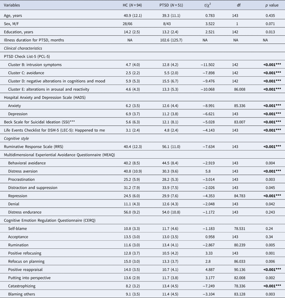
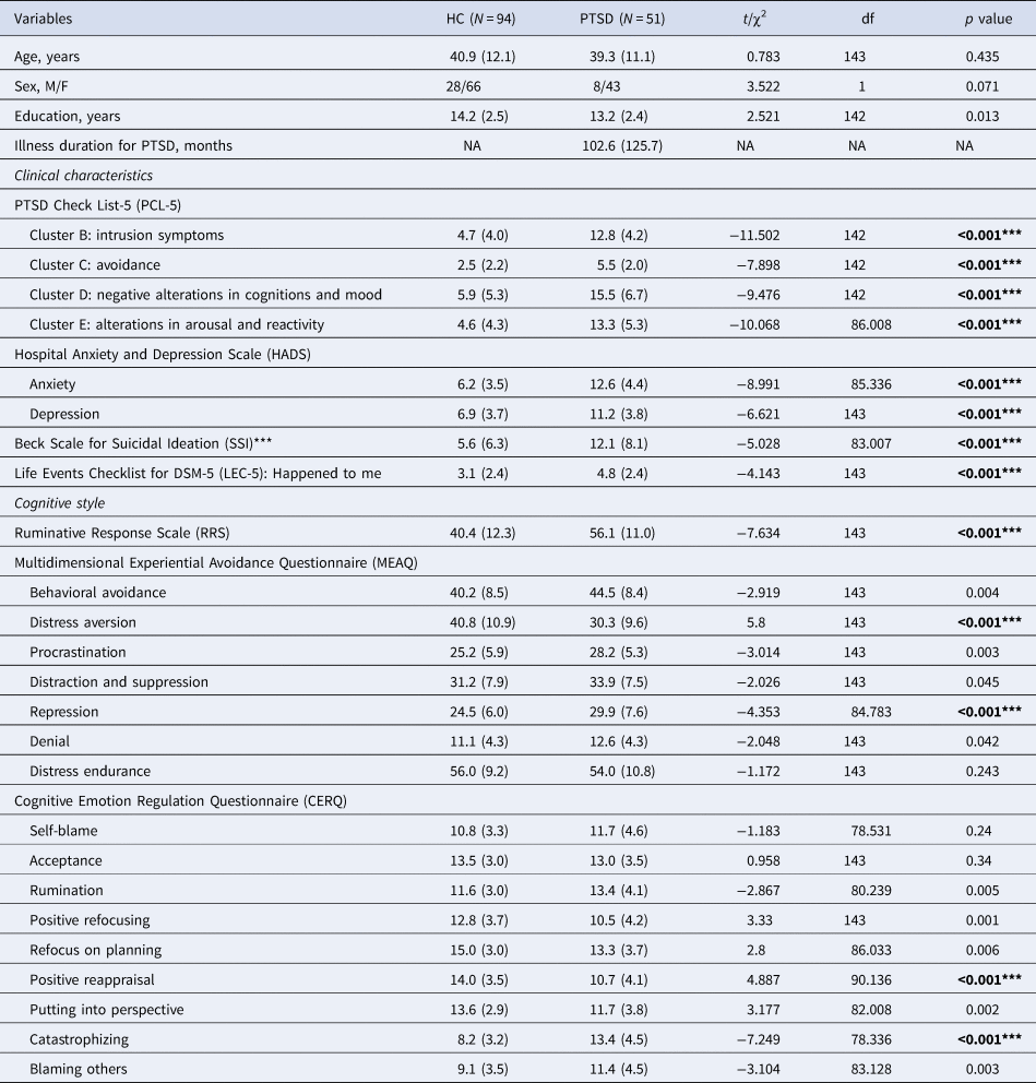
Between-group differences in sex ratio were analyzed using χ2 tests (Table 1). Differences of age, education years, clinical characteristics (illness duration for PTSD, four sub-scores of the PCL-5, anxiety and depression sub-scores of the HADS, total score of the SSI, and the ‘happened to me’ sub-score on the LEC-5), and stress-related cognitive characteristics (total score of the RRS, seven sub-scores on the MEAQ, and nine sub-scores on the CERQ) between the PTSD and HC were calculated using independent t tests (Table 1).
Demographic, trauma-related, cognitive, and clinical characteristics
*** P < 0.001
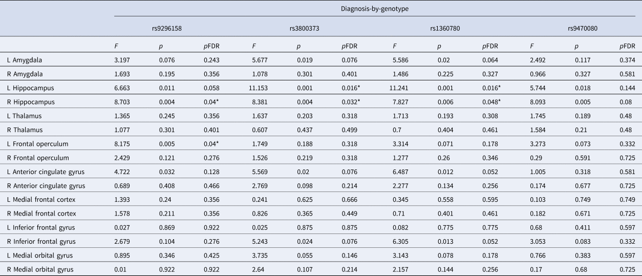
Two-way analysis of covariance (ANCOVA; with the covariates of age, sex, education and total intracranial volume adjusted) examined the main effect of diagnostic groups, main effect of FKBP5 genotype (four FKBP5 SNPs including the rs9296158 [GG and (AG + AA)], rs3800373 [AA and (AC + CC)], rs1360780 [CC and (CT + TT)], and rs9470080 [CC and (CT + TT)]), and the interaction of diagnosis-by-genotype (each SNP separately) for the regional brain gray matter volumes in 16 cortical and subcortical ROIs. All analyses were corrected for multiple comparisons using a false discovery rate (FDR) threshold of p < 0.05 based on the Benjamini–Hochberg (BH) procedure (Afifi et al., Reference Afifi, Asmundson, Taylor and Jang2010).
Among these 16 cortical and subcortical ROIs, those showing statistically significant diagnosis-by-genotype interactions were subjected to the partial correlation coefficients calculation (adjusted for age, sex, education, and total intracranial volume) with stress-related cognitive styles for which significant differences found between HC and PTSD groups. Issues of multiple correlation testing were statistically adjusted with a 5000-bootstrap resampling technique for HC and PTSD separately (Haukoos & Lewis, Reference Haukoos and Lewis2005; Pernet, Wilcox, & Rousselet, Reference Pernet, Wilcox and Rousselet2013; Ruscio, Reference Ruscio2008). Statistical analyses were conducted using IBM SPSS version 23.0 (IBM Corp., Armonk, NY, USA).
Results
Demographic and clinical characteristics
A summary of demographics, clinical characteristics, and stress-related cognitive styles for PTSD and HC groups is provided in Table 1. Compared with HC, patients with PTSD reported more previous exposure to stressful life events (LEC-5: ‘happened to me’) and demonstrated more severe psychiatric symptoms scored using the PCL-5 (intrusion symptoms, avoidance, negative alterations in cognitions and mood, alterations in arousal and reactivity), more severe anxiety-depressive mood (sub-scores of HADS) as well as suicidal ideation (total score of SSI). Notably, patients with PTSD reported more ruminative response (total score of the RSS) and experiential avoidance (MEAQ sub-scores of distress aversion and repression) in stressful events confrontation. Patients with PTSD demonstrated a stronger tendency of catastrophizing and weaker positive reappraisal when attempting to create reason of the stressful events (CERQ sub-scores).
Diagnosis-by-FKBP5 genotype interactions for gray matter volumes
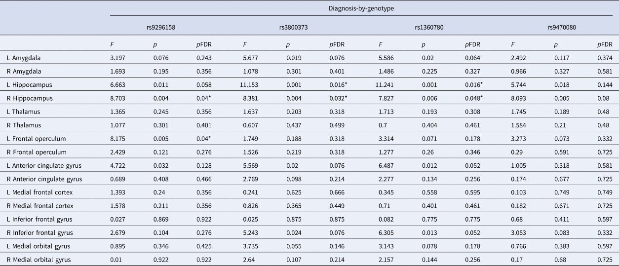
The minor allele frequencies of four FKBP5-related SNPs were greater than 5% in both groups. Additionally, the genotypic distributions of the four FKBP5 SNPs did not deviate from the Hardy–Weinberg equilibrium for both groups (all ps > 0.05; online Supplementary Table S1). Two-way ANCOVA was calculated to examine the effects of PTSD diagnosis and FKBP5 SNPs genotype on the regional brain volumes. Among the two-way ANCOVA results in Table 2, only for those with statistically significant diagnosis-by-genotype interactions [p < 0.05 (FDR-corrected)], simple main effects of diagnosis and genotype for regional brain volumes were examined [with statistical significance of p < 0.025 (Bonferroni-corrected)] as described below.
PTSD diagnosis-by-FKBP5 genotype interactions (adjusted for age, sex, education years, and intracranial volume) for regional brain volumes
*p < 0.05 (FDR-corrected).
FKBP5 SNP rs3800373 genotype and left hippocampal volume
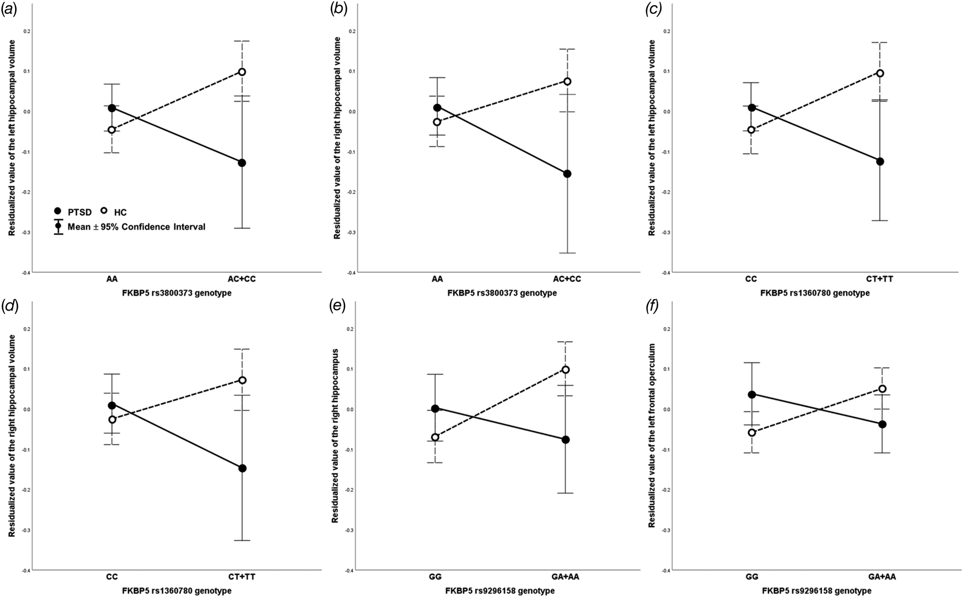
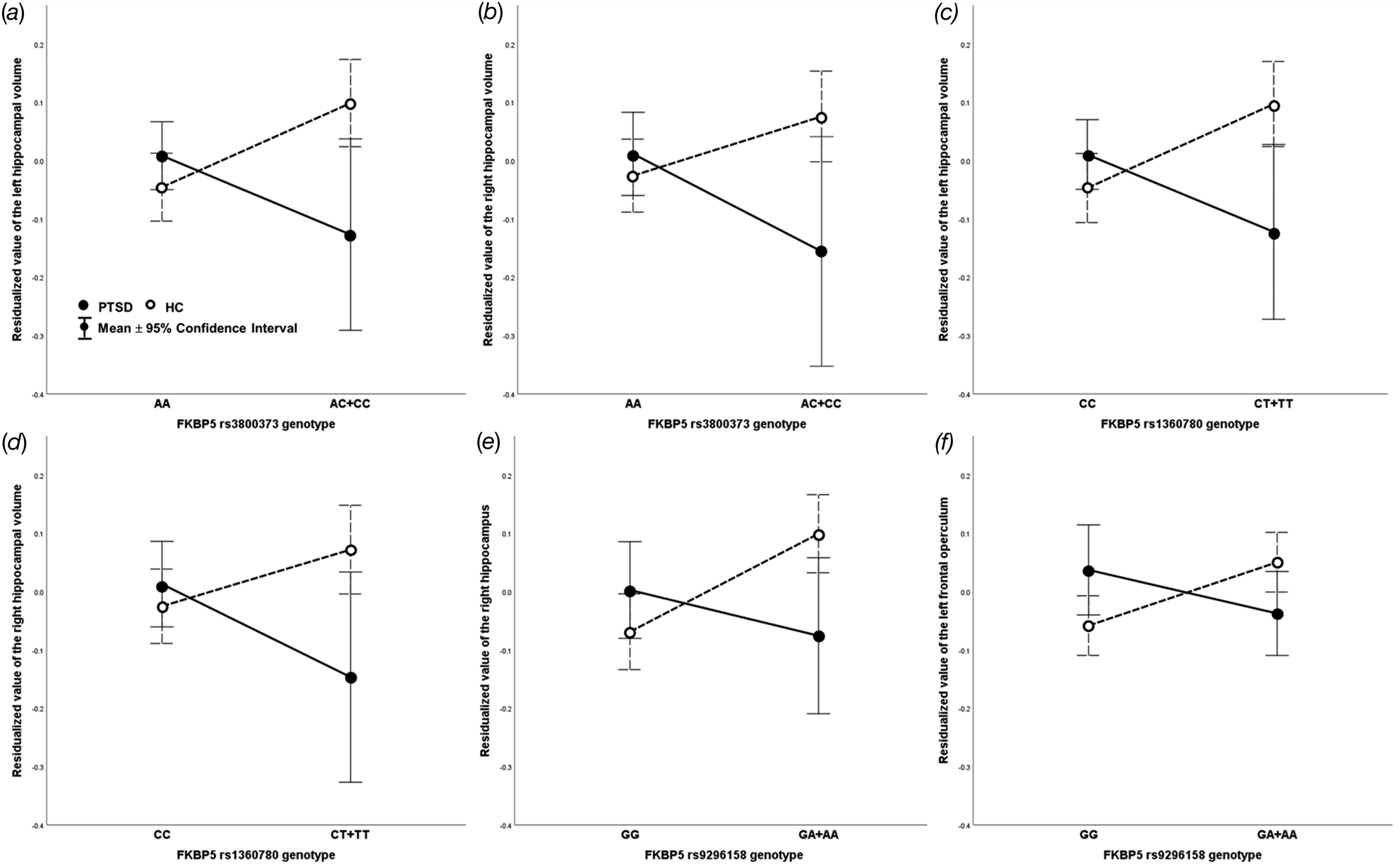
A two-way ANCOVA showed statistically significant diagnosis-by-rs3800373 genotype interaction on the regional volume of the left hippocampus [F = 11.153, p = 0.016 (FDR-corrected); Table 2 and Fig. 1a]. First, the effect of PTSD diagnosis was significant for the rs3800383 AC or CC genotype (F = 10.176, p = 0.002) but not for the AA genotype (F = 1.278, p = 0.260). With the rs3800373 AC or CC genotype, left hippocampal volume was larger in HC than PTSD (p = 0.002). Second, the effect of rs3800373 genotype was significant in HC (F = 10.315, p = 0.002) but not in PTSD (F = 3.651, p = 0.058). For HC, left hippocampal volume was larger in the rs3800373 AC or CC than in the AA genotype (p = 0.002).
Interactions between the post-traumatic stress disorder (PTSD) diagnosis and FKBP5 single nucleotide polymorphism (SNP) genotype for regional brain volumes. (a) FKBP5 rs3800373 and left hippocampus: with the rs3800373 genotype of AC or CC, left hippocampal volume was larger in healthy controls (HC) than in PTSD. In HC, left hippocampal volume was larger for the rs3800373 AC or CC genotype than for the rs3800373 AA genotype. (b) FKBP5 rs3800373 and right hippocampus: with the rs3800373 AC or CC genotype, right hippocampal volume was larger in HC compared to PTSD. (c) FKBP5 rs1360780 genotype and left hippocampus: with the rs1360780 CT or TT genotype, the left hippocampal volume was larger in HC compared to PTSD. In HC, the left hippocampal volume was larger for the rs1360780 CT or TT genotype than for the CC genotype. (d) FKBP5 rs1360780 genotype and right hippocampus: for the rs1360780 genotype of CT or TT, the right hippocampal volume was larger in HC than in PTSD. (e) FKBP5 rs9296158 genotype and right hippocampus: for the rs9296158 genotypes of GA or AA, the right hippocampal volume was larger in HC compared to PTSD. In HC, the right hippocampal volume was larger for the rs9296158 GA or AA genotype than for GG genotype. (f) FKBP5 rs9296158 genotype and left frontal operculum: for HC, left frontal operculum volume was larger in the rs9296158 GA or AA genotype compared to the rs9296158 GG genotype. Y scale depicts residual of regional brain volume (y-axis) calculated by regressing the covariates (age, sex, education years, and total intracranial volume) out.
FKBP5 SNP rs3800373 genotype and right hippocampal volume
In addition, another statistically significant diagnosis-by-rs3800373 genotype interaction was found in the regional volume of the right hippocampus [F = 8.381, p = 0.032 (FDR-corrected); Table 2 and Fig. 1b]. First, the effect of diagnosis was significant for the rs3800383 AC or CC genotype (F = 8.748, p = 0.004) but not for the rs3800373 AA genotype (F = 0.462, p = 0.498). Specifically, for the rs3800373 AC or CC genotype, right hippocampal volume was larger in HC than in PTSD (p = 0.004). Second, the effect of rs3800373 genotype for the right hippocampal volume was not significant both in HC (F = 4.159, p = 0.043) and PTSD (F = 4.544, p = 0.035).
FKBP5 SNP rs1360780 genotype and left hippocampal volume
For the rs1380780 genotype, a statistically significant diagnosis-by-genotype interaction on the regional volume of the left hippocampus [F = 11.241, p = 0.016 (FDR-corrected); Table 2 and Fig. 1c] was found. First, the effect of diagnosis was significant with the rs1360780 CT or TT genotype (F = 10.157, p = 0.002) but not with the rs1360780 CC genotype (F = 1.389, p = 0.241). For the rs1360780 CT or TT, the left hippocampal volume was larger in HC than PTSD (p = 0.002). Second, the effect of rs1360780 genotype in the left hippocampal volume was significant in HC (F = 10.253, p = 0.002) but not in PTSD (F = 3.625, p = 0.059). The left hippocampal volume in HC was larger for the rs1360780 CT or TT genotype than for CC genotype (p = 0.002).
FKBP5 SNP rs1360780 genotype and right hippocampal volume
Moreover, diagnosis-by-rs1360780 genotype interaction was also statistically significant for the right hippocampal volume [F = 7.827, p = 0.048 (FDR-corrected); Table 2 and Fig. 1d]. First, the effect of diagnosis was significant for the rs1360780 CT or TT genotype (F = 8.233, p = 0.005) but not for the rs1360780 CC genotype (F = 0.449, p = 0.504). With the rs1360780 CT or TT genotype, right hippocampal volume was larger in HC than in PTSD (p = 0.005). Second, the effect of rs1360780 genotype for the right hippocampal volume was not significant both in HC (F = 3.750, p = 0.055) and in PTSD (F = 4.277, p = 0.041).
FKBP5 SNP rs9296158 genotype and right hippocampal volume
In terms of the rs9296158 genotype, diagnosis-by-genotype interaction was statistically significant for the right hippocampal volume [F = 8.703, p = 0.040 (FDR-corrected); Table 2 and Fig. 1e]. First, the effect of PTSD diagnosis was significant for the rs9296158 GA or AA genotype (F = 7.583, p = 0.007) but not for the rs9296158 GG genotype (F = 1.560, p = 0.214). Specifically, for the rs9296158 GA or AA, the right hippocampal volume was larger in HC than in PTSD (p = 0.007). Second, the effect of rs9296158 genotype for the right hippocampal volume was significant in HC (F = 11.937, p = 0.001) but not in PTSD (F = 1.275, p = 0.261). In HC, the right hippocampal volume was larger for the rs9296158 GA or AA genotype than for GG genotype (p = 0.001).
FKBP5 SNP rs9296158 genotype and volume of the left frontal operculum
Further, another significant diagnosis-by-rs9296158 genotype interaction was found for the regional volume of the left frontal operculum [F = 8.175, p = 0.040 (FDR-corrected); Table 2 and Fig. 1f]. First, the effect of diagnosis for the regional volume of left frontal operculum was not significant with the rs9296158 GA or AA genotype (F = 3.053, p = 0.083) and also with the rs9296158 GG genotype (F = 5.097, p = 0.026). Second, the effect of rs9296158 genotype for the left frontal operculum volume was significant in HC (F = 8.587, p = 0.041) but not in PTSD (F = 1.948, p = 0.165). For HC, the left frontal operculum volume was larger in the rs9296158 GA or AA genotype than for GG genotype (p = 0.004).
FKBP5 SNP rs9470080 genotype
For the rs9470080 genotype, no statistically significant diagnosis-by-genotype interactions of regional brain volumes were found (all p > 0.05; Table 2).
Correlations between the regional brain volumes and stress-related cognitive styles
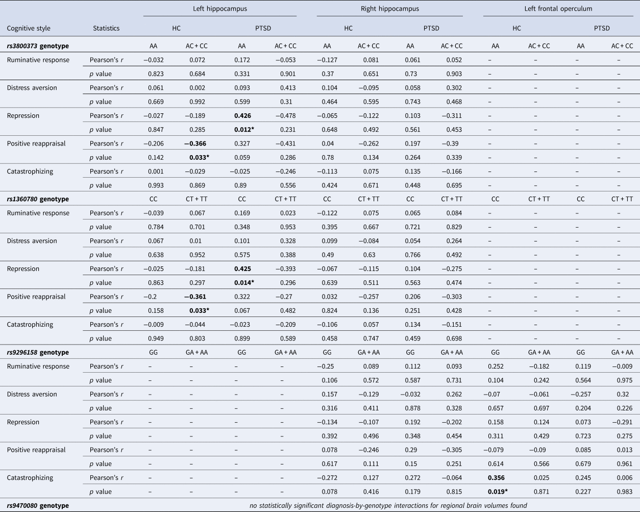
Partial correlation coefficients (adjusted for age, sex, education, and total intracranial volume) between five stress-related cognitive styles (ruminative response, distress aversion, repression, positive reappraisal, catastrophizing) with significant between-group (HC and PTSD) differences and three brain regions with significant diagnosis-by-genotype interactions for regional brain volumes [left hippocampus (rs3800373 and rs1360780), right hippocampus (rs9296158, rs3800373, and rs1360780), and left frontal operculum (rs9296158)] were calculated for each subgroup segregated according to the diagnosis-by-FKBP5 SNP genotypes.
FKBP5 SNP rs3800373 genotype
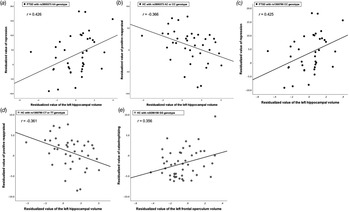
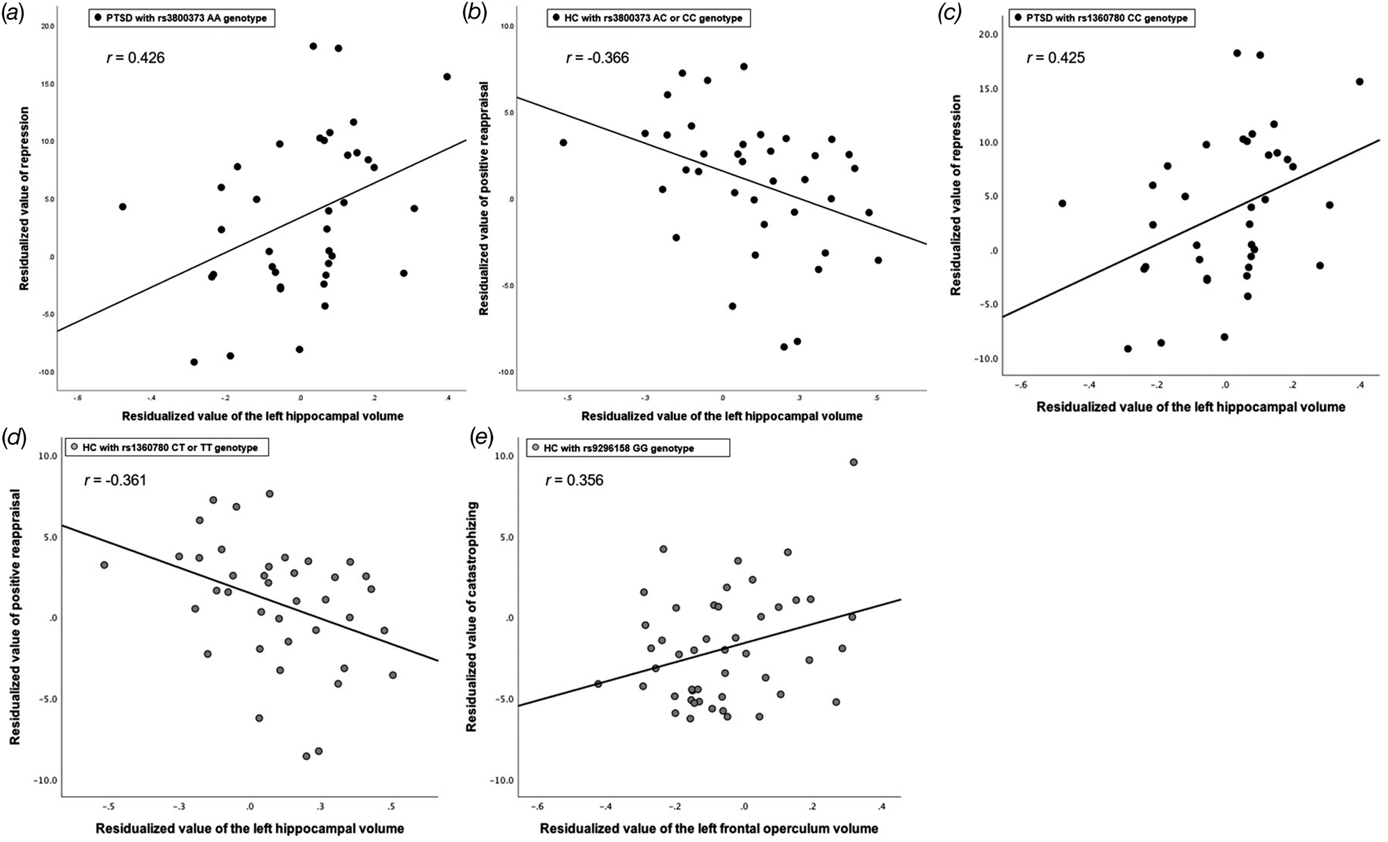
For the rs3800373 genotype, there was a significant positive correlation between repression and left hippocampal volume (r = 0.426, p = 0.012) in PTSD with rs3800373 AA genotype (Fig. 2a). On the other hand, there were no significant correlations found between the repression and left hippocampal volume in PTSD with rs3800373 C carriers (r = −0.478, p = 0.231), in HC with rs3800373 AA genotype (r = −0.027, p = 0.847), or in HC with rs3800383 C carriers (r = −0.189, p = 0.285; Table 3).
Partial correlations between cognitive styles and regional brain volumes (adjusted for age, sex, education year, and total intracranial volume). (a) Positive correlation between repression and the left hippocampal volume (r = 0.426, p = 0.012) in post-traumatic stress disorder (PTSD) with rs3800373 AA genotype. (b) Negative association between the left hippocampal volume and positive reappraisal in healthy controls (HC) with rs3800373 AC or CC genotype (r = −0.366, p = 0.033). (c) Positive correlation between the repression and left hippocampal volume in PTSD with rs1360780 CC genotype (r = 0.425, p = 0.014). (d) Negative relationship between the left hippocampal volume and positive reappraisal in HC with rs1360780 CT or TT genotype (r = −0.361, p = 0.033). (e) Positive correlation between regional volume of the left frontal operculum and the severity of catastrophizing in HC with rs9296158 GG genotype (r = 0.356, p = 0.019). X and Y scales depict residuals of cognitive style (y-axis) and regional brain volume (x-axis) calculated by regressing the covariates (age, sex, education years, and total intracranial volume) out.
Partial correlations between the regional brain volumes and stress-related cognitive styles (adjusted for age, sex, education years, and intracranial volume)
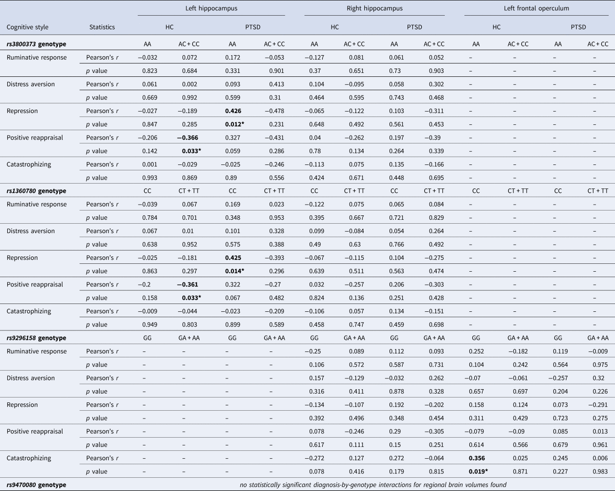
*p < 0.05 (adjusted with a 5000-bootstrap resampling technique).
Moreover, left hippocampal volume also showed a statistically significant negative correlation with the strength of positive reappraisal in HC with rs3800373 AC or CC genotype (r = −0.366, p = 0.033; Fig. 2b) which was not evident in HC with AA genotype (r = −0.206, p = 0.142), in PTSD with AA genotype (r = 0.327, p = 0.059), nor in PTSD with AC or CC genotype (r = −0.431, p = 0.286; Table 3).
FKBP5 SNP rs1360780 genotype
Regarding the rs1360780 genotype, there was a significant positive association between the repression and left hippocampal volume in PTSD with rs1360780; CC (r = 0.425, p = 0.014; Fig. 2c). On the contrary, association between the repression and the left hippocampal volume did not show statistical significance for PTSD with rs1360780 CT or TT genotype (r = −0.393, p = 0.296), HC with rs1360780 CC genotype (r = −0.025, p = 0.863), nor HC with rs1360780 CT or TT genotype (r = −0.181, p = 0.297; Table 3).
Further, for HC with rs1360780 CT or TT genotype, regional volume of the left hippocampus and strength of positive reappraisal showed statistically significant negative association (r = −0.361, p = 0.033; Fig. 2d), which was not statistically significant in other subgroups of HC with CC genotype (r = −0.200, p = 0.158), PTSD with CC genotype (r = 0.322, p = 0.067), nor PTSD with CT or TT genotype (r = −0.270, p = 0.482; Table 3).
FKBP5 SNP rs9296158 genotype
In terms of the rs9296158 genotype, a significant positive correlation between catastrophizing and left frontal operculum volume was found in HC with rs9296158 GG genotype (r = 0.356, p = 0.019; Fig. 2e). On the contrary, associations between the catastrophizing and volume of the left frontal operculum were not statistically significant in other subgroups of HC with rs9296158 GA or AA genotype (r = 0.025, p = 0.871), PTSD with rs9296158 GG genotype (r = 0.245, p = 0.227), nor PTSD with rs9296158 GA or AA genotype (r = 0.006, p = 0.983; Table 3).
FKBP5 SNP rs9470080 genotype
As no statistically significant diagnosis-by-genotype interactions of regional brain volumes were found regarding the rs9470080 genotype (refer to the ‘Diagnosis-by-FKBP5 genotype interactions for gray matter volumes’ section in the ‘Results’ above), differential partial correlations between the stress-related cognitive styles v. the regional brain volumes regarding the rs8470080 genotype and PTSD diagnosis were not calculated in the current study.
Discussion
To the best of our knowledge, this is the first study that unraveled the FKBP5 genotype-related neural underpinning of stress-related cognitive style in PTSD. In this study, patients with PTSD showed more severe catastrophizing, rumination, and repression in addition to the weaker tendencies of distress aversion and positive reappraisal compared to HC. Significant diagnosis-by-genotype interactions were found for bilateral hippocampi and left frontal operculum. Moreover, the significant positive correlations between the cognitive style (repression) and the left hippocampal volume were uncovered in patients with PTDS who had homozygous genotypes for the FKBP5 SNPs of rs3800373 (AA genotype) or rs1360780 (CC genotype). These results suggest that stress-related cognitive style exists in neural circuitry including the hippocampus under the influence of FKBP5 genotype in patients with PTSD.
The hippocampus has been regarded as a major region involved in the pathology of PTSD. A meta-analysis showed that hippocampal volumes were smaller in the PTSD group and trauma-exposed group without PTSD compared with those of the trauma-unexposed group (Woon, Sood, & Hedges, Reference Woon, Sood and Hedges2010). The significant negative correlation between the hippocampal volumes and PTSD symptomatology was shown only in PTSD patients (Nelson & Tumpap, Reference Nelson and Tumpap2017), but not in malingering subjects (Butler et al., Reference Butler, Herr, Willmund, Gallinat, Zimmermann and Kuhn2018) or in HC (Logue et al., Reference Logue, van Rooij, Dennis, Davis, Hayes, Stevens and Morey2018). In a resting-state fMRI study, a successful distinction between patients with PTSD and HC was reported from the effective connectivity of the amygdala to the right hippocampus and of the right hippocampus to the left striatum-precuneus-insula (Rangaprakash et al., Reference Rangaprakash, Dretsch, Venkataraman, Katz, Denney and Deshpande2018). Likewise, the resting-state functional connectivity between the hippocampus and other regions – as reflected in the values of nodal efficiency and degree centrality – was elevated in patients with PTSD exposed to a major earthquake (Zhang et al., Reference Zhang, Yin, Hu, Duan, Qi, Xu and Li2017).
In the current study, the regional volume of bilateral hippocampi demonstrated a significant interaction between the PTSD diagnosis and FKBP5 genotype. This result is consistent with prior studies that reported the influence of genetic factors on the morphology of the hippocampus in patients with PTSD. Indeed, a positive correlation between the hippocampal volume and degree of methylation in the exon 1F promoter region of the glucocorticoid receptor gene was found in patients with PTSD (McNerney et al., Reference McNerney, Sheng, Nechvatal, Lee, Lyons, Soman and Adamson2018).
Furthermore, in the current study, positive correlations between the left hippocampal volume and severity of repression (a form of experiential avoidance) were also found in PTSD patients homozygous for FKBP5 SNPs rs3800373 (AA genotype) or rs1360780 (CC genotype). Increased experiential avoidance was reported to be related to the decreased functional activation of the hippocampus in healthy adults in an fMRI study (Schlund, Magee, & Hudgins, Reference Schlund, Magee and Hudgins2011). Moreover, increased functional activation of the hippocampus during encoding of episodic memory is associated with re-experiencing symptoms of PTSD in these patients (Stevens et al., Reference Stevens, Reddy, Kim, van Rooij, Ely, Hamann and Jovanovic2018). All these previous evidences support the validity of our results.
Previous research has shown that two FKBP5 SNPs (rs3800373 and rs1360780) interact with child abuse severity (Binder et al., Reference Binder, Bradley, Liu, Epstein, Deveau, Mercer and Ressler2008) or adult natural disaster (Hawn et al., Reference Hawn, Sheerin, Lind, Hicks, Marraccini, Bountress and Amstadter2019) as predictors of PTSD symptoms in adult age. Previous studies showed an association between the genotype of FKBP5 SNP rs1360780 and resting-state functional connectivity of the hippocampus-anterior cingulate (Fani et al., Reference Fani, King, Shin, Srivastava, Brewster, Jovanovic and Ressler2016) and between increased expression of FKBP5 protein, increased hippocampal volume, and clinical improvement after cognitive-behavioral therapy in patients with PTSD (Levy-Gigi, Szabo, Kelemen, & Keri, Reference Levy-Gigi, Szabo, Kelemen and Keri2013).
The present study uncovered a cognitive style and brain volume in patients with PTSD. Repression is a kind of mental defense mechanism that prevents overwhelming PTSD symptoms such as flashback, and re-experience and has been considered as a secondary distorted cognitive style to abnormal neocortical and hippocampal arousal and corticosteroid and enkephalin secretion, which can induce functional, cellular, and anatomical abnormality within the hippocampus (Joseph, Reference Joseph1998). Hulbert, Henson, and Anderson (Reference Hulbert, Henson and Anderson2016) showed that when people suppress retrieval of unwanted memory, stopping retrieval engages a suppression mechanism that is related to the hippocampal processes. A cognitive style, repression, through mediation of frontal cortex, could operate to protect the brain from stressful symptoms. Neuroimaging data revealed that trauma-exposed individuals showed reduced activation in the right middle frontal gyrus during memory suppression. Difficulty in active suppression of memories may contribute to the development of PTSD (Sullivan et al., Reference Sullivan, Marx, Chen, Depue, Hayes and Hayes2019). Current study showed a positive correlation between the left hippocampal volume and severity of repression in patients with PTSD homozygous for FKBP5 SNPs rs3800373 (AA genotype) or rs1360780 (CC genotype). We hypothesized that homozygous for FKBP5 SNPs rs3800373 (AA genotype) or rs1360780 (CC genotype) could be protective factors for symptoms aggravation through protective cognitive style (repression) in patients with PTSD compared with other genotypes. The hypothesis is in line with the results of Binder et al. (Reference Binder, Bradley, Liu, Epstein, Deveau, Mercer and Ressler2008) who suggested a protective genotype effect of FKBP5 toward traumatic stress.
In addition, significant diagnosis-by-genotype interactions for regional brain volumes were also found in the left frontal operculum, which showed significant correlations with cognitive style in HC. The regional grey matter volume of the left frontal operculum showed a positive association with sensitivity to negative life events as mediated by rumination (Qiao et al., Reference Qiao, Wei, Li, Chen, Che, Li and Liu2013). Activation of the frontal operculum is positively correlated with the intensity of trait rumination (Kocsel et al., Reference Kocsel, Szabo, Galambos, Edes, Pap, Elliott and Kokonyei2017) and with rewards of food intake in youth at risk for obesity (Stice, Yokum, Burger, Epstein, & Small, Reference Stice, Yokum, Burger, Epstein and Small2011). Likewise, different genotypes of catechol-I-methyltransferase gene are associated with functional connectivity between frontal operculum and other posterior regions (Jaspar et al., Reference Jaspar, Manard, Dideberg, Bours, Maquet and Collette2016). Furthermore, patients with major depressive disorder having a risk allele (T) for FKBP5 rs1360780 SNP showed reduced activation of frontal operculum during emotional attention task compared to patients without the T allele (Tozzi et al., Reference Tozzi, Carballedo, Wetterling, McCarthy, O'Keane, Gill and Frodl2016).
Limitations
This study has some limitations. First, this study compared the HC and PTSD using the cross-sectional design and did not assess the longitudinal trajectories of regional brain volumes and stress-related cognitive styles. Accordingly, the current study results might indicate not causation but association among the PTSD diagnosis, FKBP5 genotype, regional brain volumes, and cognitive styles. Second, possible distinctive profiles of diagnosis-by-genotype interactions for cortical surface area or cortical thickness separately (Panizzon et al., Reference Panizzon, Fennema-Notestine, Eyler, Jernigan, Prom-Wormley, Neale and Kremen2009) were not explored.
Conclusion
Collectively, this study is the first to unravel the FKBP5 genotype-related neural underpinning of stress-related cognitive style in PTSD. The FKBP5 gene, one of the risk factors for PTSD development or symptoms, is involved with the hypothalamic-pituitary-adrenal axis and associated with the stress response. This study showed that the FKBP5 genotype-by-PTSD diagnosis interaction for grey matter volumes of the hippocampus is also associated with cognitive style in PTSD, including the elevated experiential avoidance and lowered cognitive-emotional regulation. Our work shows the neurobiological support for the inclusion of the hippocampus and left frontal operculum as possible candidate brain regions of the neuromodulation of patients with PTSD homozygous for risk alleles of FKBP5-associated SNPs, targeting the improvement of stress-related cognitive styles in PTSD.
Supplementary material
The supplementary material for this article can be found at https://doi.org/10.1017/S0033291720002949
Data availability
The corresponding author S.H.L. has full access to the study data. For more information regarding the request for access to the study data, please contact the corresponding author by way of e-mail: lshpss@paik.ac.kr.
Author details
Je-Yeon Yun, M.D., Ph.D., Associate Professor, Seoul National University Hospital, Seoul, Republic of Korea and Yeongeon Student Support Center, Seoul National University College of Medicine, Seoul, Republic of Korea; Min Jin Jin, M.S., Ph.D. Student, Clinical Emotion and Cognition Research Laboratory, Inje University, Republic of Korea and Department of Psychology, Chung-Ang University; Sungkean Kim, Ph.D., Postdoctoral Fellow, Clinical Emotion and Cognition Research Laboratory, Inje University, Republic of Korea and Department of Biomedical Engineering, Hanyang University, Seoul, Republic of Korea; Seung-Hwan Lee, M.D., Ph.D., Professor, Clinical Emotion and Cognition Research Laboratory, Inje University, Republic of Korea and Department of Psychiatry, Ilsan Paik Hospital, Inje University College of Medicine, Goyang, Republic of Korea.
Author contributions
J.Y.Y. contributed to the overall conceptualization and study design, statistical analysis and interpretation of the findings, and writing of the manuscript. M.J.J. contributed to the collection of data and interpretation of the findings. S.K.K. contributed to the collection of data and interpretation of the findings. S.H.L. was the chief investigator and contributed to the overall design and conduct of the study, collection of data, and interpretation of the findings. All authors reviewed, revised, and approved the final version of the manuscript.
Financial support
This work was supported by the Brain Research Program through the National Research Foundation of Korea (NRF) funded by the Ministry of Science, ICT and Future Planning (S.H.L., NRF-2015M3C7A1028252); and by the Korea Science and Engineering Foundation (KOSEF) funded by the Korean government (S.H.L., NRF-2018R1A2A2A05018505).
Conflict of interest
None.