


Introduction
Megaspora verrucosa (Ach.) L. Arcadia & A. Nordin var. verrucosa is a common, nearly cosmopolitan terricolous lichen that primarily colonizes dead mosses and other plant debris on calcareous rocks and soils in arcto-alpine and related habitats (Smith et al. Reference Smith, Aptroot, Coppins, Fletcher, Gilbert, James and Wolseley2009; Nimis Reference Nimis2026). Corticolous and lignicolous specimens of M. verrucosa are usually assigned to var. mutabilis (Ach.) Nimis & Cl. Roux (Nimis Reference Nimis2026). After Wirth (Reference Wirth1987), M. verrucosa was long treated as the sole species in the genus Megaspora; currently it comprises five species (Index Fungorum Partnership 2026). The genus belongs to the family Megasporaceae (Pertusariales, Lecanoromycetes), together with Aspicilia, Circinaria, Lobothallia, Sagedia, and Teuvoa (Lücking et al. Reference Lücking, Hodkinson and Leavitt2017). Nordin et al. (Reference Nordin, Savić and Tibell2010) demonstrated a close phylogenetic relationship between Megaspora and Circinaria.
Examination of Megaspora verrucosa var. verrucosa specimens from the Arctic and the mountains of Central Asia, together with additional Antarctic and subantarctic material, revealed a number of lichenicolous fungi, most of which proved to be new to science. The aims of the present paper are to document the diversity of lichenicolous fungi occurring on Megaspora verrucosa var. verrucosa and to provide a key for their identification.
Materials and Methods
This study is based on 49 specimens of lichenicolous fungi, mainly from the Holarctic (primarily Mongolia and Russia), but also from Antarctica and Chile. Of these, 46 specimens are deposited in the Mycological Herbarium of the Komarov Botanical Institute (LE), Russian Academy of Sciences, St Petersburg, Russia, and three specimens in the private herbarium of J. Etayo (hb. Etayo).
Microscopic observations and imaging were carried out using a Zeiss Axio Zoom V16 stereomicroscope equipped with a Zeiss AxioCam 712 colour digital camera and a Zeiss Axio Imager A1 compound microscope fitted with Nomarski differential interference contrast (DIC) optics and a Zeiss AxioCam 807 colour digital camera. Microscopic characters were examined using hand-cut sections prepared with a razor blade and mounted in water, brilliant cresyl blue (BCr), 10% aqueous KOH solution (K), lactophenol cotton blue, 0.5% Lugol’s iodine directly (I) or after a K pretreatment (K/I), or phloxine directly or after a K pretreatment. Unless otherwise stated, measurements and descriptions refer to water mounts. When the number of measurements (n) exceeded 10, dimensions of measured structures, including the length-to-width ratios (l/w) of ascospores and conidia, are presented as (min–)(x̄ – SD) – (x̄ + SD)(–max), where ‘min’ and ‘max’ denote the observed extremes, x̄ the arithmetic mean, and SD the standard deviation. Dimensions of the microstructures, both newly obtained and taken from the literature, were rounded to the nearest 0.5 μm. Colours were mainly named according to Kornerup & Wanscher (Reference Kornerup and Wanscher1978).
Taxonomy
Arthonia aspiciliae Alstrup & E. S. Hansen var. megasporae Zhurb. var. nov.
MycoBank No.: MB 864910
Distinguished from Arthonia aspiciliae Alstrup & E. S. Hansen var. aspiciliae by the I+ brownish orange to reddish brown versus I± purple hymenium, epihymenium and hypothecium without a blackish tint, constantly heteropolar ascospores, and a different host selection: Megaspora versus Aspicilia (both in Megasporaceae).
Type: Kyrgyzstan, Issyk-Kul Region, Central Tyan’-Shan’ mountain system, southern slope of Terskey Ala-Too Range, Ottuk River (right tributary of the Saryjas River), c. 42°19′00″N, 79°03′37″E, c. 3000 m, mountain steppe-tundra, on Megaspora verrucosa var. verrucosa (thalline margin of apothecia, occasionally thallus) growing on the tussock of a flowering plant, 31 July 1979, L. I. Bredkina 3063 (LE F-355757—holotype).
(Fig. 1)
Arthonia aspiciliae var. megasporae (holotype). A, ascomata growing on apothecial thalline margin of Megaspora verrucosa var. verrucosa. B, ascoma in median vertical section. C, ascoma in median vertical section. D, hymenium. E, asci. F, ascospores. B, C (left), E (left), F (top left) in water; C (right), F (bottom left) in K; D (left), E (middle), F (top right) in I; D (right), E (right), F (bottom right) in K/I. Scales: A = 200 μm; B = 50 μm; C–F = 10 μm. In colour online.
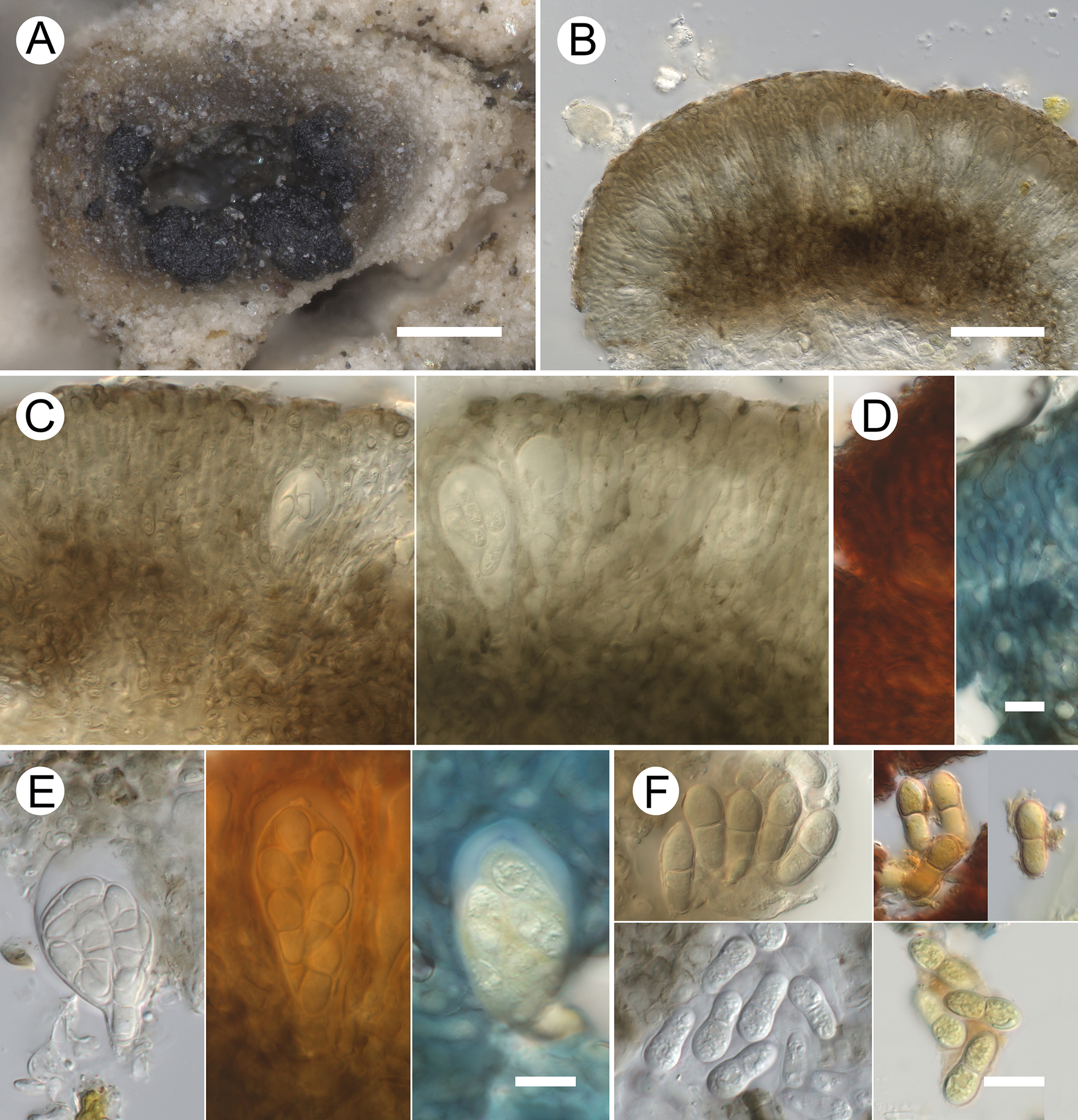
Figure 1. Long description
A multi-panel micrograph series labeled A through F.
* Panel A: A macroscopic photo of dark, granular ascomata clustered on the pale, textured thalline margin of Megaspora verrucosa. Scale bar equals 200 micrometers.
* Panel B: A vertical cross-section of an ascoma in water, showing a curved, brownish hymenial layer. Scale bar equals 50 micrometers.
* Panel C: Two side-by-side microscopic views of an ascoma section. The left side is in water and the right side is in K, showing the internal hyphal structure and developing asci. Scale bar equals 10 micrometers.
* Panel D: Two vertical strips of the hymenium. The left strip is stained red in I and the right strip is stained blue in K / I. Scale bar equals 10 micrometers.
* Panel E: Three individual asci shown in different reagents. From left to right: water, I (stained orange-brown), and K / I (stained blue). The asci are pear-shaped and contain multiple ascospores. Scale bar equals 10 micrometers.
* Panel F: Four sub-panels showing individual ascospores. Top-left shows spores in water; top-right shows spores in I; bottom-left shows spores in K; bottom-right shows spores in K / I. The spores are multi-celled and oblong. Scale bar equals 10 micrometers.
Lifestyle lichenicolous. No gall formation or discoloration of the infected host parts observed. Vegetative hyphae not observed.
Ascomata apothecioid, black, slightly glossy, roughened, non-pruinose, irregularly rounded or elongated in surface view, slightly convex and flattened at the top to nearly hemispherical, (95–)120–260(–350) μm diam. (n = 32), up to 130 μm tall; erumpent and becoming superficial, usually aggregated, sometimes confluent into clusters up to 750 μm long. Hymenium 45–65 μm tall (including epihymenium), pale brownish grey, sometimes darker in the upper part, I+ brownish orange to reddish brown, K/I+ blue, not inspersed with oil or crystals; epihymenium medium brown, K+ greyish brown to greyish olivaceous, 3–7 μm tall, locally rather indistinct. Paraphysoids ±straight, 2.5–4 μm diam. in the middle, apically usually thickened (3.5–7 μm diam.) but not capitate, and often bearing a thin, dark brown, occasionally scabrous hood; frequently septate (cells 4–9 μm long), occasionally branched and anastomosing. Hypothecium medium brown, K+ greyish brown to greyish olivaceous, pigmentation indistinctly reticulate due to darker cell walls and intercellular spaces, 40–60 μm tall, composed of ±isodiametric or elongated cells, 3.5–11 × 3–7.5 μm. Asci broadly clavate, short-stalked, rounded at the apex, with a wall 1–6 μm thick apically, rarely with a distinct ocular chamber, (32–)38–50(–53) × (15–)16–22(–26) μm (n = 11, in water or I), 8-spored; epiplasm turning orange in I and K/I; a faint, poorly distinct, blurred K/I+ blue ring occasionally observed in the ascus tholus near the apex of the ocular chamber. Ascospores hyaline, narrowly obovate to cylindrical-clavate (with a wider upper cell relative to its position in the ascus), with rounded ends, (12.5–)13–15.5(–16.5) × (4.5–)5.5–7(–7.5) μm, l/w = (1.8–)2.1–2.5(–3.5) (n = 61, in water or I), 1-septate (septum median), not constricted at the septum, with a smooth wall c. 0.5 μm thick, without a gelatinous sheath, irregularly 2–3-seriate in the ascus. Note: in two of 12 thin apothecial sections examined (totalling c. 100 ascospores), two pale brown, verrucose, 1–2-septate spores of similar shape and size were observed; their origin is uncertain and they may not belong to this species.
Anamorph not observed.
Etymology
The varietal epithet refers to the host lichen, Megaspora verrucosa.
Distribution and host
The new variety is described here based on a single specimen collected in Central Asia (Kyrgyzstan). It was found on the thallus and apothecia of the terricolous lichen Megaspora verrucosa var. verrucosa growing in mountain steppe-tundra.
Notes
The Arthonia specimen presented here closely matches descriptions of the morphologically very similar A. aspiciliae (described from Greenland on saxicolous Aspicilia elevata) and A. hertelii (Calat. et al.) Hafellner & V. John (described from steppe/continental regions of Eurasia on vagrant Aspicilia and Circinaria) that was initially treated as a subspecies of A. aspiciliae (Alstrup & Hansen Reference Alstrup and Hansen2001; Calatayud et al. Reference Calatayud, Barreno and Rico2004). Notably, both species also occur on lichens in the Megasporaceae, the family that includes Megaspora. They differ from the fungus we examined, in part because they occupy different host genera, and most conspicuously by the hymenial iodine reaction: I± purple in those species (Calatayud et al. Reference Calatayud, Barreno and Rico2004) versus I+ brownish orange to reddish brown in my material. Additional distinguishing characters include ascospore morphology and pigmentation of hymenial layers. In A. aspiciliae var. aspiciliae the ascospore cells are sometimes almost equal in size and shape and the epihymenium and hypothecium have a blackish tint. Arthonia hertelii has slightly narrower, more elongate ascospores, rarely 2-septate, measuring (12–)14–17 × (4–)5–6 μm, l/w = (2–)2.5–3.4(–3.5), and is reported to cause necrosis in infected host tissue (Calatayud et al. Reference Calatayud, Barreno and Rico2004).
To my knowledge, the only previous report of an Arthonia species on Megaspora is that of Kondratyuk & Kudratov (Reference Kondratyuk and Kudratov2002: 167), who reported A. epiphyscia Nyl. from Tajikistan ‘on thallus of Physcia dubiana and Megaspora verrucosa var. mutabilis, 4.06.1974, I. Kudratov no. 1501 (KW)’. Arthonia epiphyscia, usually found on Physcia, is morphologically similar to my specimen but differs in having a thicker epihymenium (10–15 μm) and occasionally pale brown, smaller ascospores measuring (9.5–)10.5–13(–15) × (3.5–)4–5(–5.5) μm, l/w = (2–)2.3–2.9(–3.8) (n = 88). The Tajikistan collection may represent the same taxon as described here.
Specimens of Arthonia epiphyscia re-examined for comparison
Russia: Arkhangelsk Region: Franz Josef Land, Hooker Island, 80°20′N, 52°52′E, on Physcia dubia (Hoffm.) Lettau, 2 viii 1930, V. P. Savicz 9a (LE 206918). Chukotka Autonomous Area: Vesnovannaia River, 65°20′N, 174°26′E, on P. cf. dubia, 14 viii 1980, I. I. Makarova (LE 233518). Karachayevo-Circassian Republic: 6 km E of Teberda town, 43°27′13″N, 41°49′11″E, on P. aipolia (Ehrh. ex Humb.) Fürnr., 22 viii 2012, M. P. Zhurbenko 12207 (LE 261454). Krasnoyarsk Territory: Taimyr Peninsula, Byrranga Mts, Levinson-Lessinga Lake, 74°32′N, 98°36′E, on P. caesia, (Hoffm.) Hampe ex Fürnr. 29 vii 1994, M. P. Zhurbenko 94142b (LE F-206917b).
Cercidospora verrucosaria (Arnold) Arnold
The material examined from Megaspora verrucosa var. verrucosa corresponds closely with the species description of Navarro-Rosinés et al. (Reference Navarro-Rosinés, Calatayud and Hafellner2009). The only notable differences are that 4-spored asci are not exceptional but occur in six of 16 specimens examined (although most asci were 8-spored), and that ascospores are slightly longer: (13–)16.5–21(–25.5) × (4–)5–6.5(–7) μm, l/w = (2.3–)2.9–3.7(–4.8) (n = 238) versus (14.5–)15.5–19.5(–23) × (4.5–)5–6(–7) μm, l/w = (2.1–)2.8–3.8(–4.4) (n = 113). These data cast doubt on the hypothesis that Cercidospora verrucosaria s. lat., growing on corticolous M. verrucosa var. mutabilis represents a distinct taxon characterized by mainly 6-spored asci and slightly larger ascospores, (15.5–)18–24(–25) × 5–7 μm (Navarro-Rosinés et al. Reference Navarro-Rosinés, Calatayud and Hafellner2009: 16).
It is worth noting that the observed ascomata mainly grew on the thallus and thalline margin of the apothecia of Megaspora, where they were fully immersed, but were also found on the disc of host apothecia, where they were usually protruding up to one-half of their height.
Specimens examined (all on the thallus and apothecial thalline margin of Megaspora verrucosa var. verrucosa)
Mongolia: Arkhangai Region: Ikhtamir Somon, Khoid Tamir Gol River, near Khokh Nuur Lake, 47°05′47.4″N, 102°28′49.7″E, 2670 m, 29 vii 2023, M. P. Zhurbenko 23110 (LE F-355747). Bayan Khongor Region: Zhargalant Somon, NE of Zhargalant settlement, near Egiin Davaa Pass, 47°11′11.3″N, 99°52′13.9″E, 2700 m, 4 viii 2023, O. Enkhtuya & S. Javkhlan (LE F-355749).—Russia: Krasnoyarsk Territory: Severnaya Zemlya Archipelago, Bol’shevik Island, peninsula with Cape Baranova, 79°16′N, 101°40′E, 20 m, 15 vii 1996, M. P. Zhurbenko 96357d (LE F-355756d).
Specimens of Cercidospora verrucosaria re-examined for comparison (all on the thallus and apothecial thalline margin of Megaspora verrucosa var. verrucosa)
Canada: Northwest Territories: Ellef Ringnes Island, Isachsen Bay, 78°47′N, 103°33′W, 40 m, 22 vii 2005, N. V. Matveeva (LE 260702).—Mongolia: Arkhangai Region: Khul Sayayn Davaa Pass, 46°49′52″N, 100°48′45″E, 9 vii 2019, M. P. Zhurbenko 19328 (LE F-309954). Khuvsugul Region: Khordolyn Sardig Nuruu Range, Khordolyn Davaa Pass, 50°52′47″N, 99°56′24″E, 2200 m, 17 vii 2018, M. P. Zhurbenko 1880 (LE 309693). Ubsunur Region: Mt Tsagan-Khairkhan-Ula, 49°23′N, 94°20′E, 2511 m, 5 vii 1976, L. G. Biazrov 2304 (LE 260223).—Russia: Chukotka Autonomous Area: Vesnovannaya River, 65°20′N, 174°26′E, 14 viii 1980, I. I. Makarova (LE 261552). Komi Republic: Eletz River, 67°02′N, 64°26′E, 100 m, 29 vi 1993, M. P. Zhurbenko 93197 (LE 260053); Pechora River, Yanypupuner Range, 62°05′N, 59°06′E, 600 m, 5 vii 1997, M. P. Zhurbenko 97167 (LE 207748). Krasnodar Territory: Lagonaki Upland, between Mts Fisht and Pshekho-Su, 43°58′N, 39°54′E, 2050–2300 m, 21 viii 2014, M. P. Zhurbenko 14409 (LE F-309541). Krasnoyarsk Territory: Severnaya Zemlya Archipelago, Bol’shevik Island, Cape Antseva, 78°13′N, 103°15′E, 30 m, 21 viii 1998, N. V. Matveeva (LE 233026); same island, Ostantsovaya River, 79°13′N, 102°02′E, 40 m, 12 vii 1996, M. P. Zhurbenko 96871 (LE 232646); same island, peninsula with Cape Baranova, 79°16′N, 101°40′E, 20 m, 15 vii 1996, M. P. Zhurbenko 96905 (LE 232966); Eastern Sayan Mts, Kryzhina Range, Belyi Kitat River, 53°59′N, 95°28–29′E, 1400–1500 m, 15 vii 2009, M. P. Zhurbenko 0926 (LE 260152). Republic of Sakha (Yakutia): Indigirka River, 65°48′N, 142°53′E, 250 m, 20 vii 1992, M. P. Zhurbenko 92555 (LE 233236).—USA: Alaska: Toolik Lake, 68°37′21″N, 149°39′03″W, 850 m, 1 viii 2001, M. P. Zhurbenko 01293 (LE 261542).
Leptosphaeria verrucosae Zhurb. sp. nov.
MycoBank No.: MB 864911
Distinguished from Leptosphaeria clarkii D. Hawksw. mainly by the 2-spored versus 8-spored asci, larger ascospores, (36–)42–50(–55) × (12.5–)14–16.5(–18.5) μm versus 19–22 × 4.5–6.5 μm, and a different host selection: Megaspora (Pertusariales) versus Peltigera (Peltigerales).
Type: Russia, Krasnoyarsk Territory, Taimyr Peninsula, Byrranga Mts, northern shore of Levinson-Lessinga Lake, Krasnaya River mouth, 74°34′N, 98°47′E, 250 m, limestone rocks among tundra, on Megaspora verrucosa var. verrucosa (disc and thalline margin of apothecia, thallus) growing on moribund mosses, 20 August 1995, M. P. Zhurbenko 95633a (LE F-355746a—holotype).
(Fig. 2)
Leptosphaeria verrucosae (holotype). A, ascomata growing on thallus of Megaspora verrucosa var. verrucosa. B, ascoma in median vertical section. C, squashed exciple in surface view. D, exciple in median vertical section: at the top (left), in the middle (centre), and at the base (right). Note hamathecial filaments and subhymenium. E, periphyses. F, asci. G, ascospores. B, C & G in water; D in phloxine after K; E in lactophenol cotton blue; F in K/I (left) and I (right). Scales: A = 200 μm; B = 50 μm; C–G = 10 μm. In colour online.
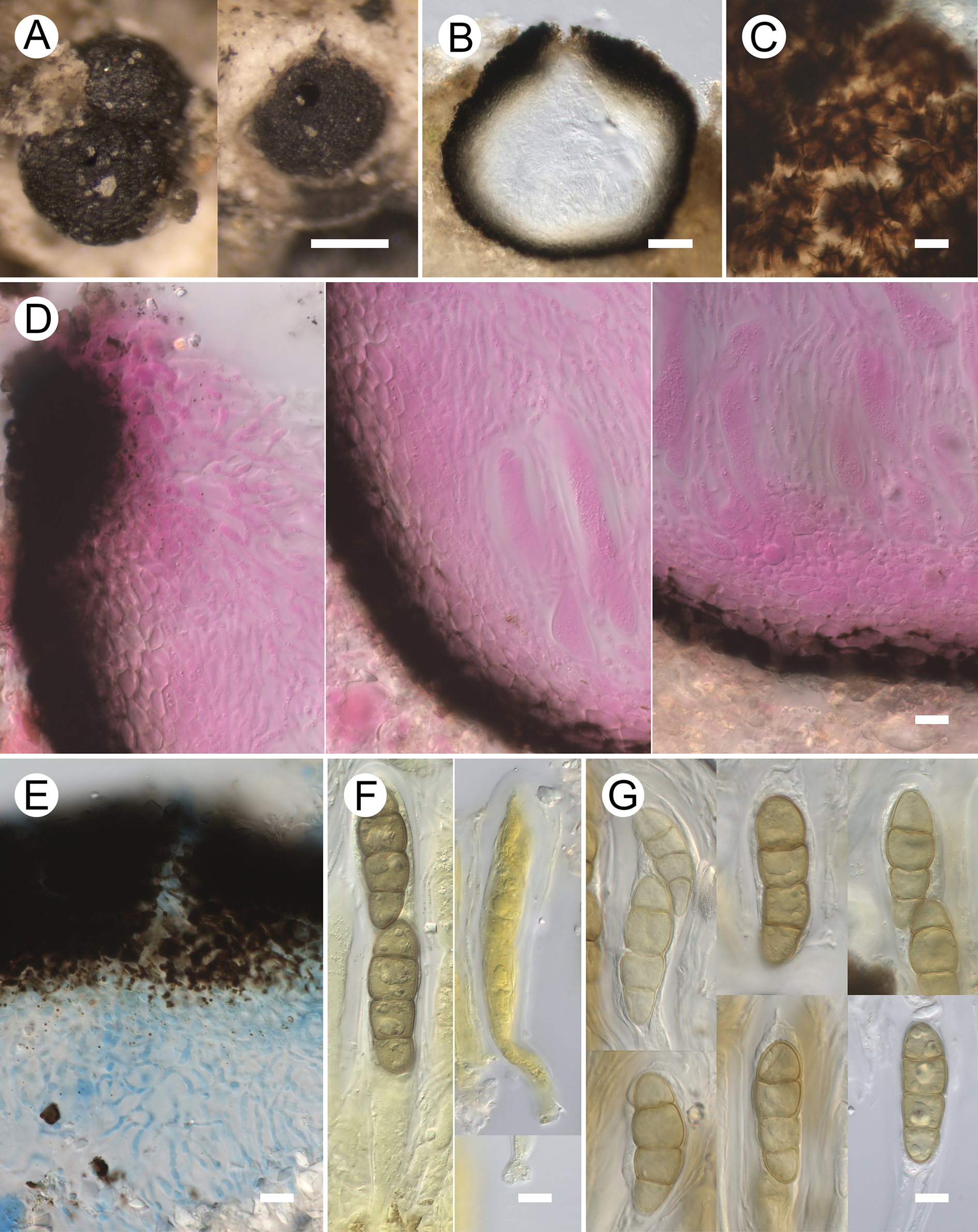
Figure 2. Long description
The figure consists of seven panels labeled A through G.
* Panel A: Two macro photos showing black, globose ascomata emerging from the pale, textured thallus of Megaspora verrucosa. A white scale bar represents 200 micrometers.
* Panel B: A vertical cross-section of an ascoma showing a dark, thick outer wall and a clear, pear-shaped inner cavity. A white scale bar represents 50 micrometers.
* Panel C: A high-magnification surface view of the squashed exciple showing dark, radiating fungal hyphae. A white scale bar represents 10 micrometers.
* Panel D: Three sub-panels stained pink with phloxine showing the exciple structure. The left image shows the top section, the center shows the middle, and the right shows the base. These reveal the cellular arrangement of the wall and the subhymenium. A white scale bar represents 10 micrometers.
* Panel E: A section stained with lactophenol cotton blue showing blue-tinted periphyses near the dark ostiole. A white scale bar represents 10 micrometers.
* Panel F: Two micrographs of asci. The left shows an ascus in K / I stain containing brown ascospores; the right shows an ascus in I stain. A white scale bar represents 10 micrometers.
* Panel G: A collection of six micrographs showing individual brown, multi-celled ascospores with distinct septa. A white scale bar represents 10 micrometers.
Lifestyle lichenicolous. No gall formation or discoloration of the infected host parts observed. Vegetative hyphae not observed.
Ascomata perithecioid, black, slightly glossy, surface of exposed parts roughened by protruding outer excipular cells (6–8 μm tall), subglobose to broadly ovoid, sometimes basally flattened, 300–420 μm diam. (n = 10), lacking a distinct papilla, with a central ostiole 15–45 μm wide, sometimes surrounded by several vertical cracks, erumpent, up to half exposed, solitary to often aggregated, sometimes contiguous. Exciple uniformly pigmented above and below, pale to dark brown, K+ greyish brown. In surface view medium to dark brown, pigmentation uneven, mottled, probably at least partially localized in intercellular spaces; composed of roughly isodiametric polygonal cells (4–10 μm diam.) with darker walls and intercellular spaces 1–5 μm wide; textura obscured because of highly variable thickness and uneven staining of intercellular spaces. Munk pores absent. In median vertical section composed of 2–10 dark to medium brown outer cell layers (cells isodiametric to tangentially elongated, c. 3.5–11 × 2–6 μm) and 2–6 pale brown to hyaline inner cell layers (cells isodiametric to strongly radially compressed); thickness at the base 20–30 μm (dark portion 10–15 μm), at the sides 25–35 μm (dark portion 10–20 μm), and at the top 45–70 μm (dark portion 20–45 μm). Subhymenium colourless, 10–15 μm tall. Hymenial gel I−, K/I−. Ostiolar canal irregular, without distinct periphyses. Periphysoids present in the upper part of the ascomatal cavity, hyaline, not apically enlarged, 20–50 × 1–2.5 μm, septate, densely branched, anastomosing. Long interascal filaments attached to the base and lateral walls of the ascomatal cavity, forming a dense layer higher than the asci; filiform, 1–2.5 μm wide, not apically enlarged, septate with cells c. 5–15 μm long, moderately branched and anastomosing, embedded in a gelatinous matrix. Asci bitunicate, elongate-clavate to subcylindrical, rounded at the apex; wall 2–5 μm thick laterally and 3–9 μm thick apically, sometimes exhibiting an irregular ocular chamber, gradually tapering below, rather short-stalked, with a short swollen, sometimes furcate base of the foot (c. 4.5–9 μm tall × 5.5–10 μm wide); (109–)115–145(–160) × (19–)20–25(–26) μm (n = 18, in water or K/I), consistently 2-spored, I−, K/I− (except for epiplasm turning yellow); croziers not observed. Ascospores soon becoming pale greyish yellow then pale to medium olivaceous buff (sensu Petersen Reference Petersen1996), ellipsoid, clavate (widest in the upper half relative to its position in the ascus) or oblong, with rounded to slightly acute ends, (36–)42–50(–55) × (12.5–)14–16.5(–18.5) μm, l/w = (2.6–)2.9–3.3(–3.6) (n = 64), 3-septate, septal pores not observed, often slightly to strongly constricted at the septa (especially at the median septum), without a torus; wall c. 1 μm thick, finely reticulate (barely visible at ×1000, DIC), with many rather inconspicuous guttules, without a gelatinous sheath, uniseriate in the ascus, sometimes slightly overlapping.
Anamorph not observed.
Etymology
The epithet refers to the host lichen, Megaspora verrucosa.
Distribution and host
The new species is described here based on a single specimen collected in the Asian Arctic (Russia). It was found on the thallus and apothecia of the terricolous lichen Megaspora verrucosa var. verrucosa growing in zonal tundra.
Notes
The new species does not fully match any of the morphologically closest genera of lichenicolous pyrenomycetes, which are characterized by non-stromatic ascomata, the absence of a clypeus, an ascomatal wall without Munk pores, persistent branched and anastomosing interascal filaments, bitunicate asci, and pigmented septate ascospores lacking distosepta. Such genera include Dacampia, Didymocyrtis, Leptosphaeria, Pleospora, Polycoccum, Pseudopyrenidium, and Pyrenidium. Dacampia differs mainly by having 8-spored asci and muriform ascospores (Ertz et al. Reference Ertz, Diederich, Lawrey, Berger, Freebury, Coppins, Gardiennet and Hafellner2015). Didymocyrtis is characterized by 8-spored, narrowly cylindrical asci with a distinct ocular chamber (Ertz et al. Reference Ertz, Diederich, Lawrey, Berger, Freebury, Coppins, Gardiennet and Hafellner2015). Pleospora is defined by papillate ascomata, 8-spored asci and muriform ascospores (Zhang et al. Reference Zhang, Crous, Schoch and Hyde2012). Polycoccum has asci with a small internal apical beak and typically 1-septate, often conspicuously ornamented ascospores (Ertz et al. Reference Ertz, Diederich, Lawrey, Berger, Freebury, Coppins, Gardiennet and Hafellner2015). Pseudopyrenidium is set apart by interascal filaments that are unbranched or only rarely branched and do not anastomose, asci with a distinct ocular chamber and submuriform, markedly verruculose ascospores (Navarro-Rosinés et al. Reference Navarro-Rosinés, Zhurbenko and Roux2010). Pyrenidium differs in having brown to dark brown ascospores, often paler at the tips, frequently with a central pore in the septa (Navarro-Rosinés & Roux Reference Navarro-Rosinés, Roux, Nash, Gries and Bungartz2007).
Although Leptosphaeria differs in having papillate ascomata, lacking periphyses in the type species L. doliolum Ces. & De Not. and bearing 8-spored asci (Zhang et al. Reference Zhang, Crous, Schoch and Hyde2012), we provisionally place the new species in this genus because: 1) some lichenicolous members of Leptosphaeria exhibit a similar, relatively uncommon ascospore colouration, subhyaline to pale golden brown in L. clarkii, yellowish-greenish to very light brown in L. oxneriae Cl. Roux & S. Y. Kondr., and 2) L. oxneriae possesses periphysoids (Hawksworth Reference Hawksworth1980; Kondratyuk et al. Reference Kondratyuk, Lőkös, Halda, Roux, Upreti, Schumm, Mishra, Nayaka, Farkas and Park2017). Three previously described lichenicolous Leptosphaeria species differ clearly from L. verrucosae in host preference, 8-spored asci, and much smaller ascospores: 19–22 × 4.5–6.5 μm in L. clarkii (Hawksworth Reference Hawksworth1980), 13–27 × 5.5–8.5 μm in L. oxneriae (Kondratyuk et al. Reference Kondratyuk, Lőkös, Halda, Roux, Upreti, Schumm, Mishra, Nayaka, Farkas and Park2017) and 20–24 × 8–9 μm in L. pycnostigma (Nyl.) Sacc. & D. Sacc. (Vouaux Reference Vouaux1913).
Leptosphaeria verrucosae should also be compared with Zwackhiomacromyces constrictocarpus Etayo & van den Boom, which likewise occurs on Megaspora verrucosa var. verrucosa and has perithecioid ascomata, interascal filaments and 3-septate ascospores. However, the latter differs by its papillate, smaller ascomata (200–300 μm diam.), the absence of periphysoids, (6–)8-spored asci that are larger (c. 150–185 × 35–40 μm), and hyaline, shorter ascospores (32–46 × 13.5–20 μm), sometimes with a torus (van den Boom & Etayo Reference van den Boom and Etayo2014).
Lichenostigma subgen. Lichenogramma sp.
(Fig. 3)
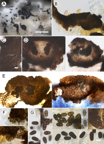
Lichenostigma subgen. Lichenogramma sp. (LE F-355750a). A, ascomata and vegetative hyphal strands growing on thallus of Megaspora verrucosa var. verrucosa. B, vegetative hyphal strand in median vertical section. C, ascomatal wall in surface view. D & E, ascomata in median vertical section. Note macroconidia developing on the ascoma surface (E right). F, thallus-borne macroconidia. G, ascospores. B, E (left), F, G (right) in K; C, D, G (left) in water; E (right) in K/I. Scales: A = 200 μm; B–G = 10 μm. In colour online.
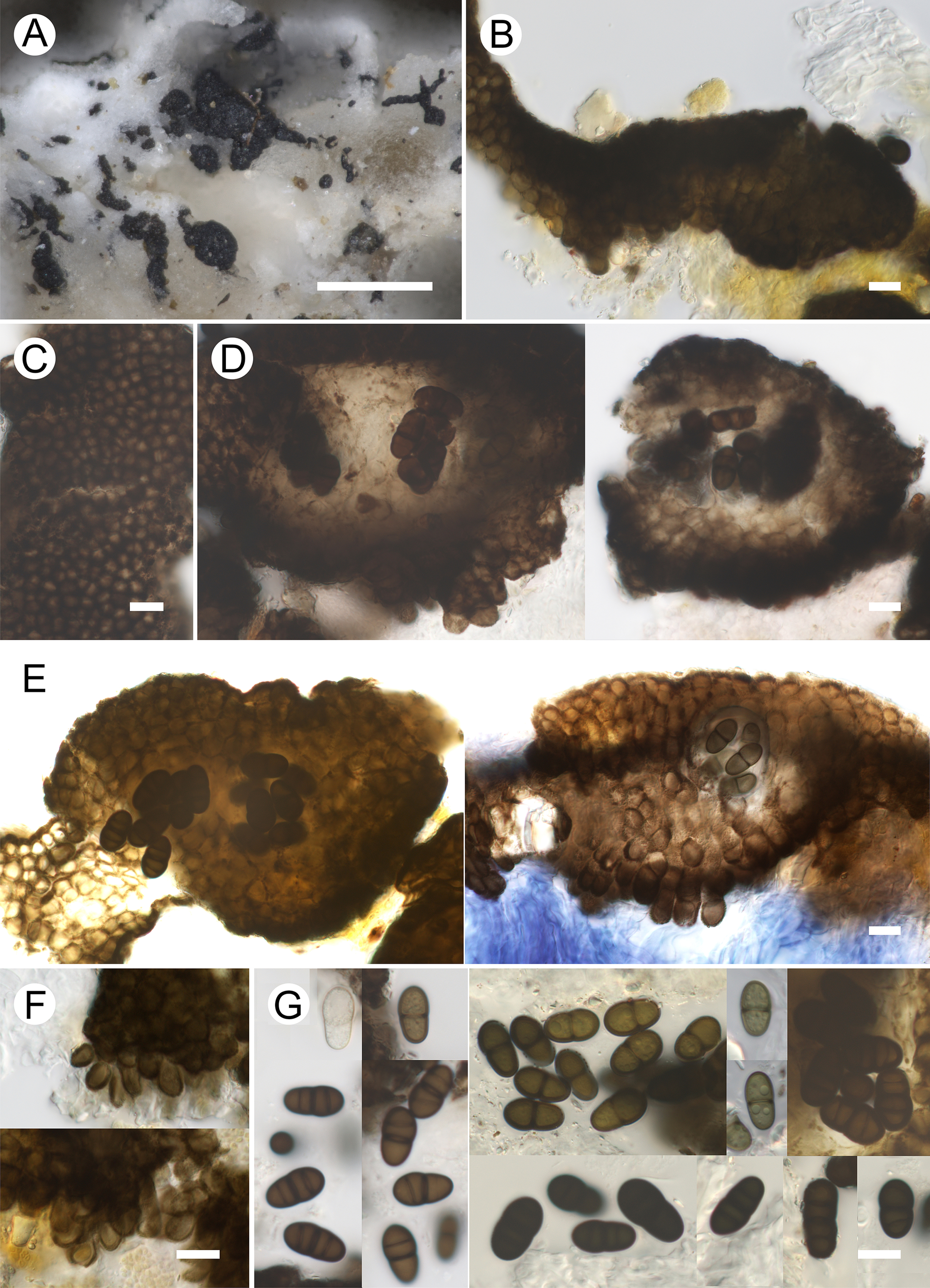
Figure 3. Long description
A multi-panel micrograph series labeled A through G.
* Panel A: A macro photo showing dark, branched vegetative hyphal strands and rounded ascomata scattered across the pale, textured thallus of Megaspora verrucosa. A white scale bar at the bottom right represents 200 micrometers.
* Panel B: A median vertical section of a dark, irregular vegetative hyphal strand.
* Panel C: A high-magnification surface view of the ascomatal wall showing a dense, tessellated pattern of dark cells.
* Panel D: Two micrographs showing ascomata in median vertical section. The left image shows dark internal structures, while the right image reveals several brown, multi-celled ascospores clustered in the center.
* Panel E: Two detailed vertical sections. The left section shows dark ascomata with distinct macroconidia on the surface. The right section, treated with K / I, shows a blue-stained base with macroconidia developing on the upper surface.
* Panel F: Two close-up views of thallus-borne macroconidia, appearing as dark, rounded clusters at the edge of the fungal tissue.
* Panel G: A composite of several micrographs showing individual ascospores. The spores are brown, mostly two-celled with a central septum, though some appear multi-celled. They vary from translucent to dark brown.
Scale bars for B through G represent 10 micrometers.
Lifestyle lichenicolous. No gall formation or discoloration of the infected host parts observed. Vegetative hyphal strands superficial, sparce, scattered, not markedly radiating, sometimes formed of a chain of discrete segments; black, somewhat flexuose, occasionally branched, c. 30–190 μm long, 10–45 μm wide, consisting of 3–10 rows of ±isodiametric cells.
Ascomata stromatic, black, slightly glossy, superficial, developing as swellings of vegetative hyphal strands; convex, more or less circular, elongate-ellipsoid or irregular in top view, 60–130 μm long, 30–85 μm thick, lacking an ostiole, occurring singly or in groups of 2–8, sometimes confluent. Ascomatal wall in surface view composed of medium brown, granular-pigmented, K+ olivaceous, K/I−, isodiametric to slightly elongated cells, 3.5–7.5 × 3–6 μm, with a dark brown wall 1–1.5 μm thick. In median vertical section the ascomata are paraplectenchymatous throughout, medium to dark brown externally and subhyaline to pale brown internally (inner cells 5–8.5 × 4–7 μm, with a wall 0.5–1.5 μm thick). Hamathecium not observed. Asci develop within the paraplectenchymatous tissue, 1–3 per ascoma, c. 30–40 × 22–26 μm, 6–8-spored, evanescent; wall K/I+ weak blue. Ascospores initially subhyaline, becoming medium olive brown to dark brown, K+ olivaceous, with darker walls and septa; somewhat pyriform (slightly wider in one half), occasionally oblong or ellipsoid, with rounded ends, (12.5–)13.5–16(–19.5) × (6.5–)7.5–9(–10.5) μm, l/w = (1.4–)1.6–2(–2.3) (n = 73, in water or K), 1–3-septate, sometimes slightly constricted at the median septum, sometimes with a distinct darker torus, with a smooth wall 0.5–1 μm thick, often containing many conspicuous guttules, lacking a gelatinous sheath, irregularly 2–3-seriate in the ascus.
Thallus-borne macroconidia develop on the surface of the vegetative hyphal strands and ascomata; oblong, ellipsoid or subglobose, 6–10 × 4–7 μm (aseptate) or 12.5–17 × 6.5–8 μm (1-septate).
Notes
The fungus examined belongs to the genus Lichenostigma subgen. Lichenogramma (Calatayud et al. Reference Calatayud, Navarro-Rosinés and Hafellner2002). It differs from most species of that subgenus by the combination of relatively sparse, scarcely ramified plurihyphal vegetative strands, 1–3-septate ascospores and the presence of thalloconidia. Morphologically, it is most comparable to Lichenostigma cupreogriseae P. Pinault & Cl. Roux (on Circinaria, Megasporaceae), L. rouxii Nav.-Ros. et al. (on Squamarina, Lecanorales), L. spermatomanis P. Pinault & Cl. Roux (on Aspicilia, Megasporaceae), L. svandae Vondrák & Šoun (on Acarospora, Acarosporales) and L. triseptatum Halıcı & D. Hawksw. (on Aspicilia and Circinaria, Megasporaceae). Lichenostigma cupreogriseae mainly differs by having well-developed vegetative hyphal strands and wider ascospores, (9.5–)11.5–16.5(–20) × (5.5–)7–10.5(–13) μm, l/w = (1.2–)1.4–1.8(–2), which may become submuriform, and by producing larger macroconidia (20–53 × 7–16 μm) composed of a single row of 3–6 cells or 1–2 rows of 6–13 cells (Roux & Pinault Reference Roux and Pinault2021). Lichenostigma rouxii is readily distinguished by its longer and thinner vegetative hyphal strands (mainly 400–1000 μm long, 8–13 μm thick, formed by only 1–3 rows of cells) and by smaller ascospores, (10–)10.5–11.9–13.5(–15.5) × (5.5–)6–6.5–7(–8) μm (Calatayud et al. Reference Calatayud, Navarro-Rosinés and Hafellner2002). Lichenostigma spermatomanis has often submuriform, larger ascospores, (12.5–)14–19.5(–22.5) × (7–)8–11(–14) μm (Roux & Pinault Reference Roux and Pinault2021). Lichenostigma svandae mainly differs in having shorter, finely granular ascospores with 1(–2) septa, (11–)11.5–13.5(–15) × (6–)6.5–8.5(–10) μm (Vondrák & Soun Reference Vondrák and Soun2007). Lichenostigma triseptatum is chiefly distinguished by its unbranched vegetative hyphal strands and by the occasionally submuriform, somewhat verruculose ascospores bearing a distinct gelatinous sheath (Halıcı & Hawksworth Reference Halıcı, Atienza and Hawksworth2007).
The fungus described here does not fully match any currently known species of Lichenostigma subgen. Lichenogramma and was found on a previously unreported host genus. However, available material is limited and may not reflect the full range of intraspecific variation. Therefore, we provide an informal description pending the collection of additional specimens that should clarify its taxonomic status.
Specimen examined
Mongolia: Arkhangai Region: Ikhtamir Somon, Khoid Tamir Gol River, near Khokh Nuur Lake, 47°05′47.4″N, 102°28′49.7″E, 2670 m, steppe-tundra, on Megaspora verrucosa var. verrucosa (thallus and thalline margin of apothecia, rarely apothecial disc), 29 vii 2023, Ochirbat Enkhtuya & Samiya Javkhlan (LE F-355750a).
Merismatium subnigritellum Zhurb. sp. nov.
MycoBank No.: MB 864912
Distinguished from Merismatium nigritellum (Nyl.) Vouaux mainly by the smaller ascospores, (9.5–)14–18(–28) × (6–)8–9.5(–13) μm versus (14–)15–24.5(–32) × (6.5–)8–12(–15) μm.
Type: Russia, Krasnoyarsk Territory, Taimyr Peninsula, Byrranga Mts, northern shore of Levinson-Lessinga Lake, unnamed Mt ‘348.5’, 74°33′N, 98°33′E, 300 m, sandstone rocks among tundra, on Megaspora verrucosa var. verrucosa (prothallus) growing on moribund moss, 22 August 1995, M. P. Zhurbenko 95632a (LE F-355755a—holotype). The host is co-infected with Polycoccum megasporae.
(Fig. 4)
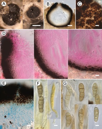
Merismatium subnigritellum (A & D, LE 233186; B, C & E (far right), holotype; E (except the far right), LE F-355756c). A, ascomata growing on prothallus of Megaspora verrucosa var. verrucosa. B, squashed exciple in surface view. C, periphysoids. D, exciple in median vertical section: at the top (left), in the middle (centre), and at the base (right). Note periphysoids and subhymenium. E, asci and ascospores. B, C (above), E (left) in water; C (below), E (centre) in K; E (second from the right) in I; E (far right) in K/I. Scales: A = 200 μm; B–E = 10 μm. In colour online.
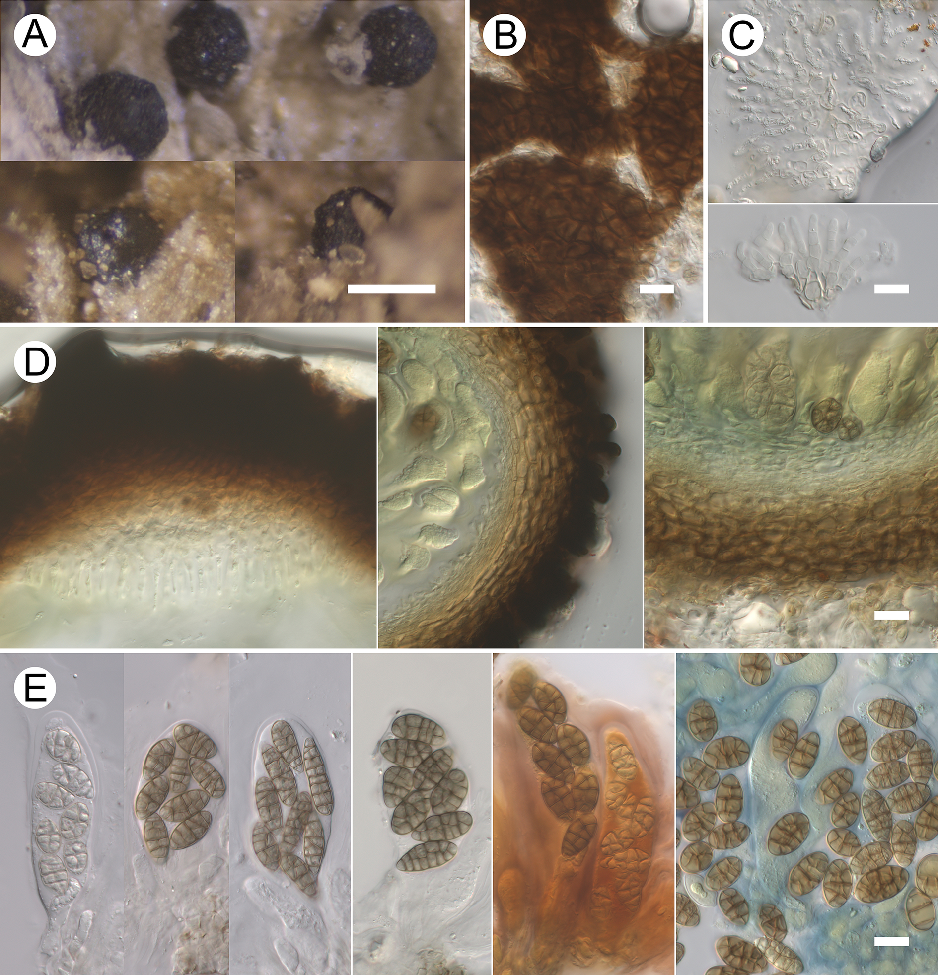
Figure 4. Long description
Panel A shows three macro photos of black, globose ascomata embedded in the pale, crustose prothallus of Megaspora verrucosa. A white scale bar represents 200 micrometers.
Panel B displays a high-magnification surface view of a squashed exciple, revealing a dark brown, cellular texture. A white scale bar represents 10 micrometers.
Panel C contains two vertical sub-panels showing periphysoids. The top sub-panel is in water, showing translucent, filamentous structures. The bottom sub-panel is in K, showing a more defined, fan-like arrangement of these filaments.
Panel D consists of three sub-panels showing vertical sections of the exciple. The left sub-panel shows the dark, thickened top portion. The center sub-panel shows the middle section with periphysoids and developing asci. The right sub-panel shows the base with a distinct subhymenium layer. A white scale bar represents 10 micrometers.
Panel E is a composite of six vertical strips showing asci and ascospores under different chemical treatments. From left to right, the first three strips in water and K show translucent asci containing brown, multi-celled, muriform ascospores. The fourth strip in I shows spores with a slightly darker contrast. The fifth strip in K/I shows a reddish-brown reaction in the hymenial gel. The far-right strip shows a cluster of liberated brown ascospores against a blue-stained background in K/I. A white scale bar represents 10 micrometers.
Lifestyle lichenicolous to probably partly saprobic. No gall formation or discoloration of the infected host parts observed. Vegetative hyphae immersed, pale brown, 2–3.5 μm diam., sparingly branched, sparsely septate.
Ascomata perithecioid, black, occasionally slightly glossy, surface of exposed parts roughened by protruding outer excipular cells or their protuberances (2–8 μm tall), subglobose, ovoid or slightly ampulliform, occasionally somewhat truncate above, (100–)150–220(–255) μm diam. (n = 82), with a sometimes inconspicuous central ostiole 10–35 μm wide, occasionally surrounded by several vertical cracks/folds, erumpent, mostly half-exposed to almost sessile, solitary to loosely aggregated. Exciple uniformly pigmented above and below, pale to dark brown, K+ greyish brown; in surface view composed of medium brown, isodiametric to somewhat elongated polygonal cells (3–12 μm long) with darker walls 1–1.5 μm thick; in median vertical section composed of 8–15 layers of tangentially elongated cells (c. 5–10 × 2–5 μm), dark brown externally and pale brown internally; thickness at the base 20–30 μm, at the sides 25–35 μm, and at the top 30–45 μm. Subhymenium colourless. Interascal gel I+ blue then dull red, K/I+ blue. Hamathecium composed of periphysoids developing in the upper part of the ascomatal cavity; these are hyaline, ±subulate, 7–25 μm long, 2.5–4.5 μm wide at the base and 1.5–3 μm wide at the apex, 0–2(–4)-septate, occasionally branched and anastomosing (near the base), embedded in a gelatinous matrix. Asci elongate-clavate to subcylindrical, rounded at the apex; wall 0.5–5 μm thick apically and 0.5–4 μm thick laterally, rarely with an indistinct ocular chamber (c. 1 μm tall × 2.5 μm wide); rather short-stalked, (60–)67–84(–92) × (14–)16–25 μm (n = 28, in water or I), 8-spored, I−, K/I− (except for ascoplasm turning yellow or orange), croziers not observed. Ascospores pale brownish grey to medium brown, ellipsoid to narrowly ellipsoid, sometimes broadly ellipsoid, oblong, (ob)ovate, occasionally somewhat inequilateral or irregular (±triangular etc.), with rounded apices, (9.5–)14–18(–28) × (6–)8–9.5(–13) μm, l/w = (1.1–)1.6–2.2(–4.1) (n = 233, in water or I) ((10.5–)15–20(–27) × (6–)7.5–9.5(–10) μm, l/w = (1.3–)1.7–2.5(–3.7) (n = 171, in K)), (sub)muriform, with 3–8 trans-septa, 0–6 oblique septa, and 0–6 longisepta in central segments, evidently including A- and B-septa sensu Eriksson (Reference Eriksson1981: 20–21), not constricted at the septa, with (6–)8–11(–18) cells visible in optical section through the spore centre (n = 136), with a smooth wall c. 0.5 μm thick, without a gelatinous sheath, overlappingly uniseriate to biseriate in the ascus. Goniocysts not observed.
Anamorph not observed.
Etymology
The epithet alludes to the morphological similarity to Merismatium nigritellum.
Distribution and host
The new species is described here based on nine specimens collected in the European and Asian parts of Russia. These were found on the prothallus (mostly) and thallus of the terricolous lichen Megaspora verrucosa var. verrucosa growing in polar desert and zonal and mountain tundras.
Notes
Contemporary taxonomy in Merismatium relies heavily on ascospore characters (e.g. Triebel Reference Triebel1989; Hafellner Reference Hafellner2011). In terms of ascospore dimensions, the species treated here most closely resembles M. cladoniicola Alstrup (ascospores 13–16.5 × 5.5–7 μm; Alstrup Reference Alstrup1997), M. coccisporum (Norm.) Vouaux ((8.5–)11–17(–19) × (5–)6.5–8(–9.5); Triebel Reference Triebel1989), M. corae (Pat.) Etayo & R. Sant. (14–22 × 6–7.5 μm; Etayo Reference Etayo2010a), M. decolorans (Arnold) Triebel ((10.5–)13.5–16.5(–17.5) × (3.5–)4–5.5(–6) μm; Triebel Reference Triebel1989), M. nigritellum ((14–)15–24.5(–32) × (6.5–)8–12(–15) μm; Triebel Reference Triebel1989, (13–)16.5–25(–31) × (6.5–)8.5–12.5(–16) μm; Zhurbenko Reference Zhurbenko2009a), and M. peregrinum (Flot.) Triebel ((12–)14–16(–17) × (4.5–)5–6.5(–7) μm; Hertel & Rambold Reference Hertel and Rambold1990). However, as the cited measurements demonstrate, all of these differ markedly from the new species in ascospore dimensions. With respect to cell numbers, all of the cited species except M. nigritellum have substantially fewer cells per ascospore: up to four in M. cladoniicola, up to eight in M. coccisporum, up to eight in M. corae, up to five in M. decolorans, and up to four in M. peregrinum. The sole Merismatium species exhibiting ascospores with a similar number of cells is M. nigritellum, with ascospores reported as (8–)10–18-celled by Hafellner (Reference Hafellner2011) and as reaching up to 24 cells by Triebel (Reference Triebel1989). Nevertheless, M. nigritellum differs by having substantially larger ascospores (mainly 15–25 × 8–12.5 μm vs 14–18 × 8–9.5 μm), somewhat larger ascomata (mainly 200–250 μm vs 150–220 μm diam.), and larger asci (65–115 × 20–27 μm vs 60–92 × 14–25 μm) (Triebel Reference Triebel1989). Additionally, goniocysts, commonly developed near ascomata in M. nigritellum (Triebel op. cit.), were absent in all specimens examined here. Merismatium nigritellum agg. has been reported on at least 35 lichen genera, predominantly terricolous taxa (von Brackel Reference von Brackel2014). These included specimens on Megaspora verrucosa (Zhurbenko Reference Zhurbenko2002, Reference Zhurbenko2009a, Reference Zhurbenko b), which are reidentified here as M. subnigritellum.
Additional specimens examined (all on the prothallus and thallus of Megaspora verrucosa var. verrucosa)
Russia: Chukotka Autonomous Area: Wrangel Island, Krasnyi Flag River, 71°30′N, 178°55′W, 100 m, 27 vii 1997, S. S. Kholod (LE 233367); formerly published as M. nigritellum (Zhurbenko Reference Zhurbenko2009a). Krasnodar Territory: Lagonaki Upland, Mt Fisht, 43°58′N, 39°53′E, 2300 m, 21 viii 2014, P. M. Zhurbenko (LE 309544). Krasnoyarsk Territory: Severnaya Zemlya Archipelago, Bol’shevik Island, peninsula with Cape Baranova, 79°16′N, 101°40′E, 20 m, 15 vii 1996, M. P. Zhurbenko 96357c (LE F-355756c), 961004 (LE 260103), formerly published as M. nigritellum (Zhurbenko Reference Zhurbenko2009b); same island, Akhmatova Bay coast, 79°04′N, 102°45′E, 60 m, 16 vii 1996, M. Zhurbenko 96929 (LE 233347), formerly published as M. nigritellum (Zhurbenko Reference Zhurbenko2009a); Taimyr Peninsula, Byrranga Mts, N of Levinson-Lessinga Lake, 74°31′N, 98°36′E, 50–180 m, 6 viii 1995, M. P. Zhurbenko 95372 (LE 233186), formerly published as M. nigritellum (Zhurbenko Reference Zhurbenko2009a); S of Levinson-Lessinga Lake, 74°24′N, 98°46′E, 150–250 m, 9 viii 1995, M. P. Zhurbenko 95371 (LE 233166), formerly published as M. nigritellum (Zhurbenko Reference Zhurbenko2009a). Yamal-Nenets Autonomous Area: Polar Urals, Mt Rai-Iz, 66°56′N, 65°35′E, 600 m, 26 vi 1993, M. P. Zhurbenko 9362 (LE 309026), formerly published as M. nigritellum (Zhurbenko Reference Zhurbenko2002).
Polycoccum megasporae Zhurb. sp. nov.
MycoBank No.: MB 864913
Distinguished from Polycoccum opulentum (Th. Fr. & Almq.) Arnold mainly by its longer asci, 100–170 μm versus 35–90 μm long, longer ascospores, (26–)31.5–39.5(–44) μm versus (19–)22–27(–30) μm long, and a different host selection: Megasporaceae (Lecanoromycetes) versus Verrucariaceae (Eurotiomycetes).
Type: Russia, Krasnoyarsk Territory, Taimyr Peninsula, Byrranga Mts, N of Levinson-Lessinga Lake, Zamknutaya River, 74°37′N, 98°33′E, 150 m, rocks in tundra, on Megaspora verrucosa var. verrucosa (disc and thalline margin of apothecia) growing on moribund moss, 6 August 1995, M. P. Zhurbenko 95631 (LE F-355751—holotype).
(Fig. 5)
Polycoccum megasporae (A, C–F, H, holotype; B & G, LE F-355758). A, ascomata growing on apothecium of Megaspora verrucosa var. verrucosa. B, ascoma in median vertical section. C, exciple in median vertical section: at the base (left), at the top from the side (centre), and around the ostiolar canal (right). D, asci and interascal filaments. E, upper part of the ascus. Note ocular chamber. F, interascal filaments. G, exciple in surface view. H, ascospores. B, C & H (left and centre) in water; D & F in I; E in K/I; G & H (right) in K. Scales: A = 200 μm; B = 50 μm; C, E–H = 10 μm; D = 20 μm. In colour online.
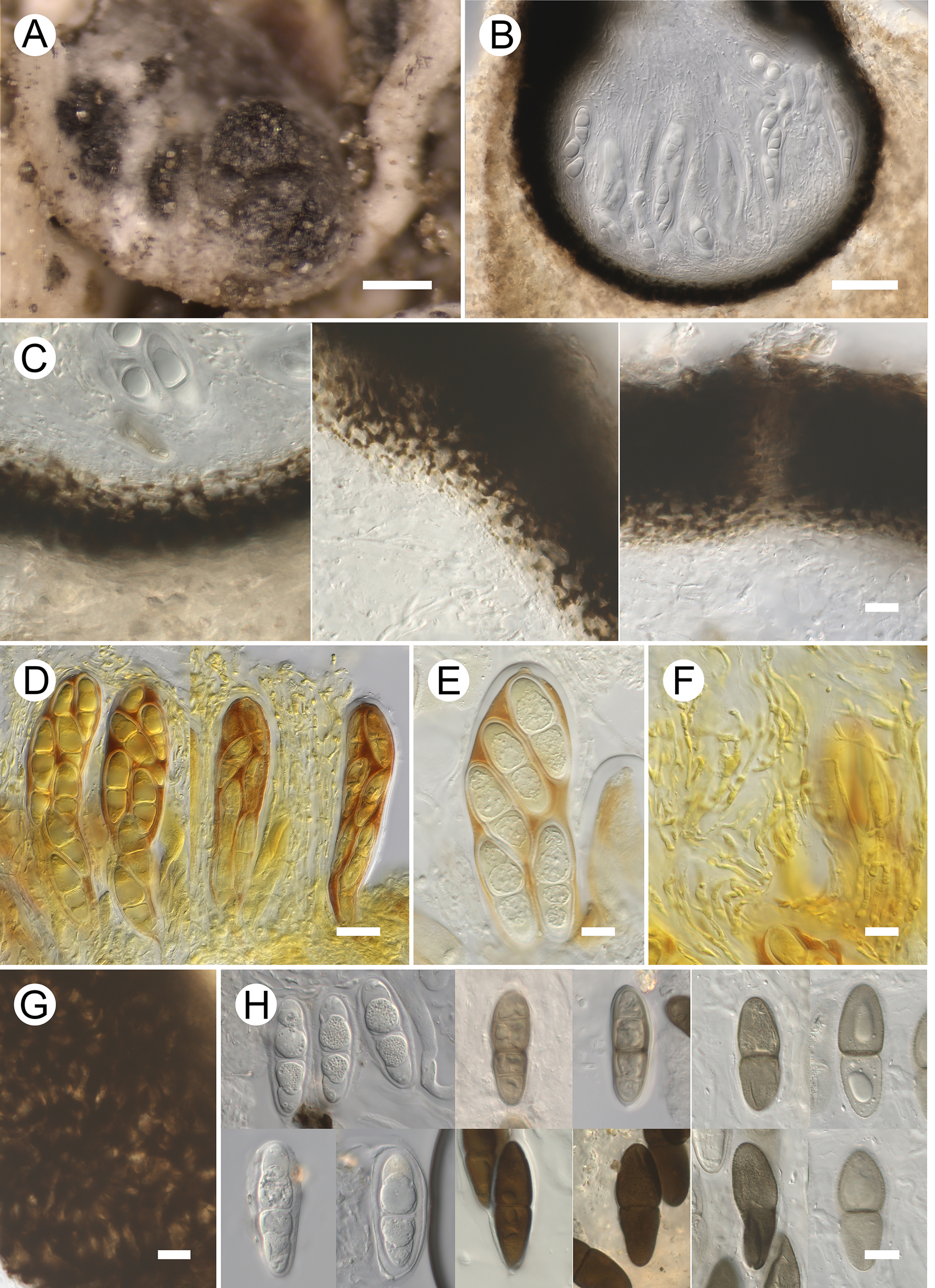
Figure 5. Long description
A multi-panel micrograph series organized in four rows.
Panel A at the top left shows a macro photo of dark, granular ascomata growing on the pale surface of a Megaspora verrucosa apothecium. Scale bar is 200 micrometers.
Panel B at the top right is a vertical cross-section of a single globose ascoma embedded in host tissue, containing numerous asci. Scale bar is 50 micrometers.
Panel C consists of three sub-panels showing the exciple structure: the base on the left, the side wall in the center, and the ostiolar canal at the top right. Scale bar is 10 micrometers.
Panel D shows a cluster of elongated, sac-like asci containing developing ascospores, stained yellow-brown. Scale bar is 20 micrometers.
Panel E provides a close-up of the upper portion of an ascus, highlighting a distinct apical ocular chamber. Scale bar is 10 micrometers.
Panel F displays thin, branched interascal filaments. Scale bar is 10 micrometers.
Panel G shows the surface view of the dark, textured exciple. Scale bar is 10 micrometers.
Panel H at the bottom right is a composite of twelve smaller frames showing individual ascospores in various stages and chemical stains. The spores are two-celled, ranging from hyaline to dark brown, with some showing a distinct septum and granular contents. Scale bar is 10 micrometers.
Lifestyle lichenicolous. No gall formation or discoloration of the infected host parts observed. Vegetative hyphae not observed.
Ascomata perithecioid, black, slightly glossy, faintly roughened on exposed surfaces, subglobose, obpyriform or broadly ovoid, (180–)240–370(–480) μm diam. (n = 34), occasionally shortly papillate, with a central ostiole 10–40 μm wide, erumpent, often protruding only in the ostiolar region, occasionally up to half exposed; solitary to loosely aggregated, sometimes contiguous. Exciple uniformly pigmented above and below, pale to dark brown, K−; in surface view medium to dark brown; composed of ±polygonal cells (3–12 μm long) with darker walls 1.5–4 μm thick; in median vertical section composed of 3–10 dark to medium brown outer cell layers (cells isodiametric or somewhat elongated, 3–11 μm long, walls 1.5–4 μm thick) and 3–6 pale brown or hyaline inner cell layers (cells strongly radially compressed); thickness at the base 20–40 μm (dark portion 15–25 μm), at the sides 20–40 μm (dark portion 10–20 μm), and at the top 40–65 μm (dark portion 40–50 μm). Subhymenium colourless. Hymenial gel I−, K/I−. Hamathecium composed of well-developed long interascal filaments filling the entire ascomatal cavity and protruding above asci, hyaline, 1–3.5 μm wide, septate, frequently branched, anastomosing. Asci bitunicate, elongate-clavate to broadly cylindrical, rounded at the apex; wall 1–2.5 μm thick laterally and 2–3.5 μm thick apically, often with a distinct small ocular chamber (1–3 μm tall × 2.5–5 μm wide); short-stalked, with a slightly swollen base of the foot (c. 5.5 μm wide), (100–)120–155(–170) × 26–32(–36) μm (n = 23, in water or I), (rarely 4–)8-spored, I−, K/I− (except for epiplasm turning orange); croziers not observed. Ascospores ellipsoid to often clavate (soleiform), with a wider and longer upper cell relative to its position in the ascus, with rounded to often rather acute ends, (26–)31.5–39.5(–44) × (8–)10.5–13.5(–16.5) μm, l/w = (2.1–)2.6–3.4(–5.2) (n = 165), 1(–3)-septate, usually constricted at the septa, sometimes with 1(–2) large guttules per cell, diagonally uniseriate to irregularly biseriate in the ascus; initially long remaining hyaline (some ascomata contain only hyaline spores), 1-septate, smooth, usually with a gelatinous sheath 1–3.5 μm thick; eventually pale to dark brown or greyish brown, K+ greying, 1(–3)-septate, distinctly verrucose.
Possible anamorph. Inside a single ascocarp of specimen Zhurbenko 95632b a light brown, Phoma-like, subglobose conidioma c. 80 μm diam. was observed. Conidiogenous cells arise from the conidiomatal wall, holoblastic. Conidia hyaline, aseptate, smooth-walled, oblong, with a rounded apex and a truncate base, 3.5–5 × 2–2.5 μm.
Etymology
The epithet refers to the host lichen, Megaspora verrucosa.
Distribution and host
The new species is described here based on four specimens collected in the Asian Arctic (Russia). These were found on the thallus and apothecia of the terricolous lichen Megaspora verrucosa var. verrucosa growing in zonal tundra.
Notes
The new species generally fits the concept of Polycoccum as circumscribed by Ertz et al. (Reference Ertz, Diederich, Lawrey, Berger, Freebury, Coppins, Gardiennet and Hafellner2015). It matches the generic core group in features such as an exciple composed of ±isodiametric cells (in median vertical section), relatively thick interascal filaments, broadly cylindrical asci, and distinctly ornamented, distichously arranged ascospores. Polycoccum megasporae differs from almost all known species of the genus, which are characterized by consistently 1-septate ascospores, in that its ascospores sometimes bear up to three septa, a phenomenon previously observed only in P. versisporum (Bagl. & Carestia) D. Hawksw. (Gaya & Navarro-Rosinés Reference Gaya and Navarro-Rosinés2008). In addition, it is distinguished by having the longest ascospores (up to 44 μm) reported for the genus.
Comparable ascospore lengths (up to 35.5 μm) occur in Polycoccum acarosporicola Halıcı & D. Hawksw. (on Acarospora cervina), P. anatolicum Halıcı & E. Akgül (on Lepraria incana), P. cartilaginosum (Arnold) D. Hawksw. (on an unidentified saxicolous lichen), P. crassum Vězda (on Peltigera), P. dzieduszyckii (Boberski) D. Hawksw. (on Bagliettoa), P. longisporum Etayo (on Pseudocyphellaria), P. marmoratum (Kremp.) D. Hawksw. (on Clauzadea, Polyblastia, Thelidium, and Verrucaria), P. opulentum (on Verrucariaceae), and P. versisporum (on Rhizocarpon). In addition to differences in host selection, they can be distinguished as follows: P. acarosporicola has shorter asci, 85–96 μm long, and non-halonate, much narrower ascospores, (7–)8–9.5 μm wide (Halıcı et al. Reference Halıcı, Atienza and Hawksworth2007); P. anatolicum has smaller ascomata, 75–90 μm diam., shorter asci, 71–103 μm tall, and monostichous, non-halonate ascospores (Halıcı et al. Reference Halıcı, Akgül, Oztürk and Kiliç2013); the insufficiently known P. cartilaginosum appears to have smaller ascomata, to c. 200 μm diam., and dark brown to almost black ascospores up to 34 μm long (Hawksworth & Diederich Reference Hawksworth and Diederich1988); P. crassum has I+ reddish hymenial gel, 4-spored asci, and narrower ascospores, 8–10(–11) μm wide (Hawksworth & Diederich Reference Hawksworth and Diederich1988); P. dzieduszyckii has smaller ascomata, 50–100 μm diam., and 2-spored, shorter asci, 55–70 μm tall (Hawksworth & Diederich Reference Hawksworth and Diederich1988); P. longisporum has much narrower ascospores, 4.5–6 μm wide (Etayo & Sancho Reference Etayo and Sancho2008); P. marmoratum has I+ blue hymenial gel and shorter asci, 50–90 μm tall (Hawksworth & Diederich Reference Hawksworth and Diederich1988); P. opulentum has shorter asci, 35–90 μm tall, and smooth ascospores (Navarro-Rosinés & Roux Reference Navarro-Rosinés and Roux1990); and P. versisporum has consistently or mainly 4-spored, smaller asci, (90–)110–140 × 16–21 μm, and homopolar or slightly heteropolar, shorter ascospores, mainly 26.5–33 μm long (Gaya & Navarro-Rosinés Reference Gaya and Navarro-Rosinés2008).
Additional specimens examined (all on the thallus and apothecia of Megaspora verrucosa var. verrucosa)
Russia: Nenets Autonomous Area: Malozemelskaya Tundra, Seduyakha River, 68°23′N, 53°15′E, 3 viii 1998, O. V. Lavrinenko (LE F-355752a). Krasnoyarsk Territory: Taimyr Peninsula, Byrranga Mts, S of Levinson-Lessinga Lake, 74°25′N, 98°48′E, 50 m, 31 vii 1995, M. P. Zhurbenko 95630 (LE F-355758); N of Levinson-Lessinga Lake, unnamed Mt ‘348.5’, 74°33′N, 98°33′E, 300 m, 22 viii 1995, M. P. Zhurbenko 95632b (LE F-355755b).
Pyrenidium actinellum Nyl. s. lat.
Ascomata subglobose to broadly obpyriform, 230–420 μm diam., mostly slightly protruding above the host surface, aggregated to contiguous, not causing gall formation. Hymenial gel in the ostiolar region blue-green. Asci 4-spored. Ascospores pale to medium brown, ellipsoid to oblong, with rounded ends, (very rarely 1–2–)3-septate, seldom with paler end cells, usually strongly constricted at the septa, (17.5–)20–23(–25.5) × (7–)8.5–9.5(–11) μm, l/w = (2.1–)2.2–2.6(–2.9) (n = 126), smooth, non-halonate, often bearing a large guttule per cell, overlappingly uniseriate in the ascus.
Though the narrow concept of this species matches the examined specimen well, we assign it to the aggregate because the species was originally characterized by ascomata that are sessile or rarely semi-immersed in the host thallus, and it was described from a lichen of a different order, Scytinium teretiusculum (Wallr.) Otálora et al. (Peltigerales) (Huanraluek et al. Reference Huanraluek, Ertz, Phukhamsakda, Hongsanan, Jayawardena and Hyde2019).
Specimen examined
Mongolia: Arkhangai Region: Ikhtamir Somon, Khoid Tamir Gol River, near Khokh Nuur Lake, 47°05′47.4″N, 102°28′49.7″E, 2670 m, on Megaspora verrucosa var. verrucosa (thallus), 29 vii 2023, Ochirbat Enkhtuya & Samiya Javkhlan (LE F-355744a).
Sclerococcum deminutum (Th. Fr.) Ertz & Diederich
This is a widespread species reported on a wide range of lichen genera (von Brackel Reference von Brackel2014). It was previously reported on Megaspora verrucosa from Great Britain (Fryday & Coppins Reference Fryday and Coppins2012; as Dactylospora urceolata (Th. Fr.) Arnold).
Specimen examined
Russia: Nenets Autonomous Area: Malozemelskaya Tundra, Seduyakha River, 68°23′N, 53°15′E, on Megaspora verrucosa var. verrucosa (thallus), 3 viii 1998, O. V. Lavrinenko (LE F-355752b).
Stigmidium kudratovii Zhurb. sp. nov.
MycoBank No.: MB 864914
Distinguished from Stigmidium buelliae Zhurb. & Himelbr. mainly by the shorter ascospores, (9.5–)10.5–12(–13) × (4–)4.5–5 μm versus (12.5–)14.5–17.5(–22.5) × 3–4(–5) μm, and a different host selection: Megaspora (Pertusariales) versus Buellia (Caliciales).
Type: Mongolia, Khubsugul Region, Ulaan Uul Somon, Mungash River valley, Ulaan Taiga state special protected area, 50°40′55.2″N, 99°19′53.4″E, 1760 m, Larix forest and open boulder scattering, on Megaspora verrucosa (apothecial discs) growing on moribund mosses, 30 July 2024, M. P. Zhurbenko 2447 (LE F-355742—holotype).
(Fig. 6)
Stigmidium kudratovii (holotype). A, infected apothecia of Megaspora verrucosa. B, ascomata and vegetative hyphae of the parasite in the hymenium of the host apothecium. C, ascoma in median vertical section. Note hyphae framing the ostiole and exciple structure. D, ascomata in median vertical section. Note structure of exciple. E, ascoma in median vertical section. Note periphyses and rudimentary pseudoparaphyses. F, asci. G, ascospores. B & G in water; C in phloxine after K; D & E in lactophenol cotton blue; F in weak BCr (left) and K/I (right). Scales: A = 200 μm; B = 50 μm; C–G = 10 μm. In colour online.
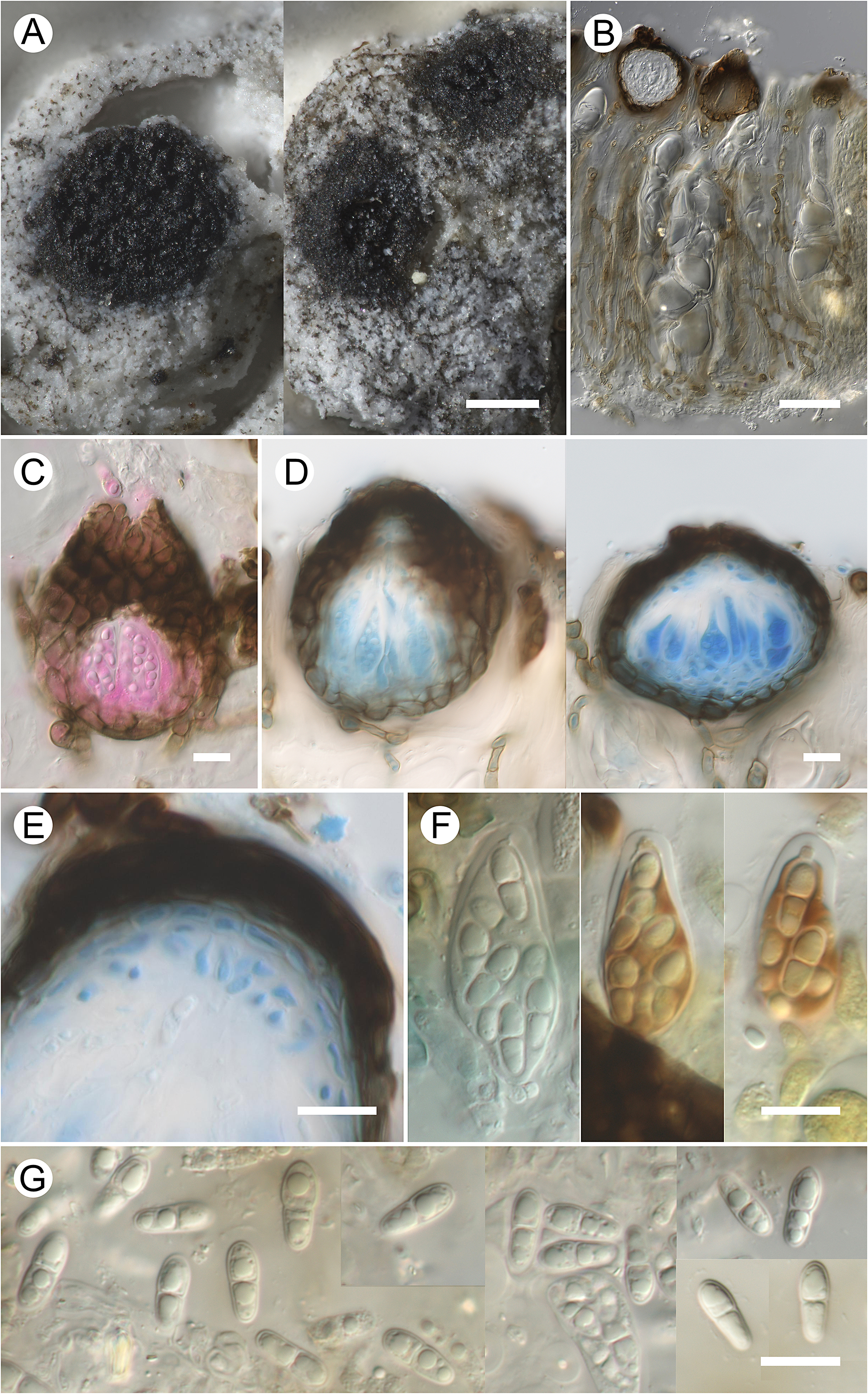
Figure 6. Long description
A multi-panel micrograph series organized as follows.
* Panel A. Two macro photos showing dark, circular infected apothecia of Megaspora verrucosa on a white, crustose substrate. Scale bar is 200 micrometers.
* Panel B. A vertical section showing dark, globose ascomata and vegetative hyphae of the parasite embedded within the host hymenium. Scale bar is 50 micrometers.
* Panel C. A median vertical section of an ascoma stained pink with phloxine, showing the dark exciple and hyphae framing the ostiole at the top. Scale bar is 10 micrometers.
* Panel D. Two median vertical sections of ascomata stained with lactophenol cotton blue, highlighting the dark, thick-walled exciple structure and blue-stained internal contents. Scale bar is 10 micrometers.
* Panel E. A high-magnification vertical section showing the upper portion of an ascoma with periphyses and rudimentary pseudoparaphyses stained blue. Scale bar is 10 micrometers.
* Panel F. Three individual asci. The left ascus is in weak B C r, while the two on the right are in K / I, showing the sac-like structure containing developing ascospores. Scale bar is 10 micrometers.
* Panel G. A collection of several transparent, two-celled ascospores with a slight constriction at the septum. Scale bar is 10 micrometers.
Lifestyle lichenicolous. No gall formation or discoloration of the infected host parts observed; however, when the parasite’s ascomata develop profusely they may completely occupy the host hymenium. Vegetative hyphae abundant, permeating the entire hymenium of host apothecia and often also developing around apothecia on the host thallus, then (sub)superficial, blackish, sinuous, sparingly branched, and clearly visible under a stereomicroscope. In water mounts they are uniformly light brown when immersed and medium brown at the host surface, with smooth (not sculptured) walls c. 0.5 μm thick, sparingly branched (often at nearly right angles), frequently septate and divided into cells 4.5–15 × 4.5–10 μm, sometimes noticeably constricted near the septa and partly toruloid.
Ascomata perithecioid, brownish black, slightly shiny, subglobose to broadly ovoid, rarely shortly papillate, (35–)55–85(–100) μm diam. (n = 39), 45–80 μm tall, with a central ostiole 5–15 μm wide, lacking setae, appendages or projections; immersed to half-protruding, usually densely crowded to contiguous and filling the entire hymenium of the host apothecia. Exciple medium to pale brown (darker above) with dark brown cell walls, K+ deep olive; in surface view mainly composed of ±rectangular cells (7–14 × 5–10 μm) with walls c. 1–2 μm thick; in median vertical section composed of 1) an outer brown layer one cell thick (3–5 μm), formed by tangentially elongated cells 3–11 × 3–5 μm and 2) an inner colourless layer 1–4 cells thick (2–5 μm), composed of tangentially elongated cells 3.5–7.5 × 1–2.5 μm. Subhymenium colourless. Interascal gel I−, K/I−. Ostiolar canal indistinct. External periphyses light brown, bacilliform, 5–11 × 2–3.5 μm. Internal periphyses difficult to distinguish from rudimentary pseudoparaphyses of ‘type a’ sensu Roux & Triebel (Reference Roux and Triebel1994), developing in the ostiolar region; hyaline, bacilliform, 1(–rarely 2)-celled, 3.5–7.5 × 1.5–2.5 μm. Paraphysoids absent. Asci narrowly ovoid, obpyriform (wider below) or ±ellipsoid, rounded at the apex, with an apically thickened wall (up to 2.5 μm), sometimes with a tiny ocular chamber (1.5–2.5 μm tall × 1.5–3 μm wide, as seen in K/I), sessile or short-stalked, 27–39 × 13–17 μm (n = 10), 8-spored, I−, K/I− (except for epiplasm turning reddish brown), BCr−. Ascospores hyaline, cylindrical-clavate (with a slightly wider upper cell relative to its position in the ascus), (9.5–)10.5–12(–13) × (4–)4.5–5 μm, l/w = (2–)2.3–2.7(–2.9) (n = 73), 1-septate (septum median), sometimes slightly constricted at the septum, often with 1–2 conspicuous guttules per cell, with a smooth wall c. 0.5 μm thick, without a gelatinous sheath, irregularly 2–3-seriate in the ascus, BCr−.
Anamorph not observed.
Etymology
The species is named in honour of Dr Imomnazar Kudratov, an eminent researcher of Central Asian lichens, on the occasion of his 80th birthday.
Distribution and host
The new species is described here based on three specimens collected in the centre of Asia (Mongolia, Russia). These were found at the upper limit of the mountain forest belt, growing on the apothecial discs of the terricolous lichen Megaspora verrucosa var. verrucosa.
Notes
The studied pyrenomycete closely matches the concept of the genus Stigmidium but its abundant, relatively thick, dark and macroscopically conspicuous vegetative hyphae recall species of Sphaerellothecium (Roux & Triebel Reference Roux and Triebel1994). However, it differs from the latter by the absence of paraphysoids (Roux & Triebel op. cit.).
Stigmidium kudratovii belongs to the group of species that inhabit the hymenium of lichen apothecia. All previously known Stigmidium species in this group, except S. grex Alstrup & Olech, occur on lichens belonging to other orders. These species can be further distinguished from the new species as follows: Stigmidium acetabuli Calat. & Triebel (on Pleurosticta, Lecanorales) has mainly colourless vegetative hyphae and somewhat smaller ascospores, (7.5–)8–10.5(–11) × 3–4(–4.5) μm, which eventually become golden brown (Calatayud & Triebel Reference Calatayud and Triebel2001); S. ahtii Etayo & Palice (on Cladonia, Lecanorales) differs by its inconspicuous vegetative hyphae, branched-anastomosed paraphysoids, and narrower ascospores, 11–13 × 3–4.5 μm (Etayo Reference Etayo2017); S. buelliae (on Buellia, Caliciales) has BCr+ and much longer ascospores, (12.5–)14.5–17.5(–22.5) × 3–4(–5) μm (Zhurbenko et al. Reference Zhurbenko2012); S. cartilagineae Calat. & Triebel (on Squamarina, Lecanorales) differs in its colourless vegetative hyphae and an I+ violet hymenial gel (Calatayud & Triebel Reference Calatayud and Triebel2003); S. cerinae Cl. Roux & Triebel (on Caloplaca, Teloschistales) has colourless, inconspicuous vegetative hyphae and BCr+, slightly narrower ascospores, 3–4 μm wide (Roux & Triebel Reference Roux and Triebel1994); S. collematis Cl. Roux & Triebel (on Collema, Peltigerales) is distinct in its smaller ascomata (30–45 μm diam.), smaller asci (21–23 × 8–10 μm), and BCr+, smaller ascospores, (7–)7.5–9.5(–10.5) × (2–)2.5–3 μm (Roux & Triebel Reference Roux and Triebel1994); S. congestum (Körb.) Triebel (on Lecanora, Lecanorales) has BCr+, longer and narrower ascospores, (11–)12–14(–16) × (2.5–)3–4 μm (Roux & Triebel Reference Roux and Triebel1994); the poorly known S. cupulare (Pat.) D. Hawksw. (on Sticta, Peltigerales) readily differs by having much larger ascomata, 200–400 μm diam. (Vouaux Reference Vouaux1912); S. disconephromeum Etayo (on Nephroma and Lobariella, Peltigerales) has BCr+, narrower ascospores, 10–13.5 × 3–3.5 μm (Etayo & Sancho Reference Etayo and Sancho2008); S. exasperatum Etayo (on Melanohalea, Lecanorales) lacks pseudoparaphyses, has smaller asci (25–28 × 10–12 μm), and slightly brownish mature ascospores (Etayo Reference Etayo2010b); the poorly known S. grex (on Ochrolechia?, Ochrolechiaceae, Pertusariales; probably not restricted to host apothecia) was described with slightly narrower ascospores, 9.5–11.5 × 3–4 μm (Alstrup & Olech Reference Alstrup and Olech1996; Zimmermann & Berger Reference Zimmermann and Berger2024); S. lecidellae Triebel et al. (on Lecidella, Lecanorales) differs in its mainly colourless, rather inconspicuous vegetative hyphae and BCr+, longer and narrower ascospores, (11–)12–15(–16) × 3–4 μm (Roux & Triebel Reference Roux and Triebel1994); S. microsporum Etayo & Osorio (on Teloschistes, Teloschistales) readily differs in its minute ascospores, 5.5–6 × 2–2.5 μm (Etayo & Osorio Reference Etayo and Osorio2004); and S. petri Hafellner (on Clauzadea, Lecideales) has smaller asci (20–30 × 9–12 μm) and BCr+, slightly narrower ascospores, 9–12 × 3–4 μm (Hafellner et al. Reference Hafellner, Herzog and Mayrhofer2008).
Comparison with Sphaerellothecium species confined to lichen apothecial discs is also relevant. Sphaerellothecium atryneae (Arnold) Cl. Roux & Triebel (on Glaucomaria and Lecanora, Lecanorales) has BCr+, halonate ascospores that become brownish with age (Roux & Triebel Reference Roux and Triebel1994); S. epilecanora Zhurb. (on Lecanora) differs by its smaller ascomata, 20–40(–60) μm diam., and the presence of a compact halo around the ascospores (Zhurbenko & von Brackel Reference Zhurbenko and von Brackel2013); S. nimisii von Brackel & Puntillo (on Xanthoparmelia, Lecanorales) has brown, granulose-verruculose, (0–)1(–3)-septate mature ascospores (von Brackel & Puntillo Reference von Brackel and Puntillo2023); and S. propinquellum (Nyl.) Cl. Roux & Triebel (on Glaucomaria) has BCr+ and slightly larger ascospores, (10–)11.5–15(–16) × (3–)4–5.5(–6) μm, which eventually become brown (Roux & Triebel Reference Roux and Triebel1994).
Additional specimens examined (both on apothecial discs of Megaspora verrucosa var. verrucosa)
Mongolia: Khubsugul Region: Ulaan Uul Somon, Mungash River valley, Ulaan Taiga state special protected area, 50°40′55.2″N, 99°19′53.4″E, 1780 m, 1 viii 2024, M. P. Zhurbenko 2456 (LE F-355743).—Russia: Krasnoyarsk Territory: Eastern Sayan Mts, Kryzhina Range, Belyi Kitat River, 5 km SE of Stal’nov Glacier, 53°59′N, 95°28′E, 1500 m, 15 vii 2009, M. P. Zhurbenko 0925a (LE F-355753a).
Talpapellis ochirbatae Zhurb., Etayo & U. Braun sp. nov.
MycoBank No.: MB 864915
Morphologically close to Talpapellis solorinae Zhurb. et al. but distinguished by forming abundant superficial hyphae (vs lacking superficial hyphae), wider internal hyphae, 3–5 μm, and external hyphae, 4–5.5 μm wide (vs 1.5–4 μm), larger stromatic cells, 5–8 μm diam. (vs up to 6 μm), wider conidiophores, 4–7.5 μm (vs 3–5(–6) μm), a rugose-squamulose wall (vs verruculose to verrucose), rare aseptate conidia (vs common aseptate conidia), and a non-related host lichen, Megaspora verrucosa (Perusariales) versus Solorina crocea (L.) Ach. (Peltigerales).
Type: Mongolia, Bayan Khongor Region, Zhargalant Somon, NE of Zhargalant settlement, near Egiin Davaa Pass, 47°11′11.3″N, 99°52′13.9″E, 2700 m, mountain steppe-tundra, on Megaspora verrucosa var. verrucosa (disc and thalline margin of apothecia, thallus) growing on plant remains, 4 August 2023, Ochirbat Enkhtuya & Samiya Javkhlan (LE F-355745—holotype).
(Fig. 7)
Talpapellis ochirbatae (holotype). A, colonies on thallus of Megaspora verrucosa var. verrucosa. B, immersed and superficial vegetative hyphae. C, conidiophores arising from stroma. D, conidiophores arising from swollen hyphal cells. E, conidiophores. Note percurrent monopodial proliferations. F, branched conidiophore. G, conidial chains and conidia. A, B, D–G in water; C in K. Scales: A = 20 μm; B–G = 10 μm. In colour online.
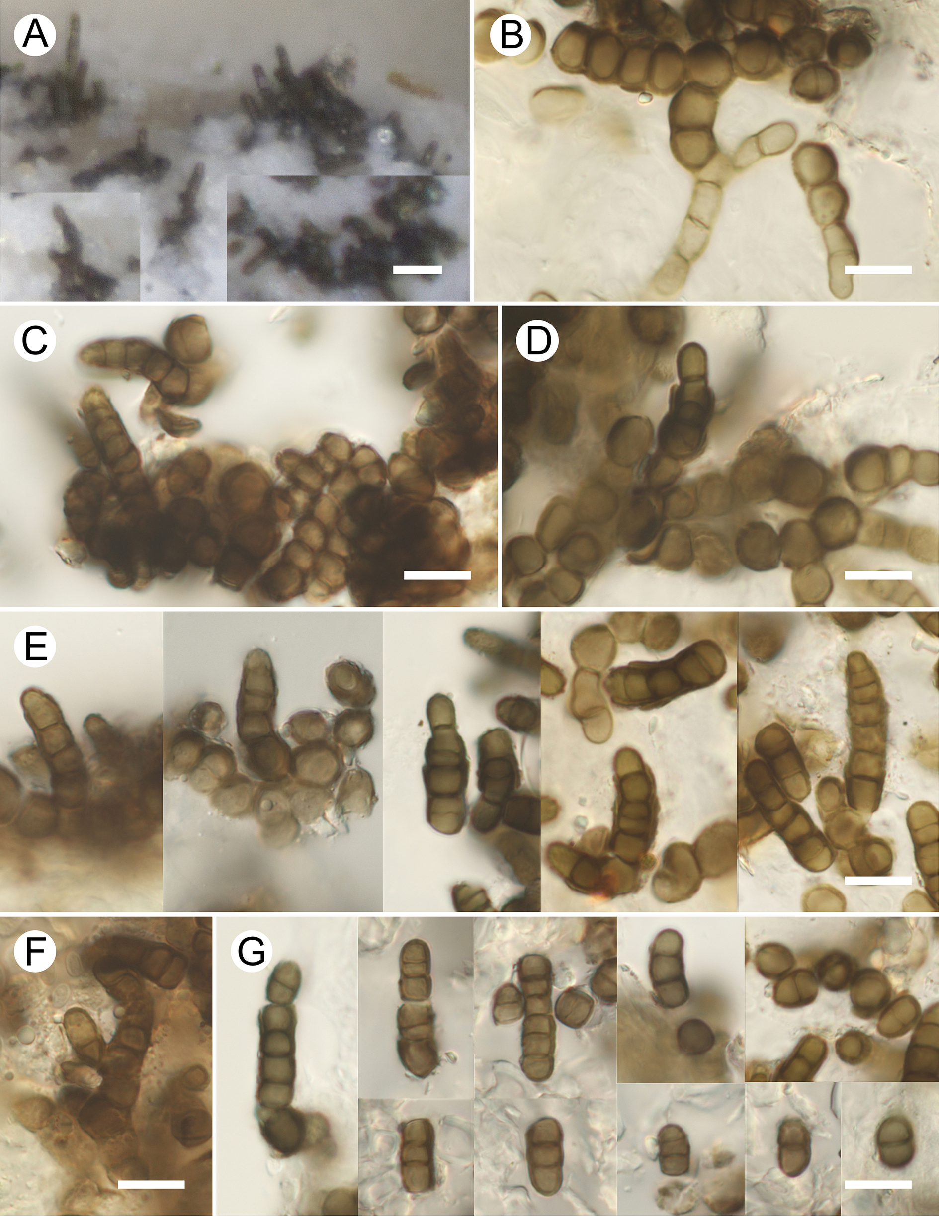
Figure 7. Long description
A multi-panel micrograph containing seven sections labeled A through G.
* Panel A: A low-magnification photo showing dark, tufted colonies of the fungus growing on the pale, crustose thallus of Megaspora verrucosa. A white scale bar represents 20 micrometers.
* Panel B: High-magnification view of vegetative hyphae composed of chains of rounded, brown, thick-walled cells. Scale bar represents 10 micrometers.
* Panel C: A dense cluster of conidiophores emerging from a dark, multicellular stroma. Scale bar represents 10 micrometers.
* Panel D: Conidiophores arising directly from swollen, globose hyphal cells. Scale bar represents 10 micrometers.
* Panel E: A composite of five vertical frames showing various conidiophores. Some exhibit percurrent monopodial proliferations, where the new growth extends through the tip of the previous cell. Scale bar represents 10 micrometers.
* Panel F: A single branched conidiophore with multiple brown, septate segments. Scale bar represents 10 micrometers.
* Panel G: A composite showing conidial chains and individual conidia. The conidia are dark brown, mostly two-to-four-celled with thick transverse septa. Scale bar represents 10 micrometers.
Lifestyle lichenicolous. No gall formation or discoloration of the infected host parts observed. Colonies effuse to loosely caespitose, blackish. Immersed mycelium consisting of light brown, straight to sinuous-torulose, branched, septate hyphae, sometimes constricted at the septa, hyphal cells 6.5–12.5 × 3–5 μm, walls smooth, c. 0.5 μm thick. Superficial mycelium consisting of brown hyphae, composed of non-swollen cells, 4–5.5 μm diam., or ±subglobose cells, 5–8 μm diam., with walls 1–1.5 μm thick, commonly forming prostrate catenate chains or stromatic aggregations up to c. 30 × 40 μm.
Conidiophores semi-macronematous, mononematous, solitary, arising from superficial swollen hyphal cells or from stromatic hyphal aggregations; brown, erect to ascending, subcylindrical or slightly tapering from base to top, straight to somewhat curved, 8–40 × 4–7.5 μm, up to 11-septate, often somewhat constricted at the septa, rarely singly branched; walls at first almost smooth, later becoming rimulose or rugose-squamulose, c. 0.5–1 μm thick; with up to five enteroblastic-percurrent monopodial rejuvenations (proliferations) leaving distinct, often rather coarse annellations (common in the holotype but rarely observed in other specimens examined). Conidiogenous cells integrated, terminal, ±doliiform, c. 4.5–6 μm wide, 2.5–4 μm tall, determinate with a single broad, truncate conidiogenous locus, c. 3.5–5 μm wide; wall of the locus undifferentiated (neither thickened nor darkened); conidiogenesis holoblastic, percurrently proliferating. Conidia formed in short, unbranched, disarticulating acropetal chains; brown, ellipsoid-ovoid, doliiform or short subcylindrical, with rounded (primary conidia) or truncate (secondary conidia) apex and truncate base; hilum unthickened, neither distinctly refractive nor darkened; (0–)1(–2)-septate, 4–6.5 × 4–4.5 μm (aseptate), 6–10.5 × 4.5–6.5 μm (1-septate), 7–15 × 4.5–7 μm (2-septate), broken off fragments of not yet disarticulated conidial chains longer and with up to four septa, occasionally slightly constricted at the septa, with smooth, rimulose or rugose-squamulose wall, c. 0.5–1 μm thick; ramoconidia not observed.
Teleomorph not observed.
Etymology
The species is named in honour of Dr Ochirbat Enkhtuya, a distinguished Mongolian lichenologist who collected the holotype.
Distribution and host
The new species is described here based on seven specimens collected in Antarctica, Asia (Mongolia, Russia) and South America (Chile). These were found in arctic and antarctic tundras, as well as mountain forest-tundra and steppe-tundra habitats, growing on the thallus (rarely the apothecial discs) of the terricolous lichen Megaspora verrucosa var. verrucosa.
Notes
The new lichenicolous hyphomycete fully conforms to the concept of Talpapellis as circumscribed by Heuchert et al. (Reference Heuchert, Braun, Diederich, Ertz, Diederich, Ertz and Braun2024). With its relatively short conidiophores, up to 40 μm long, and (0–)1(–2)-septate conidia in short chains, it is morphologically comparable only to two species of this genus, viz. T. lendemeri Diederich (on Lecania) and T. solorinae (on Solorina). It differs from both by the different host selection; in addition, it can be distinguished from the former species by its occasionally branched conidiophores and from the latter by forming abundant superficial hyphae.
Additional specimens examined (all on the thallus of Megaspora verrucosa var. verrucosa)
Antarctica: South Shetlands Archipelago: Livingston Island, Caleta Argentina, 62°40′20″S, 60°24′43″W, 0–10 m, 2 iii 2018, J. Etayo 31537 (hb. Etayo); same island, above Punta Polaca, 62°39′48.8″S, 60°23′40″W, 20–50 m, 5 iii 2018, J. Etayo 31374 (hb. Etayo).—Chile: XII Región de Magellanes: Isla Navarino, near Paso Mladineo, 55°01′14.8″S, 68°05′53.8″W, 630–700 m, 25 i 2005, J. Etayo 22913 (hb. Etayo).—Russia: Krasnoyarsk Territory: Taimyr Peninsula, Byrranga Mts, N of Levinson-Lessinga Lake, unnamed Mt ‘451.9’, 74°32′N, 98°33′E, 350 m, 29 viii 1994, M. P. Zhurbenko 9417a (LE F-355754a); ibid., N of Levinson-Lessinga Lake, Krasnaya River mouth, 74°34′N, 98°47′E, 250 m, 20 viii 1995, M. P. Zhurbenko 95633b (LE F-355746b); Eastern Sayan Mts, Kryzhina Range, Belyi Kitat River, 5 km SE of Stal’nov Glacier, 53°59′N, 95°28′E, 1500 m, 15 vii 2009, M. P. Zhurbenko 0925b (LE F-355753b).
Key to the species of lichenicolous fungi growing on Megaspora verrucosa
All records refer to var. verrucosa unless stated otherwise. References cite the main publications on each species’ taxonomy and on their occurrence on Megaspora verrucosa.
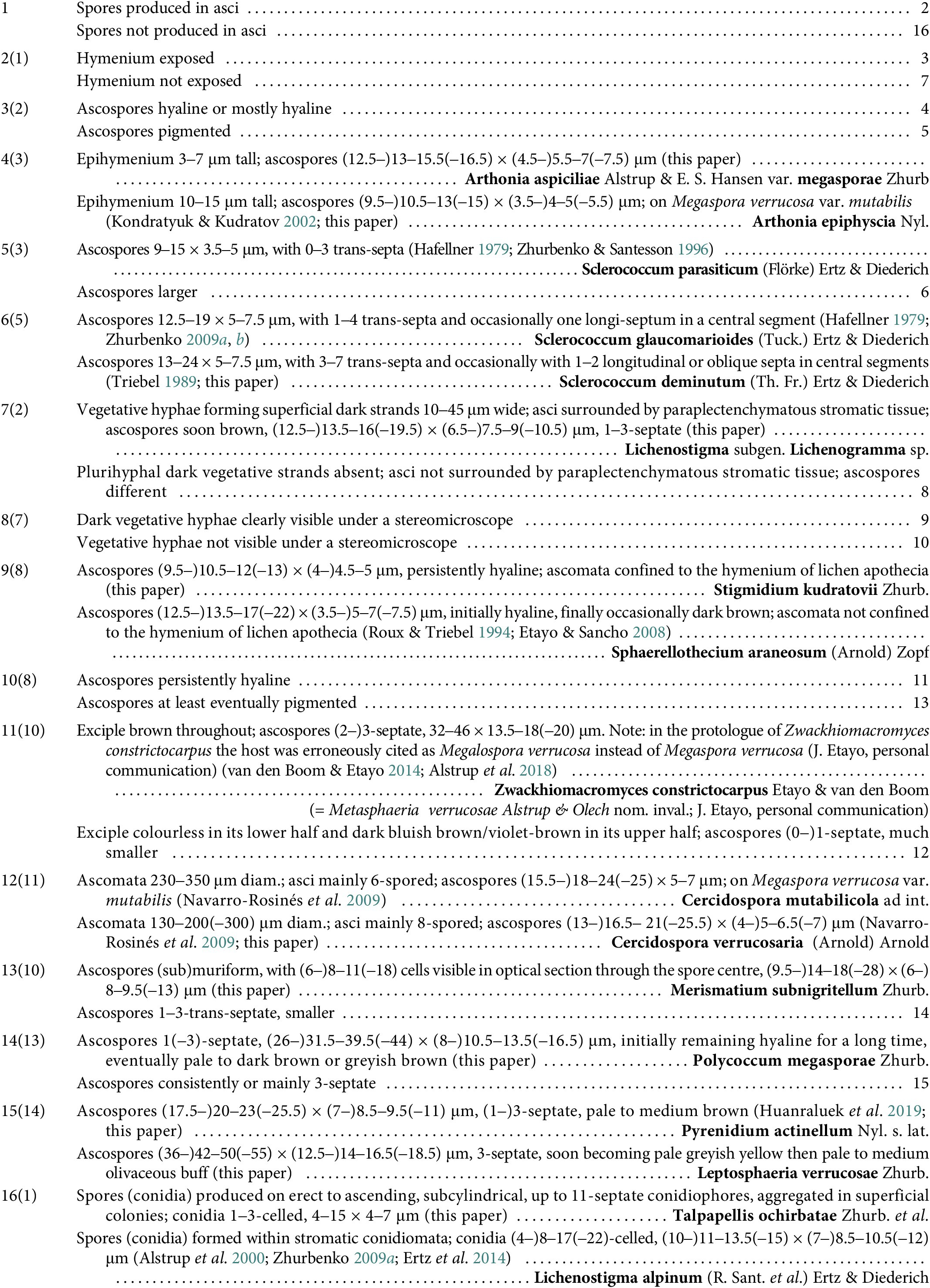
Long description
The identification key is structured as follows:
* Step 1: Spores produced in asci leads to step 2. Spores not produced in asci leads to step 16.
* Step 2: Hymenium exposed leads to step 3. Hymenium not exposed leads to step 7.
* Step 3: Ascospores hyaline or mostly hyaline leads to step 4. Ascospores pigmented leads to step 5.
* Step 4: Epihymenium 3 to 7 micrometers tall with ascospores 13 to 15.5 by 5.5 to 7 micrometers identifies Arthonia aspiciliae variety megasporae. Epihymenium 10 to 15 micrometers tall with ascospores 10.5 to 13 by 4 to 5 micrometers identifies Arthonia epiphyscia.
* Step 5: Ascospores 9 to 15 by 3.5 to 5 micrometers with 0 to 3 trans-septa identifies Sclerococcum parasiticum. Larger ascospores lead to step 6.
* Step 6: Ascospores 12.5 to 19 by 5 to 7.5 micrometers with 1 to 4 trans-septa identifies Sclerococcum glaucomarioides. Ascospores 13 to 24 by 5 to 7.5 micrometers with 3 to 7 trans-septa identifies Sclerococcum deminutum.
* Step 7: Vegetative hyphae forming dark strands 10 to 45 micrometers wide identifies Lichenostigma subgenus Lichenogramma species. Absence of these strands leads to step 8.
* Step 8: Dark vegetative hyphae visible under stereomicroscope leads to step 9. Not visible leads to step 10.
* Step 9: Ascospores 10.5 to 12 by 4.5 to 5 micrometers identifies Stigmidium kudratovii. Ascospores 13.5 to 17 by 5 to 7 micrometers identifies Sphaerellothecium araneosum.
* Step 10: Ascospores persistently hyaline leads to step 11. Pigmented ascospores lead to step 13.
* Step 11: Exciple brown throughout with 3-septate ascospores 32 to 46 by 13.5 to 18 micrometers identifies Zwackhiomacromyces constrictocarpus. Exciple colorless in lower half and dark in upper half leads to step 12.
* Step 12: Ascomata 230 to 350 micrometers diameter identifies Cercidospora mutabilicola. Ascomata 130 to 200 micrometers diameter identifies Cercidospora verrucosaria.
* Step 13: Ascospores submuriform 14 to 18 by 8 to 9.5 micrometers identifies Merismatium subnigritellum. Ascospores 1 to 3 trans-septate and smaller leads to step 14.
* Step 14: Ascospores 1 to 3 septate 31.5 to 39.5 by 10.5 to 13.5 micrometers identifies Polycoccum megasporae. Consistently 3-septate ascospores lead to step 15.
* Step 15: Ascospores 20 to 23 by 8.5 to 9.5 micrometers identifies Pyrenidium actinellum. Ascospores 42 to 50 by 14 to 16.5 micrometers identifies Leptosphaeria verrucosae.
* Step 16: Spores produced on erect conidiophores identifies Talpapellis ochirbatae. Spores formed within stromatic conidiomata identifies Lichenostigma alpinum.
Discussion
Recent revisions of the taxonomic diversity of lichenicolous fungi indicate that the number of known obligately lichenicolous, non-lichenized species, subspecies and varieties has increased by 18% over the past seven years (Diederich et al. Reference Diederich, Lawrey and Ertz2018, Reference Diederich, Lawrey and Ertz2025), with no sign that the rate of discovery is slowing. One of the effective approaches for revealing this diversity, alongside taxonomic and floristic surveys, is the targeted revision of lichenicolous fungi on particular groups of lichens. As a result of such a revision presented here, ten taxa of lichenicolous fungi were recorded on a single lichen variety; five species and one variety (60%) were described as new to science, almost a record level of novelty for studies of this kind (Table 1).
Proportions of new species and varieties of genuinely lichenicolous fungi described during inspections of different lichen host groups.
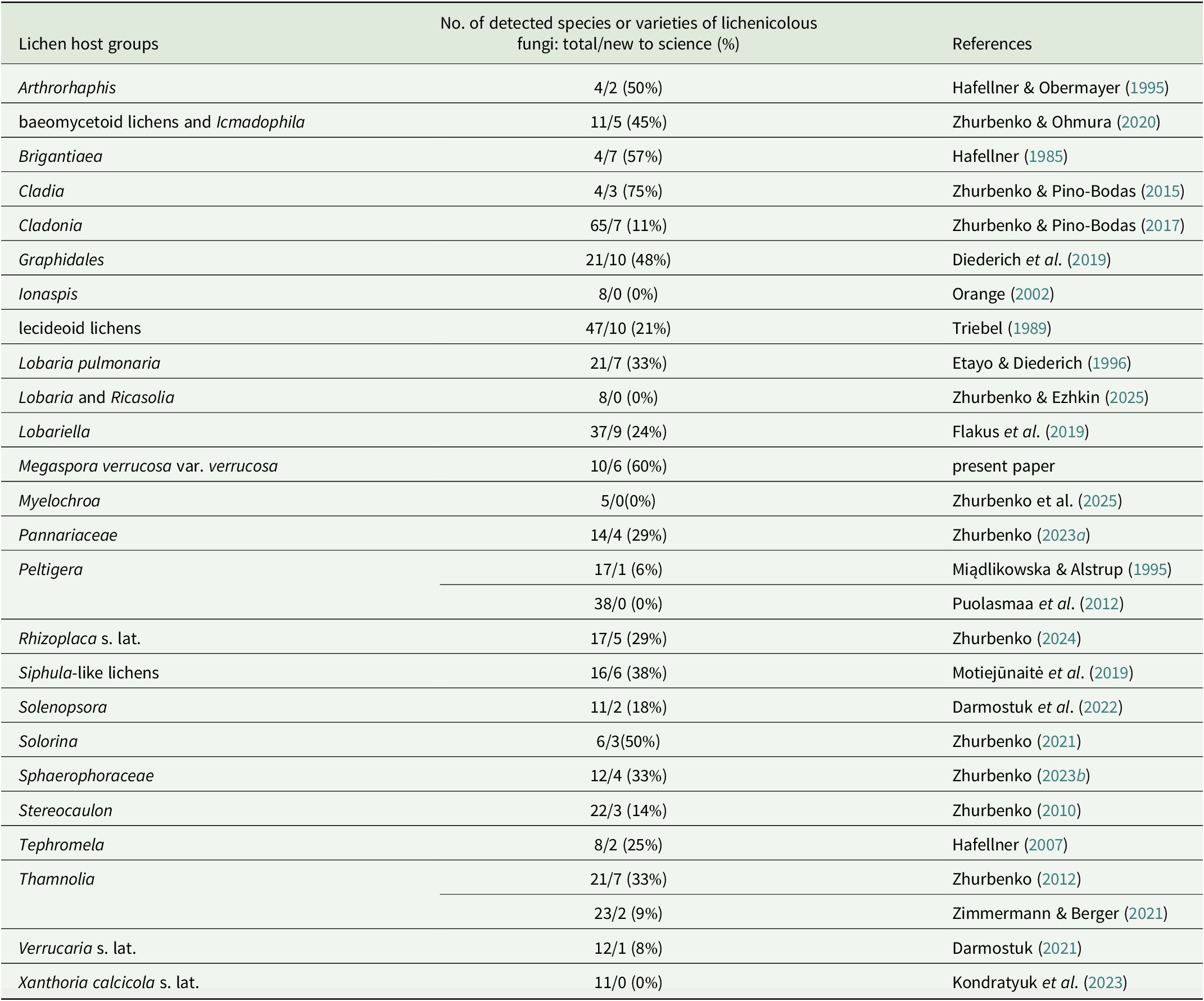
Table 1. Long description
The table consists of three columns: Lichen host groups, Number of detected species or varieties of lichenicolous fungi (total/new to science and percentage), and References.
Key data points include:
* Arthrorhaphis: 4 total, 2 new (50 percent).
* baeomycetoid lichens and Icmadophila: 11 total, 5 new (45 percent).
* Brigantiaea: 4 total, 7 new (57 percent).
* Cladia: 4 total, 3 new (75 percent).
* Cladonia: 65 total, 7 new (11 percent).
* Graphidales: 21 total, 10 new (48 percent).
* Ionaspis: 8 total, 0 new (0 percent).
* lecideoid lichens: 47 total, 10 new (21 percent).
* Lobaria pulmonaria: 21 total, 7 new (33 percent).
* Lobaria and Ricasolia: 8 total, 0 new (0 percent).
* Lobariella: 37 total, 9 new (24 percent).
* Megaspora verrucosa var. verrucosa: 10 total, 6 new (60 percent).
* Myelochroa: 5 total, 0 new (0 percent).
* Pannariaceae: 14 total, 4 new (29 percent).
* Peltigera: Two entries showing 17 total, 1 new (6 percent) and 38 total, 0 new (0 percent).
* Rhizoplaca s. lat.: 17 total, 5 new (29 percent).
* Siphula-like lichens: 16 total, 6 new (38 percent).
* Solenopsora: 11 total, 2 new (18 percent).
* Solorina: 6 total, 3 new (50 percent).
* Sphaerophoraceae: 12 total, 4 new (33 percent).
* Stereocaulon: 22 total, 3 new (14 percent).
* Tephromela: 8 total, 2 new (25 percent).
* Thamnolia: Two entries showing 21 total, 7 new (33 percent) and 23 total, 2 new (9 percent).
* Verrucaria s. lat.: 12 total, 1 new (8 percent).
* Xanthoria calcicola s. lat.: 11 total, 0 new (0 percent).
It is noteworthy that all 17 lichenicolous fungal species recorded on Megaspora were collected on M. verrucosa, and 15 of them exclusively on its var. verrucosa. Of these 17 species, nine species and one variety are currently known only from Megaspora verrucosa.
Acknowledgements
The research of M. P. Zhurbenko was conducted as part of the project ‘Taxonomic, ecological and structural-functional diversity of fungi and fungus-like protists’ (124013100829–3) at the V. L. Komarov Botanical Institute of the Russian Academy of Sciences, using equipment from the institute’s Core Facility Center ‘Cell and Molecular Technologies in Plant Science’. The author’s field research in Mongolia was supported by the joint Russian-Mongolian complex biological expedition of the Russian Academy of Sciences and the Academy of Sciences of Mongolia. The author is grateful to Ochirbat Enkhtuya and Samiya Javkhlan, who kindly made their lichen collections available to him. Javier Etayo, Uwe Braun and Claude Roux are thanked for valuable comments and assistance with taxonomic decisions.
Author ORCID
Mikhail P. Zhurbenko, 0000-0002-9839-4698.