Introduction
The stigmatization of electroconvulsive therapy (ECT) is partially based on beliefs that it has been associated with significant memory impairments (Payne & Prudic, Reference Payne and Prudic2009). These beliefs have remained even after ECT protocols were modified from sine-wave into a brief or ultra-brief stimulation, with more favorable profile of the side-effects (Sackeim, Reference Sackeim2004). The brief stimulation with the bilateral electrode placement is a currently worldwide-preferred practice, with trend towards a more frequent unilateral stimulation at some European, Australian and New Zealand sites (Leiknes et al. Reference Leiknes, Jarosh-von Schweder and Hoie2012). Despite the longstanding opinion that bilateral stimulation induces more cognitive side effects, recent data from Sienaert et al. (Reference Sienaert, Vansteelandt, Demyttenaere and Peuskens2010) showed that antidepressant efficacy was reached after both unilateral and bilateral stimulation, with no deleterious effects on cognitive function (at least in the case of ultra-brief pulse ECT with bilateral stimulus and a dose slightly above the seizure threshold).
In the extensive literature review that included 84 within-subjects designed studies on ECT-related cognitive changes, Semkovska & McLoughlin (Reference Semkovska and McLoughlin2010) found a short-term decrease in cognitive performance immediately after the ECT course, followed by subsequent recovery to the pretreatment functioning levels noticed at follow-up. Nevertheless, the authors concluded that no standardized retrograde amnesia tests were identified by this meta-analysis. We agree with many authors that improved assessment of the degree of cognitive impairment following ECT treatment has to be based on tests with good sensitivity, flexibility and resolution. According to Tsaltas et al. (Reference Tsaltas, Kalogerakou, Papakosta, Kontis, Theochari, Koutroumpi, Anyfandi, Michopoulos, Poulopoulou, Papadimitriou and Oulis2011), the degree of cognitive impairment residual to ECT cannot be reliably estimated at present, therefore definitive evaluation of ECT-related cognitive deficits calls for further research.
Methodology of the research on memory during ECT treatment of depression has to be based on tests sensitive to detect and differentiate frontal from temporal and amygdalo-hyppocampal dysfunctions, with parallel forms of tasks for countering practice effects on repeat testing. By using the Cambridge Neuropsychological Test Automated Battery (CANTAB), the group led by Professor Ian C. Reid (Falconer et al. Reference Falconer, Cleland, Fielding and Reid2010) has shown for the first time that selected tests from this battery represent an effective measure of ECT-associated memory impairments. Twenty-four severely depressed patients (aged 26–83 years) were examined before and 1 month after ECT (acute and medium-term effects), cognition was tested with an emphasis on visual and visuo-spatial memory (spatial and pattern recognition memory, visual paired associate learning and delayed matching to sample tests). Their results showed that most of the baseline cognitive impairments had disappeared 1 month following ECT, except for the impairment in spatial recognition memory, which remained present at the last follow-up visit (i.e. 1 month later). The authors emphasized that this was among the few studies which have detected anterograde memory deficits in patients with major depressive disorder (MDD) following ECT.
Recently, a group from Aberdeen published the following study, based on the 6-month follow-up of a larger group of patients with depression (mean age 61 years). For the neuropsychological assessment, authors used only the Mini Mental State Examination (MMSE) and SRM. Regarding the long-term post-ECT effects, the SRM task revealed reversible cognitive deficiencies (relative to a pre-ECT baseline) for at least 3 months following ECT, while the MMSE score and patients’ subjective reports only improved over time. Thus, the authors concluded that the CANTAB SRM task is sensitive to detect cognitive deficits, unlike the MMSE and patients’ subjective reports (Fernie et al. Reference Fernie, Bennett, Currie, Perrin and Reid2014).
However, this field remains under-investigated. Therefore, the goal of the present study was to evaluate cognitive function in adult depressed patients treated with bilateral ECT, by using the MMSE and computerized neuropsychological tests sensitive to detect possible memory changes related to frontal and temporal lobe function before and after the ECT. Furthermore, the authors intended to analyze the correlation between the severity of depression and the cognitive performance. The authors decided to focus on middle-aged patients (<65 years), in order to minimize the effects on memory that could be related to age-associated dysfunctions of the frontal and medio-temporal lobe structures (Kemps & Newson, Reference Kemps and Newson2006; Soei & Daum, Reference Soei and Daum2008).
Method
Study sample
The study was carried out at the Clinic for Psychiatry – Military Medical Academy (Belgrade), and included in- and out-patients referred with clinical diagnosis of MDD. The diagnosis was confirmed by consensus of two psychiatrists during separate interviews, according to IDC-10 criteria. All patients (n = 30, all Caucasians with Serbian as first language) had neither history of ongoing significant medical nor neurological condition. Exclusion criteria were: alcohol/substance-related disorder (present or past), mental retardation (IQ ⩽70), education ⩽8 years, age >65 years and inability to use tests due to poor eyesight.
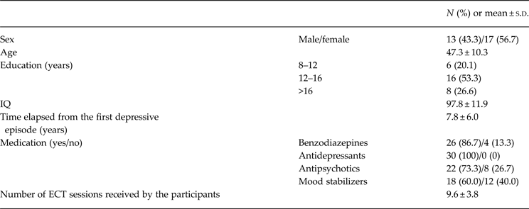
Upon admission, patients were treated with antidepressants in accordance with their doctor's choice. Alongside antidepressants, most patients were prescribed benzodiazepine, while some of them used mood stabilizers and/or antipsychotic drugs as an augmentation (for more detailed analysis of local and regional prescribing patterns see Jordanova et al. Reference Jordanova, Maric, Alikaj, Bajs, Cavic, Iosub, Mihai, Szalontay and Sartorius2011, and Maric et al. Reference Maric, Stojiljkovic, Pavlovic and Jasovic-Gasic2012). The patients were adherent to the aforementioned medications for at least 1 month before entering the study, and the treatment did not change significantly during the study period (for detailed information concerning patients’ socio-demographic and clinical characteristics see Table 1).
Demographic and clinical characteristics of the sample

The research was performed in compliance with the International Code of Medical Ethics of the World Medical Association (Declaration of Helsinki) and was approved by Institutional Review Board. Informed consent was obtained for all study participants after an explanation of the research procedures.
Instruments
Clinical scales
The Clinical Global Impression Scale (CGI), a well-established research rating tool applicable to all psychiatric disorders (Guy, Reference Guy1976) was used to assess the overall impression. For the purpose of the present study, severity of psychopathology was recorded by using scores ranging from 1 to 7.
The intensity of depressive symptoms was evaluated with the Hamilton Rating Scale for Depression (HAMD-21; Hamilton, Reference Hamilton1960). Additionally, subjects completed the self-rating Depression Anxiety Stress Scales (DASS; Lovibond & Lovibond, Reference Lovibond and Lovibond1995; Jovanovic et al. Reference Jovanovic, Gavrilov-Jerkovic, Zuljevic and Brdaric2014). The DASS – Depression subscale assesses dysphoria, hopelessness, devaluation of life, self-deprecation, lack of interest or involvement and anhedonia. The DASS – Anxiety subscale assesses autonomic arousal, skeletal muscle effects, situational anxiety and subjective experience of anxious affect. The DASS – Stress subscale captures feelings of nervous tension, difficulty relaxing and irritability. In accordance with the recommendations of McClintock et al. (Reference McClintock, Choi, Deng, Appelbaum, Krystal and Lisanby2014), both observer-rated and self-rated instruments were used to obtain a sufficiently clear view of the current emotional state.
Neuropsychological testing
The MMSE is a widely used test for general assessment of cognitive impairment (Pangman et al. Reference Pangman, Sloan and Guse2000). It measures several cognitive domains, including orientation for time, orientation for place, attention and calculation, registration, recall, repetition and ability to follow simple commands. The highest overall score is 30.
IQ was assessed by the Wechsler Adult Intelligence Scale – Revised (WAIS-R; Wechsler, Reference Wechsler1981). The scale consists of six verbal (Information, Comprehension, Arithmetic, Digit Span, Similarities, Vocabulary) and five performance subtests (Picture Arrangement, Picture Completion, Block Design, Object Assembly, Digit Symbol). A verbal IQ, performance IQ and full-scale IQ were obtained.
CANTAB tests
The Pattern Recognition Memory (PRM) is a visual memory test, primarily sensitive to dysfunction in the medial temporal brain areas and relatively insensitive to frontal lobe dysfunction. In the first phase of the test, participants need to memorize presented series of 12 colored visual patterns. Afterwards, they are presented with 12 paired new and old patterns and are required to identify the pattern they have already seen during the presentation phase. The mean percentage of correct answers served as the main outcome parameter.
The Delayed Matching to Sample (DMS) test is primarily sensitive to impairments in the medial temporal lobe area, with some input from the frontal lobe. The participants are presented with one target (complex visual pattern) and four different patterns simultaneously or 0, 4 and 12 s after the target presentation. Afterwards, the participants are required to identify the pattern which exactly matches the target. The main performance indications were mean percentages of correct answers in all time intervals.
The Paired Associates Learning (PAL) test assesses visual memory and new learning. The test is primarily sensitive to changes in the medial temporal lobe functioning. The participants are presented with randomly opened boxes, of which one or more contain a pattern. Afterwards, the patterns are one by one displayed in the middle of the screen and participants are required to identify the box where the pattern was originally located. As the task progresses, the number of patterns is gradually increasing up to eight. The total number of errors served as a main outcome parameter.
The Spatial Recognition Memory (SRM) test is primarily sensitive to dysfunction in the frontal lobe, and relatively insensitive to the temporal lobe damage. In the first phase of the test, a white square is displayed at five different locations on the screen and participants are required to memorize the locations on the screen. Afterwards, participants are presented with a series of five pairs of squares and they have to choose the square which was presented to them previously, in the presentation phase. The mean percentage of correct answers was used as outcome parameter for this test.
Procedure
Ratings for clinical scales, as well as neuropsychological testing, were made at three time points: Baseline (T0), 1–3 days before ECT; Post-ECT (T1), 1 day after the final ECT application; Follow-up (T2), 4 weeks after the final treatment.
Testing was completed within approximately 60 min. The multi-domain cognitive assessment was feasible and well-tolerated.
Description of the ECT intervention
Similarly to most sites worldwide (Leiknes et al. Reference Leiknes, Jarosh-von Schweder and Hoie2012), our site has been using bilateral (bi-temporal) ECT for all patients referred to our specialized unit. Bilateral ECT has been chosen for several reasons. Previous data has shown reduced efficiency of unilateral v. bilateral electrode placement during ECT in depression (most recent analysis was published by McCormick et al. Reference McCormick, Brumm, Benede and Lewis2009). Moreover, patients referred for ECT course are all in-patients with high hospital costs and limited length of stay. Thus, following risks and benefits calculation, a consensus was made that bilateral ECT should be the first line choice of electrode placement and patients were treated accordingly. In line with the local guidelines, pre-treatment investigations included: blood test, hepatic and renal function tests, ECG, EEG and medical, neurological and ophthalmological consultation. All patients were required to sign a separate informed consent forms for ECT and anesthesia.
The ECT was administered three times a week. In all cases and all sessions, the Thymatron System IV machine and brief pulses (0.5 ms pulsewidth) with bilateral (bi-temporal) electrode placement and atropine premedication (dose 0.5 mg) were used. Anesthesia was induced with propofol (2–2.5 mg/kg) combined with the muscle relaxant succinylcholine (1 mg/kg). (For detailed description on ECT practice in Serbia, see Spiric et al. Reference Spiric, Stojanovic, Samardzic, Milovanovic, Gazdag and Maric2014.)
For the first treatment, stimulus dose was based on the ‘half-age’ method (Petrides & Fink, Reference Petrides and Fink1996), with pulse amplitude of 900 mA during the therapy. The first and the final delivered treatment charges were 140.26 ± 45.23 and 227.15 ± 38.07 mC (1.6 first stimulus dose), respectively. The first and the last stimulus durations were 6.2 ± 0.6 and 7.6 ± 0.3 s, respectively. The minimum seizure duration of 20 s was considered acceptable.
Statistical analyses
The data were analyzed using SPSS v. 20.0 (IBM Corp., USA) statistical software. Socio-demographic and clinical data were displayed using absolute and relative numbers, mean and standard deviation (s.d.). Neurocognitive data were presented as mean ± standard error (s.e.). Initially, all data were tested for normality, and some of them (i.e. total DASS score, DASS Depression subscale) were log-transformed prior to analysis in order to address skewed data.
Linear mixed model was used to analyze changes in depressive symptom severity and patients’ cognitive performances over time, and to assess dynamic correlations between aforementioned features. Age and gender (random effects) adjustment was applied, when applicable. An overall test of the fixed effect for the examined time points was obtained, along with the pairwise comparisons of interest (the comparison of the baseline scores with the scores obtained at T1 and T2). A mixed model was used for analysis, as it exhibited a number of advantages when repeated measurements on the same subject were performed, had great flexibility to model time effects, and adequately handled missing data. Effect size (ES) was measured using Cohen's d (repeated measurements). Baseline and the last observation score were used to calculate ES.
In order to constrain the risk of type I error, Bonferroni correction was applied. After applying the correction for multiple comparisons (two pairwise comparisons), p level was set at 0.025.
Results
Severity of depression
Severity of depressive symptoms, as measured by both clinician-rated and self-rated instruments, was significantly reduced over time (F 2,50.48 = 134.73, p < 0.01, Cohen's d = 2.92).
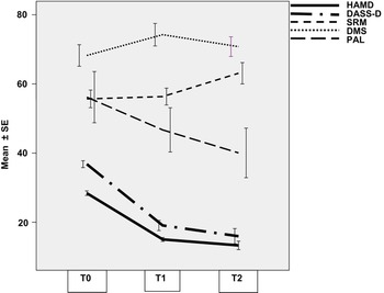
When compared to the baseline (T0), HAMD scores were significantly reduced at T1 (mean difference −13.37, s.e. = 0.87, df = 47.31, p < 0.01, 95% CI −15.39 to −11.34) and T2 (mean difference 14.99, s.e. = 1.09, df = 75.75, p < 0.01, 95% CI −17.49 to −12.50). Similarly, significant decrease in DASS scores was also observed throughout time (total DASS score: F 2,53.31 = 47.91, p < 0.01, Cohen's d = 1.93; DASS subscales – Depression: F 2,28.26 = 44.38, p < 0.01, Cohen's d = 2.37; Anxiety: F 2,27.71 = 44.19, p < 0.01, Cohen's d = 1.54; Stress: F 2,27.25 = 34.31, p < 0.01, Cohen's d = 1.41). Continuous decline in DASS scores was evident at T1 (total DASS score: mean difference −0.31, s.e. = 0.04, df = 50.67, p < 0.01, 95% CI −0.39 to −0.23; DASS subscales – Depression: mean difference −0.32, s.e. = 0.04, df = 29.00, p < 0.01, 95% CI −0.41 to −0.24; Anxiety: mean difference −0.35, s.e. = 0.05, df = 29.00, p < 0.01, 95% CI −0.45 to −0.24; Stress: mean difference −14.23, s.e. = 1.82, df = 29.00, p < 0.01, 95% CI −18.53 to −9.93) as well as at T2 (total DASS score: mean difference −0.41, s.e. = 0.05, df = 75.36, p < 0.01, 95% CI −0.51 to −0.30; DASS subscales – Depression: mean difference −0.45, s.e. = 0.06, df = 28.77, p < 0.01, 95% CI −0.59 to −0.31; Anxiety: mean difference −0.47, s.e. = 0.06, df = 27.38, p < 0.01, 95% CI −0.62 to −0.32; Stress: mean difference 14.48, s.e. = 2.13, df = 27.67, p < 0.01, 95% CI −19.54 to −9.42) in comparison to T0 (see Fig. 1).
Depressive symptom severity and CANTAB tests performance trajectory. HAMD, Hamilton Rating Scale for Depression; DASS-D, Depression subscale of the Depression Anxiety Stress Scales; SRM, Spatial Recognition Memory task; DMS, Delayed Matching to Sample task; PAL, Paired Associates Learning task; T0, baseline, before ECT; T1, 1 day after the final ECT; T2, 1 month after the final treatment.

Clinical global impression was significantly improved over the course of time (F 2,27.77 = 105.07, p < 0.01, Cohen's d = 2.88) pointing to a satisfactory response to the treatment. CGI scores were significantly lower at T1 (mean difference −1.97, s.e. = 0.15, df = 29.00, p < 0.01, 95% CI −2.32 to −1.62) and T2 (mean difference −2.55, s.e. = 0.20, df = 28.02, p < 0.01, 95% CI −3.03 to 2.08) compared to T0.
Cognition
MMSE
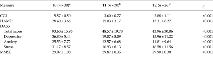
The MMSE scores varied significantly across the examined time points (F 2,27.01 = 9.14, p < 0.01, Cohen's d = 1.01). More precisely, when compared to T0, general cognition improved at T1 (mean difference 0.80, s.e. = 0.19, df = 29.00, p < 0.01, 95% CI 0.35–1.24) and T2 (mean difference 0.84, s.e. = 0.20, df = 29.08, p < 0.01, 95% CI 0.38–1.31). Nevertheless, clinical importance of that finding is questionable, since the size of the change was within one unit of the scale (see Table 2).
The clinical assessment

CGI, Clinical Global Impression Scale; HAMD, Hamilton Rating Scale for Depression; DASS, Depression Anxiety Stress Scale; MMSE, Mini Mental State Examination.
Results are presented as mean ± s.d.
a T0, before ECT.
b T1, 1 day after the final ECT.
c T2, 1 month after the final treatment.
CANTAB tests
Visual, spatial and figural memory
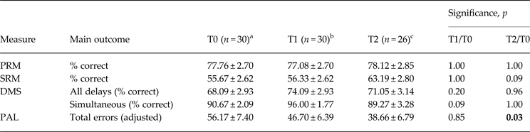
The mixed model showed no significant difference in SRM (F 2,53.64 = 2.69, p = 0.08) nor PRM (F 2,51.39 = 0.08, p = 0.92) across the examined time points (see Table 3), suggesting that participants’ visuo-spatial memory did not differ across the treatment period. Likewise, patients’ delayed matching to sample scores did not differ between baseline, 1 day after the final ECT application and 4 weeks following the treatment completion (F 2,55.22 = 1.44, p = 0.25).
Summary of the results from individual CANTAB tests

PRM, Pattern Recognition Memory, visual memory; SRM, Spatial Recognition Memory, visuo-spatial memory; DMS, Delayed Matching to Sample, visual figural memory; PAL, Paired Associate Learning, visual memory and learning.
Results are presented as mean ± s.e.
a T0, before ECT.
b T1, 1 day after the final ECT.
c T2, 1 month after the final treatment.
On the other side, we found a statistically significant difference in the simultaneous matching to sample over time (F 2,28.82 = 5.16, p = 0.01, Cohen's d = 0.51), but this finding lacked clinical importance since there was no difference between T0 and T2. Initial improvement in simultaneous matching observed at T1 returned to baseline values at T2 (see Table 3).
Visual memory and learning
There was a significant difference in the total number of errors in PAL task throughout the examined time period (F 2,26.51 = 3.90, p = 0.03, Cohen's d = 2.46). More precisely, significant reduction of total errors in PAL was observed between T0 and T2 (mean difference −0.21, s.e. = 0.08, df = 25.23, p = 0.03, 95% CI −0.41 to −0.02), while there was no significant difference between T0 and T1. Patients’ intermediate achievement in PAL at T1 was suggestive of a gradual improvement in learning ability over time (see Fig. 1).
Correlations
Linear mixed model has revealed significant dynamic correlations between depressive symptom severity and certain cognitive measures.
Since it has been shown that visual spatial and figural memory performances depended on general cognitive ability, all analyses were adjusted for IQ. PAL test showed the strongest association with depressive symptom severity (with HAMD scores: F = 17.70, p < 0.01; with total DASS scores: F = 16.00, p < 0.01; with DASS Depression subscale scores: F = 24.64; p < 0.01), referring to a marked improvement in learning ability following the remission from depression. Likewise, SRM and DMS were also significantly correlated with HAMD scores (SRM: F = 4.42, p = 0.04; DMS: F = 4.33, p = 0.04), total DASS scores (SRM: F = 6.98, p = 0.01; DMS: F = 8.54, p = 0.01) and DASS Depression subscale scores (SRM: F = 5.67, p = 0.02; DMS: F = 8.53, p = 0.01). Nevertheless, as mentioned earlier, clinical importance of the observed dynamic correlation between depressive symptoms and delayed matching to sample is questionable since the initial improvement trend observed at T1 has been lost at T2 (see Table 3).
No significant correlations were found across the treatment and follow-up period neither between the depressive symptom severity and PRM, nor in the simultaneous matching to sample.
Discussion
The present study examined the impact of ECT on memory in patients with MDD. Measurements were taken immediately following and 1 month after the last seizure was induced by bi-temporal electrode placement and brief-pulses applied three times per week (acute and medium-term treatment effects). We maximized the internal validity of the study by including patients whose age was under the usual age of onset of age-related cognitive decline and by using the fixed ECT treatment parameters. Our results have shown that ECT improves mood, as assessed by both self-reported and clinician-rated measures, which was additionally reflected through the improvement in global clinical impression. Alongside depressive symptoms, additional aspects of negative affective states, such as subjective experience of anxiety and stress, were significantly reduced during the ECT course, with the overall affective improvement still present a month later upon follow-up for majority of the patients (the large standard deviations in DASS scale, particularly at time point T2, are suggesting that for some participants, the clinical benefits of ECT were not durable). These results were expected since most of the evidence confirmed the efficacy of bilateral ECT in treatment for depression, suggesting that it was probably even more effective than drug therapy (UK ECT Review Group, 2003).
Overall improvement in MMSE scores was also observed in the present study, but the size of this change was within one single unit of the scale, and further interpretation is practically impossible. In line with the results of the recent meta-analysis (Semkovska & McLoughlin, Reference Semkovska and McLoughlin2010) showing a superiority of bilateral over unilateral electrode placement regarding MMSE scores, the improvement could have also been expected in the case of bilateral stimulation applied in our sample.
The neuropsychological tests did not detect any significant memory impairment as potential acute or medium-term effect of ECT. However, significant improvement on visual memory and learning (PAL task) has been noticed at both T1 and T2 in comparison to baseline, with positive correlation to the improvement of depression. According to the additional correlations based on the DASS subscales, increased learning and visual memory performance seemed to be exclusively related to the improvement of depression, whilst unrelated to anxiety or stress-related affective status. In studying smaller sample, with significant number of ECT-treated MDD patients lost to follow-up (17 at the beginning and nine at 1-month follow-up), Falconer et al. (Reference Falconer, Cleland, Fielding and Reid2010) also noticed the reduction in total error scores on the PAL task, but the difference did not reach statistical significance, most probably due to the insufficient sample size. On the other hand, Falconer et al. (Reference Falconer, Cleland, Fielding and Reid2010) have shown significant impairments in spatial recognition memory during ECT, which remained such during the month following ECT (apart from the patients unable to take part in the follow-up). Fernie et al. (Reference Fernie, Bennett, Currie, Perrin and Reid2014) confirmed this finding and additionally showed that these deficits have been extended to at least 3 months following the ECT course completion. Therefore, SRM task has been suggested as a sensitive instrument for detecting anterograde memory changes following ECT. However, our results on spatial recognition memory in relation to ECT did not confirm the finding of prolonged ECT-related memory deficit. We observed a slight trend towards an improvement in SRM, which did not reach significance. In opposite to PAL, this change had no correlation with depression. The question arises whether this missing improvement might be a potentially important finding?
According to the recent meta-analysis of cognitive impairment in depression assessed with CANTAB (Rock et al. Reference Rock, Roiser, Riedel and Blackwell2013), in terms of memory functions, remission from depression was characterized by a significant improvement in the PAL and SRM tasks (up to the level of healthy population), while the performances on PRM and DMS remained poorer even when depressive symptoms were negligible. Similarly, when Hungarian group evaluated cognitive function in acute MDD v. remission phase, their results also showed that depression symptom reduction was followed by improvement in PAL and SRM, but not DMS nor PRM (Egerhazi et al. Reference Egerhazi, Balla, Ritzl, Varga, Frecska and Berecz2013). On the basis of these results, our finding of missing improvement in SRM at 1 month follow-up is not quite opposite to Falconer et al. (Reference Falconer, Cleland, Fielding and Reid2010) and Fernie et al. (Reference Fernie, Bennett, Currie, Perrin and Reid2014). The missing improvement (i.e. our less robust finding) might be explained on the basis of the age-related differences between our samples. Age-related decline in SRM has been shown by several authors (Adelstein et al. Reference Adelstein, Kesner and Strassberg1992; Rabbitt & Lowe, Reference Rabbitt and Lowe2000), which could be one of the reasons for prolonged recovery of this memory domain in the samples of older adults. Older adult samples are preferably treated two times per week, since this scheme is more protective in terms of cognitive side effects (Shapira et al. Reference Shapira, Tubi and Lerer2000), which is additionally confirmed in meta-analysis by Semkovska & McLoughlin (Reference Semkovska and McLoughlin2010). Having in mind the above-mentioned findings of cognitive protection as a result of less frequent ECT application our sample could have been ‘at risk’ for cognitive impairments. However, our data yielded no more ‘risk’ in comparison to subjects from aforementioned studies, which were unexpected finding. Nevertheless, that finding increased the power of our data by reducing possible positive bias. Finally, the more detailed analysis of dosing regimen between the studies might explain the lack of the replication of earlier results, but precise comparison is difficult to make without more information regarding the stimulus dose for baseline seizure threshold (ST), the stimulus dose for subsequent treatments, the final treatment dose, etc.
To our best knowledge, the significant improvement on visual memory and learning test during and after ECT is a new and possibly important finding. The improvement on PAL, which is sensitive to the medial temporal lobe functions, might be interpreted as a part of an overall mood-improvement. However, this is not the only possible explanation. The dorsal hippocampus contains a mechanism to support paired-associate learning (Gilbert & Kesner, Reference Gilbert and Kesner2003; Langston et al. Reference Langston, Stevenson, Wilson, Saunders and Wood2010; Yoon et al. Reference Yoon, Seo, Kim and Lee2011) and, at least in animal experiments, ECT had seizure-related effects on hypocampal neurogenesis in the dentate gyrus (Rotheneichner et al. Reference Rotheneichner, Lange, O'Sullivan, Marschallinger, Zaunmair, Geretsegger, Aigner and Couillard-Despres2014). The general collective of longitudinal neuroimaging investigations in humans supported the neurotrophic effects of ECT (Abbott et al. Reference Abbott, Gallegos, Rediske, Lemke and Quinn2014), particularly in the medial temporal lobe regions, so it could be one of the possible explanations. It is also important to consider a possibility that ECT treatment could normalize the hypoactivation of medial and dorso/ventrolateral prefrontal cortices and the hyperactivation of the amygdala, hippocampal complex, anterior cingulate cortex, orbitofrontal cortex and insula, found in MDD (Beall et al. Reference Beall, Malone, Dale, Muzina, Koenig, Bhattacharrya, Jones, Phillips and Lowe2012). Inhibitory processes related to ECT treatment could be further evaluated trough serum and CSF levels of GABA, as the finding from Esel et al. (Reference Esel, Kose, Hacimusalar, Ozsoy, Kula, Candan and Turan2008) has shown GABA elevation after ECT procedures. Normalization of hypo- and hyperactivated cortico-lymbic structures (‘nodes’) included in the cognitive control network and the affective network (Sheline et al. Reference Sheline, Price, Yan and Mintun2010; Beall et al. Reference Beall, Malone, Dale, Muzina, Koenig, Bhattacharrya, Jones, Phillips and Lowe2012) following ECT is an important part of the successful treatment and improved cognition. However, the mechanisms underlying the effectiveness of ECT are many (Bolwig, Reference Bolwig2014; de Jong et al. Reference de Jong, Arts, Boks, Sienaert, van den Hove, Kenis, van os and Rutten2014), and the scope of the present research is primarily based on potential memory side effects.
Limitations
Retrograde amnesia for autobiographical information following ECT is one of the major concerns for patients and clinicians. The assessment of autobiographical amnesia was not included in our study due to the lack of methods designed to reliably and repeatedly measure the extent and progression of ECT-associated retrograde autobiographical memory (AM) over time, independently from the contribution of persistent depressive symptoms and the normal loss in AM consistency over time (Semkovska & McLoughlin, Reference Semkovska and McLoughlin2013). The cognitive assessment did not include verbal memory/learning domains, in order to avoid exhaustion of the subjects. However, these domains overlap to a certain extent (Duff et al. Reference Duff, Schoenberg, Scott and Adams2005) and need further evaluation in relation to ECT. Moreover, studies with longer follow-up and larger patients’ cohorts, not necessarily restricted to those aged <65 years are needed, as adults aged >65 years represent almost one-third of ECT-treated patients in many countries worldwide (Leiknes et al. Reference Leiknes, Jarosh-von Schweder and Hoie2012). Future studies would also benefit from controlling effects of medication in each stage of the assessment and also for the ECT frequency, dosing strategy procedures and electrode placement related parameters. Although it seemed unlikely that the ECT procedure significantly influenced our results, as it was very similar between the subjects, antidepressants and other drugs could affect the cognition, mental processing speed and motor performances, therefore we suggest that future researchers bear this in mind.
Conclusion
Good progress has been made in the search for ECT related acute and medium-term cognitive side-effects by using the tests sensitive to detect memory dysfunction, with a parallel forms to counter practice effects on repeat testing. Present research, focused on memory in adult patients with MDD (not older than 65 years) receiving bi-temporal brief pulse ECT, suggested that the aforementioned method could be used in treatment-resistant depression without significantly compromising neurocognitive function. The significant improvement on visual memory and learning in our sample after ECT course and at follow-up 1 month later might be interpreted as a part of an overall mood-improvement, but also as a direct positive effect of ECT on brain metabolism, neural processes and functional connectivity of the brain.
Acknowledgements
The authors are grateful to Selena Bouffette (King's College, London) for proofreading of the manuscript.
Declaration of Interest
None.