I. INTRODUCTION
Graphene, a two-dimensional monolayer of sp 2-hybridized carbon atoms, has attracted enormous interest in recent years due to its unusual mechanical strength, ultralarge specific surface area, and extraordinary electrical properties. Reference Qin, He, Zhao, Wang, Shi, Liu and Li1–Reference Chung, Kim, Shin, Ryoo, Hong and Min3 Several procedures, including mechanical exfoliation, liquid exfoliation, chemical vapor deposition, and the chemical method, have been developed for the preparation of graphene. Reference Kell and Billups4–Reference Coleman6 Among them, chemical method is a very good method when graphite oxide (GO) is used as the raw material because of its simplicity and mass productivity. Reference Shin, Jang, Kim, Jang and Kim7 Generally, GO can form stable dispersion in water because of its high rate of oxygen-containing groups. However, if the oxygen functionalities are removed by reduction to yield reduced graphene oxide (RGO), it would immediately lose its water dispersibility, then aggregate, and eventually precipitate due to the prominent interlayer conjugate interaction of RGO. Thus, it is essential to modify the GO and RGO surface to enhance its dispersion in different kinds of solvents. Reference Li, Young and Kinloch8
From both fundamental science and technological applications, it is beneficial to devise synthetic methodologies to add specific functionalities to graphene, thus providing them with new and tunable electrochemical and photocatalytic properties. Recent results demonstrate that doping is a good method to harvest the full potential applications of graphene sheets. Reference Shen, Li, Huang, Long, Li and Ye9 Nickel ferrite (NiFe2O4) nanocrystalline material is one of the most important ferrites because of their promising applications in power and signal transformation, storage devices, microwave devices, gas sensors, ferrofluids, and catalysts. Considerable attention has been paid on NiFe2O4 with different morphologies and properties. Reference Sivakumar, Ramesh, Ramanand, Ponnusamy and Muthamizhchelvan10
Polyelectrolytes can improve stability with a stronger repulsive force through either steric or electrosteric interactions. In addition, polyelectrolytes are often used to modify solid surfaces and colloids, exploiting the electrostatic attraction for their deposition. Poly(diallyldimethylammonium chloride) (PDDA), a water-soluble cationic polyelectrolyte, has been widely used in the synthesis of metal nanoparticles to keep the nanoparticles from aggregating in solutions. Moreover, as an ordinary polyelectrolyte, PDDA can be used both as a reducing agent and as a stabilizer for deposition of nanostructures. It is confirmed that PDDA can be absorbed on the surface of graphene nanosheets through π–π interaction and electrostatic interaction, which can prevent graphene nanosheets from aggregating in water because of the electrostatic repulsion. Reference Wang, Zhang, Du, Shao, Li, Wang, Engelhard, Li and Lin11
To the best of our knowledge, comparing with other metal oxides, as a candidate pseudocapacitor or photocatalyst, NiFe2O4 has been less studied, especially the electrochemical and catalytic properties of NiFe2O4 combined with graphene. Considering the excellent properties of graphene and NiFe2O4, in this study, a series of RGO–NiFe2O4 composites were prepared with a one-pot PDDA assisted hydrothermal method. Functionalizing RGO with NiFe2O4 nanoparticles can not only combine their advantages but also result in improved electrochemical and photocatalytic properties.
II. EXPERIMENTAL
A. Materials
Pristine graphite was purchased from Qingdao BCSM Co., Ltd. Iron chloride (FeCl3·6H2O) and nickel chloride (NiCl2·6H2O) were supplied by Sinopharm Chemical Reagent Co., Ltd. PDDA (Mw: 20,000) was purchased from Aldrich and used as received. All other reagents were at least of analytical reagent grade and used without further purification.
B. Preparation of RGO–NiFe2O4
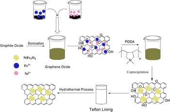
GO was obtained by the modified Hummers method as described elsewhere. Reference Shen, Li, Huang, Long, Li and Ye9 RGO–NiFe2O4 nanocomposites with different RGO contents were synthesized. In a typical synthesis process, 5 mmol (25 mL) solution of iron chloride (FeCl3·6H2O) and 10 mmol (25 mL) solution of nickel chloride (NiCl2·6H2O) were mixed in double distilled deionized water. The above mixture was stirred for 1 h to form part A. On the other hand, 5 mg of GO was added to 20 mL water. The mixture was sonicated for 30 min, followed by high-speed stirring for further 1 h. 4 mL water solution of PDDA (40 wt%) was added to the GO solution to get part B. Subsequently, part A and part B were mixed. The mixture was put into an autoclave and heated at 160 °C for 8 h. After the reduction reaction was finished, the as-synthesized products were isolated by centrifugation, washed with water and ethanol for several times, and dried at 90 °C for 12 h. The whole fabrication process is outlined in Fig. 1. The product is described as RGO-NiFe2O4-1. RGO-NiFe2O4-2, RGO-NiFe2O4-3, and RGO-NiFe2O4-4 were obtained with 10, 20, and 40 mg GO. For comparison, NiFe2O4 nanoparticle was prepared using the same solvothermal method without the addition of GO.
Procedures used in this study for preparation of RGO–NiFe2O4 nanocomposites.

C. Characterization
Fourier transform infrared spectra (FTIR) were recorded on a Nicolet IS10 spectrometer. Solid samples were imbedded in KBr disks. The spectrum was generated, collected 16 times, and corrected for the background noise. Raman spectra were recorded on a Dilor LABRAM-1B multichannel confocal microspectrometer with 514 nm laser excitation. Thermogravimetric analysis (TGA) was conducted in nitrogen atmosphere with a heating rate of 10 °C/min using a Netzsch TG 209F1. Before the tests, all the samples were carefully ground to powders to ensure sufficient diffusion of heat. X-ray diffraction (XRD) powder patterns were taken on D/max-γB diffractometer using Cu Kα radiation. Scanning electron microscopy (SEM) was performed with a Philips XL30 FEG FE-SEM instrument (Eindhoven, Netherlands) at an accelerating voltage of 25 kV. Sample was sputter-coated with gold to improve the contrast. Transmission electron microscopy (TEM) was performed with a JEOL JEM-2100F (Tokyo, Japan). To evaluate the dispersion state of the sheets in the suspensions, atomic force microscopy (AFM) images were obtained using a Multimode Nano4 in the tapping mode. Water bath sonication was performed with a JL-60 DTH sonicator (50 W). The UV-vis diffuse reflectance spectra were conducted on a UV-3600 UV-VIS-NIR spectrophotometer using an integrating sphere accessory.
D. Electrochemical measurements
To prepare the working electrode for the three-electrode system, 70 wt% active material, 20 wt% carbon black as a conductive additive, and 10 wt% polyvinylidene fluoride binder were mixed together and ground thoroughly. The mixture was further dispersed in N-methyl-2-pyrrolidone to form slurry which was pasted on nickel foams. Then the foams were dried at 120 °C for 12 h in a vacuum oven. The loading mass of each electrode was about 5 mg. Electrochemical properties of the electrodes were measured with a conventional three-electrode cell system in 6 M KOH electrolyte. The prepared electrode, platinum foil, and saturated calomel electrode (SCE) were used as the working, counter, and reference electrodes, respectively. Cyclic voltammograms (CV) and electrochemical impedance spectra (EIS) were measured on an electrochemistry workstation (Autolab PGSTAT128N).
E. Photocatalytic activity measurement
Photocatalytic experiments were conducted to photocatalytically degrade methyl orange in water. In a typical procedure, RGO–NiFe2O4 nanocomposite (10 mg) was added into 50 mL of methyl orange solution (10−5 mol/L). The mixtures were stirred moderately with a magnetic stirring apparatus at room temperature for several minutes and then maintained in the dark for 1 h to ensure the adsorption/desorption equilibrium of the organic dye. Then, the solution was exposed to an ultraviolet light lamp (60 W) which was placed 20 cm above the beaker. Samples were withdrawn regularly from the reactor and immediately centrifuged to separate any suspended solid before analysis. The concentration of methyl orange in the solution was monitored by measuring the absorbance of the solution with a UV-vis spectrophotometer. NiFe2O4 was adopted as the reference to compare the photocatalytic activity with that of RGO–NiFe2O4 nanocomposites under the same experimental conditions.
III. RESULTS AND DISCUSSIONS
It is now well known that oxygenation of graphite results in multiform functional groups on the basal planes and at the edge of sheets, which makes GO strongly hydrophilic and easily dispersible in water. These decorated functional groups reduce the interplanar forces and promote complete exfoliation of graphene oxide sheets in aqueous media. During the preparation of RGO–NiFe2O4, it is interesting to find that the addition of PDDA to GO aqueous solution induced an unexpected color change from brown to black. In comparison, no color change was observed for the GO dispersion without adding PDDA. This indicated that the formation of RGO can be attributed to the presence of ordinary polyelectrolyte PDDA. Reference Chen, Wang and Dong12
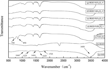
Figure 2 shows FTIR spectra of GO (a), RGO (b), NiFe2O4 (c), and RGO–NiFe2O4 nanocomposites (d–g). IR features of GO indicate the presence of oxygen-containing functional groups on its surface [Fig. 2(a)]. The characteristic bands at 1045, 1225, 1410, 1625, and 1725 cm−1 correspond to the C–O–C stretching vibrations, the C–OH stretching peak, the O–H deformation of the C–OH groups, the C–C stretching mode, and the C–O stretching vibrations of the −COOH group, respectively. The wide band at 3450 cm−1 originates from the absorption of water or O–H groups. Reference Murugan, Muraliganth and Manthiram13 As to RGO, most of the oxygen-containing groups disappeared, while only a little of them still remained. The IR spectra of the NiFe2O4 [Fig. 2(c)] and RGO–NiFe2O4 samples [Figs. 2(d)–2(g)] show small differences in the range from 4000 to 1000 cm−1. In the range from 1000 to 100 cm−1, the IR bands of solids are usually assigned to vibration of ions in the crystal lattice. It can be found that two main broad metal-oxygen bands (at 740 and 890 cm−1) are seen, which are corresponding to intrinsic stretching vibrations of the metal ions at the tetrahedral site. Reference Fu, Jiao and Zhao14
FTIR spectra of GO (a), RGO (b), NiFe2O4 (c), and RGO–NiFe2O4 nanocomposites (d–g).

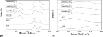
Raman spectroscopy is one of the most sensitive and informative techniques to characterize disorder in sp 2 carbon materials. Reference Goncalves, Marques, Granadeiro, Nogueira, Singh and Gracio15 Figure 3 shows Raman spectra of GO, RGO, NiFe2O4, and RGO–NiFe2O4 composites. Generally, the Raman spectrum of graphene is characterized by three main features. The G peak, which is due to the E2g vibration mode of sp 2 bonded carbon, is usually observed at 1575 cm−1; the D peak, which is arising from the doubly resonant disorder-induced mode, can be found at 1350 cm−1; and the 2D peak at 2700 cm−1, which is a second-order vibration caused by the scattering of phonons at the zone boundary. The ratio of the intensities of the D and G band (I D/I G) can be used to indicate the level of chemical functionalization in a carbon material. High I D/I G ratio usually indicates a high degree of disorder. Reference Xu, Wang, Wan, Lin and Wang16 As shown in Fig. 3, the spectrum of GO shows broadened D-band and the G-band. When GO was chemically converted to RGO, I D/I G ratio clearly increased. This change suggests that the average size of the sp 2 domains is decreased upon reduction of the GO sheets. New graphitic domains are created, which are smaller in size than the ones present in GO before reduction, but more numerous in number. In the spectra of RGO–NiFe2O4 nanocomposites, we can find that the characteristic peaks of NiFe2O4 and RGO still exist. Besides, the position and shape of the 2D peak can be used to distinguish the layer numbers of graphene. Reference Akhavan, Abdolahad, Esfandiar and Mohatashamifar17 It is known that the 2D position of single-layer graphene sheets is usually observed at 2679 cm−1, while the 2D band of multilayer shifts to higher wavenumbers. Reference Liu, Chen and Jiang18 In the spectra of RGO–NiFe2O4 nanocomposites, their 2D bands with a nearly symmetrical shape were centered at 2680 cm−1, suggesting formation of few layer sheets.
Raman spectra of GO, RGO, NiFe2O4, and RGO–NiFe2O4 nanocomposites. (a) 0–2000 cm−1; (b) 2600–2800 cm−1.

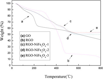
The chemical composition of the as-prepared RGO–NiFe2O4 nanocomposites is also analyzed with TGA. Figure 4 shows the TGA curves of raw graphite, GO, RGO, and RGO–NiFe2O4 nanocomposites. GO shows a relatively poor thermal stability with a rather low onset temperature for pyrolysis of the labile oxygen-containing functional groups over 180–300 °C [Fig. 4(a)]. As to the curve of RGO [Fig. 4(b)], its weight loss before 700 °C is about 85%, which is because of the strongly absorbed PDDA on RGO. Reference Shen, Hu, Shi, Lu, Qin, Li and Ye19 It is known that the weight loss of pure RGO is usually about 5%, thus the PDDA content on prepared RGO is about 80%. It can be seen that the total weight loss greatly decreased after NiFe2O4 nanoparticles were bonded onto RGO. From the TGA curve of RGO-NiFe2O4-1 [Fig. 4(c)], considering the weight loss of RGO, the weight percentage of NiFe2O4 nanoparticles of RGO-NiFe2O4-1 is about 47.1 wt%. As to RGO-NiFe2O4-2 and RGO-NiFe2O4-3, the percentages of NiFe2O4 nanoparticles of them are 49.3 and 52.7 wt%, respectively [Figs. 4(d) and 4(e)].
TGA curves of GO (a), RGO (b), and RGO–NiFe2O4 nanocomposites (c–e).

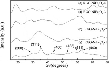
To further investigate the formation of NiFe2O4, powder XRD patterns of RGO–NiFe2O4 nanocomposites are given in Fig. 5. It is found that the RGO–NiFe2O4 nanocomposites prepared with different experimental conditions are similar with each other. Moreover, it reveals the formation of cubic NiFe2O4 as compared with the standard pattern. The characteristic peaks occur at 2θ of 30.42°, 35.77°, 44.22°, 51.47°, 57.32°, and 63.07°, which are marked by their corresponding indices (200), (311), (400), (422), (511), and (440), respectively. No diffraction peaks of other impurities such as
 ${\rm{\alpha }}$
-Fe2O3 or NiO were observed. Besides, the size of the nanoparticles is in the range from 10 to 15 nm, which is calculated using the Scherrer equation. With the support of RGO and the synergetic effect between RGO and NiFe2O4, the lattice constant of the samples may even slightly change (which is consistent with Raman results), thus gradually affect the XRD peaks of RGO–NiFe2O4 nanocomposites. As to the peak below 20° (consistently increasing in peak intensity from pattern (a) to pattern (d), which is corresponding to RGO), it hinted that the whole preparation process could influence the crystallization of the samples and part of the extensive conjugated sp
2 carbon network can be restored.
Reference Shen, Hu, Shi, Lu, Qin, Li and Ye19
XRD pattern of RGO–NiFe2O4 nanocomposites.

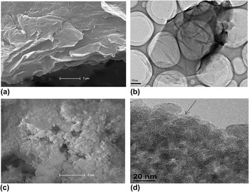
The investigation of the structure of GO and RGO-NiFe2O4-1 had been performed by SEM and TEM. From Fig. 6(a), it can be seen that GO shows flat structure with some wrinkles on the surface and edge. TEM sample of GO [Fig. 6(b)] was prepared by pipetting a few milliliters of dispersions onto holey carbon mesh grids. Large sheets are observed to be situated on the top of the grid, where they resemble silk veil waves, illustrating the flake-like shapes of graphene oxide sheets. The visible graphitic lattices revealed that the GO sheets only consisted of 1–3 graphitic layers. From Figs. 6(c) and 6(d), it can be seen that the NiFe2O4 nanoparticles are distributed on the surface of RGO. The typical NiFe2O4 nanoparticle size is around 10 nm in diameter.
SEM and TEM images of GO (a and b) and RGO-NiFe2O4-1 (c and d).

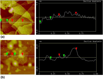
AFM characterization is one of the most direct methods for quantifying the degree of exfoliation after the dispersion of the powder in a solvent. As can be seen in Fig. 7(a), several GO sheets were randomly deposited on the mica substrate. GO consisted of flat sheets with an average thickness of about 1.2 nm. Since graphene oxide sheets are expected to be “thicker” due to the presence of oxygen-containing groups, we can expect that these sheets are uniformly graphene bilayers and trilayers. Figure 7(b) is an AFM image of the RGO-NiFe2O4-1 nanocomposite. It can be seen that the surface of the prepared hybrid material is rough. In addition, it can be seen that small NiFe2O4 nanoparticles with an average height of ∼8 nm are distributed on the surface of RGO sheets. However, agglomeration of the small nanoparticles still exists.
AFM images of GO (a) and RGO-NiFe2O4-1 (b). Image dimensions are 1.0 µm × 1.0 µm.

The above analyses confirmed the successful assembly of NiFe2O4 nanoparticles on the large RGO sheets. We propose the assembly mechanism as follows: 1) in the first step, FeCl3 and NiCl2 were attached on graphene oxide sheets, in which monolayer or bilayer graphene oxide sheets were homogeneously dispersed in water. Therefore, the mixing of RuCl3 and graphene oxide sheets can be considered as at a molecular level. In the second step, graphene oxide sheets were reduced to RGO nanosheets and NiFe2O4 nanoparticles generated simultaneously. This process can ensure the in situ formation of NiFe2O4 nanoparticles and RGO nanosheets simultaneously, with an advantage to prevent serious stacking of RGO nanosheets. 2) By functioning as a “spacer”, the NiFe2O4 nanoparticles increase the distance between the sheets, thereby making both the faces of graphene oxide sheets accessible. Reference Si and Samulski20 The introduction of NiFe2O4 nanoparticles into the dispersion of graphene oxide sheets impedes the formation of a stacked graphitic structure. As a result, the nanoparticle spacers ensure that the high specific surface area as well as other unique properties exhibited by 2D RGO is retained in its dry state. 3) From FTIR analysis, it can be found that small amount of oxygen functionalities still exists. It is reported that the nucleation and growth of nanoparticles depend on the degree of oxygen functionalization at the graphene surface. The nucleation of nanoparticles at the GO surfaces is mainly governed by the presence of oxygen groups at GO. Reference Goncalves, Marques, Granadeiro, Nogueira, Singh and Gracio15 The presence of remaining oxygen containing groups at the RGO surface can provide reactive sites for the nucleation of NiFe2O4 nanoparticles. 4) Positively charged PDDA is first absorbed onto the surface of graphene oxide sheets (negatively charged) via the electrostatic interaction, and then during the reduction of graphene oxide sheets, the adsorbed PDDA is expected to act as a stabilizer to make RGO sheets stable in water.
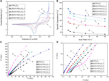
Figure 8(A) shows the CV results of NiFe2O4 and RGO–NiFe2O4 nanocomposites, which were measured in 6 M KOH at potential intervals from 0–0.4 V versus saturated calomel electrode (SCE). The specific capacitances of NiFe2O4, RGO-NiFe2O4-1, RGO-NiFe2O4-2, RGO-NiFe2O4-3, and RGO-NiFe2O4-4 nanocomposite electrodes obtained at 5 mV s−1 are 54.6 F g−1, 110.6 F g−1, 199.8 F g−1, 202 F g−1, and 139.5 F g−1, respectively. Figure 8(B) shows the specific capacitances of NiFe2O4 and RGO–NiFe2O4 nanocomposite electrodes under different scan rates. It is worth noting that the specific capacitances of the RGO–NiFe2O4 nanocomposite electrodes are much larger than that of the NiFe2O4 electrodes at the same scan rate. These results imply that the electrochemical Faradaic reactions of NiFe2O4 in RGO–NiFe2O4 nanocomposites are responsible for the great enhancement of capacitance of the electrode, and RGOs are responsible for the improvement of specific capacitance at high rates. We ascribe this improvement to (1) increase in surface area of the resultant composites and (2) the contribution of double-layer capacitance from RGO and pseudocapacitance from NiFe2O4 nanoparticles. These findings demonstrate the importance and great potential of RGO–NiFe2O4 nanocomposites in the development of high performance energy-storage systems. The electrochemical conductivity behaviors were also investigated by EIS analysis as shown in Figs. 8(C) and 8(D). The impedance plots include a high-frequency component (partial semicircle), a low-frequency component (straight slopping line along the imaginary axis), and a transition zone between two regions. Reference Justin and Rao21 The impedance curve can be explained by an equivalent Randles circuit that includes solution resistance, double layer capacitance, charge transfer resistance, and Warburg impedance. Reference Vijayakumar, Kiruthika Ponnalagi, Nagamuthu and Muralidharan22,Reference Roberts and Slade23 The intercept with the real axis at a very high frequency represents a combined resistance including intrinsic resistance of the active material and the binder in the electrodes, ionic resistance of the electrolyte, and contact resistance at the interface active material and current collector. Moving to lower frequencies, the depressed semicircle is attributable to the interfacial charge transfer resistance occurring at the electrode–electrolyte interface and double layer capacitance. The diameter of this semicircle corresponds to the interfacial charge transfer resistance, which is mainly associated with the Faradaic reactions. The line at a low frequency range called as Warburg impedance is related to the diffusive resistance of the electrolyte, which diffuses into the interior of the electrode pores, and the ion diffuses into the host materials. A near vertical line at a low frequency illustrates that the ion diffusion in the solution and the adsorption of ions onto the electrode surface occur swiftly. The Warburg impedance combined with the charge transfer resistance is often called the Faradaic resistance. As can be seen in Figs. 8(C) and 8(D), interfacial charge transfer resistance of the RGO–NiFe2O4 nanocomposites is much smaller than that of the NiFe2O4 owing to the good conductivity and the large surface area of RGO. RGO also provides easy access and more space for electrolyte diffusion, which corresponds to the small Warburg impedance.
(A) CV curves of NiFe2O4 and RGO–NiFe2O4 nanocomposites measured at 5 mV s−1; (B) The specific capacitance of NiFe2O4 and RGO–NiFe2O4 nanocomposites as a function of scan rate; (C and D) Nyquist plots of NiFe2O4 and RGO–NiFe2O4 nanocomposites.

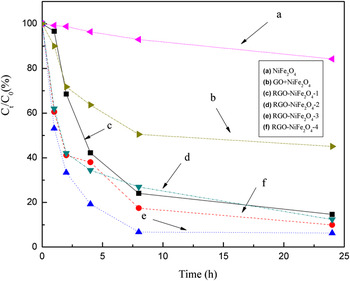
With a primary focus on titanium dioxide (TiO2), which is a durable photocatalyst, photocatalytic oxidation technology has been applied to a variety of problems of environmental interest, especially to water and air purification. Reference Lv, Mao, Wei, Zhang, Xie and Wei24–Reference Ranney, Mansfield, Sun and Schwank27 For example, Lv et al. successfully designed and synthesized pure brookite, anatase, and mixed-phase TiO2 via hydrothermal reaction by adjusting pH values. It was found that mixed-phase TiO2 exhibited the best photocatalytic activity especially when the sample was prepared under pH = 8. Reference Lv, Mao, Wei, Zhang, Xie and Wei24 However, more rational design and modification strategies for photocatalytic materials are still intriguing. Figure 9 presents the methyl orange concentration ratio (C t/C 0) of different samples and those of the direct degradation and adsorption process with irradiation time, respectively. We can find that the methyl orange conversion over the as-prepared catalyst within 24 h of reaction decreased in the order of RGO-NiFe2O4-3> RGO-NiFe2O4-4≈RGO-NiFe2O4-2>RGO-NiFe2O4-1>NiFe2O4. Bench mark experiment was done by simply mixing GO, PDDA, and NiFe2O4, which shows much lower catalytic effect as compared with RGO–NiFe2O4 nanocomposites. In addition, comparing with the commercial P25 photocatalyst (70% degradation in 3 h), Reference Oros-Ruiz, Gómez, López, Hernández-Gordillo, Pedraza-Avella, Moctezuma and Pérez28 RGO-NiFe2O4-3 even shows higher photocatalytic ability. It is known that during photocatalysis, the light absorption, charge transfer and separation are crucial factors. Reference Gao, Wang, Shang and Xu29 RGO may absorb some light and thus cause a light harvesting competition between NiFe2O4 and RGO with the increase of the RGO content, which leads to the photocatalytic performance. Reference Li, Guo, Yu, Ran, Zhang, Yan and Gong30 The enhancement of the photocatalytic performance should be ascribed to the increase of the light absorption intensity and range, and the reduction of electron–hole pair recombination. The role of RGO could act as a macromolecular “photosensitizer” for NiFe2O4, hence making it exhibit higher photoactivity. The photocatalytic process for RGO–NiFe2O4 under UV light irradiation seems to be very similar to the well-known strategy of photosensitization of semiconductors by the matched adsorption of organic dyes. Reference Goncalves, Marques, Granadeiro, Nogueira, Singh and Gracio15 The enhanced photocatalytic activity for RGO–NiFe2O4 composites could be attributed to the ability of RGO to capture and transport electrons, and to promote charge separation. It is known that the higher separation efficiency of electron–hole pairs will enhance photocatalytic activity and results in a large number of holes participated in the photocatalytic process. Reference Fan, Lai, Zhang and Wang31 However, excessive addition of black color RGO will lower the light intensity through the depth of reaction solution, thus decreasing its photoactivity.
Degradation and adsorption curves under ultraviolet irradiation of the samples for methyl orange.

Based on the experimental data, we suspect that the surface structures of RGO–NiFe2O4 may be well responsible for its improved electrochemical and photocatalytic activities. The increase in utilization efficiencies for NiFe2O4 on these RGO supports can be attributed to the increased surface areas and the well-dispersed nature of the prepared RGO. The synergetic effect of RGO and NiFe2O4 makes its electrochemical and photocatalytic properties highly increased.
IV. CONCLUSIONS
In this work, Ni–Fe ions were chemically bonded with negatively charged graphene oxide sheets via simple electrostatic interaction, forming a nanocomposite system under a template-free hydrothermal condition with PDDA. PDDA plays a dual role as a stabilizer and protecting agent in the formation of the RGO–NiFe2O4 nanocomposites. The RGO sheets provide a favorable surface for pinning the NiFe2O4 nanoparticles and prevent their agglomeration. In addition, the RGO sheets not only provide large surface areas for the deposition of NiFe2O4 nanoparticles but also facilitate electrolyte infiltration thereby helping in ion transport redox processes. The strong interactions between individual NiFe2O4 nanoparticles with the RGO sheet surface can help in the efficient collection of capacitance charge from the individual NiFe2O4 nanoparticles along the conductive RGO backbone, thereby enhancing the overall performance of RGO–NiFe2O4 nanocomposites.
ACKNOWLEDGMENTS
We gratefully acknowledge the financial sponsor of National Natural Science Foundation of China (51202034) and “Chen Guang” project (12CG02) supported by Shanghai Municipal Education Commission and Shanghai Education Development Foundation.