Endomyocardial fibrosis, although rare, is the most common cause of restrictive cardiomyopathy. The aetiology is not known. Various biological and infective agents have been associated with the disease and a link to hypereosinophilia has often been cited, but not proven. A familial occurrence and a high incidence among certain ethnic groups suggest a genetic link. The highest disease prevalence has been documented after the first decade of life and the majority of cases reported originate from sub-Saharan Africa, Latin America, Asia, and the Middle East. We present a 22-month-old child with biopsy-proven endomyocardial fibrosis, a right ventricular diverticulum, a positive Myocoplasma pneumoniae immunoglobulin M enzyme-linked immunosorbent assay test, and a familial link to an area where endomyocardial fibrosis is endemic.
Case report
A 22-month-old well-nourished male child was admitted to a tertiary care institution located in central South Africa, altitude, 1800 metres, complaining of a cough for 1 week. He had been admitted to another local hospital 1 month before with a bronchopneumonia. Treatment for tuberculosis was commenced during this admission. He was born at term with a birth weight of 2.8 kilograms in South Africa, to a South African mother and a Mozambican father. Two siblings from the same father were reported to be asymptomatic. The patient had never visited Mozambique.
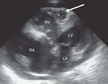
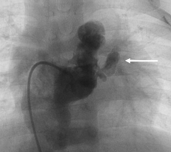
The chest radiograph showed massive cardiomegaly. Echocardiographical examination revealed a large pericardial effusion causing tamponade. Both ventricles appeared small and Doppler interrogation showed poor relaxation of the diastolic filling patterns bilaterally. The apical portion of the right ventricle was absent and retracted. There were patchy areas of echodensity at the base of the tricuspid valve and the interventricular septum with no clear cleavage plane visible. Moderate tricuspid regurgitation was present with a peak instantaneous gradient of 60 millimetres of mercury, indicating the presence of right ventricular hypertension. Both atrio-ventricular valve leaflets appeared normal with good mobility except for mild prolapse of the tip of the anterior mitral valve leaflet resulting in mild mitral regurgitation. The myocardium of the left ventricle was normal and there were no thrombi present nor was spontaneous contrast seen in any of the cardiac chambers. Other notable features were very dilated atria and inferior caval vein, paradoxical motion of the interventricular septum, and a small diverticulum in the area of the interventricular septum communicating with the right ventricle (Fig 1). Pericardiocentesis yielded 500 millilitres of serous pericardial fluid with high protein content. A myocardial biopsy showed fibrotic changes within the endocardium and myocardium associated with an inflammatory cell infiltrate comprising neutrophils, plasma cells, and eosinophils suggestive of endomyocardial fibrosis. An angiogram confirmed a diminutive bipartite right ventricular cavity with an absent apical portion and a small diverticulum (Fig 2). Pressure tracings demonstrated elevated baseline diastolic pressures with raised end-diastolic pressures of 8 millimetres of mercury in the right ventricle and 22 millimetres of mercury in the left ventricle. The peak systolic pressures in the right ventricle and the pulmonary arteries measured 35 millimetres of mercury. The patient was treated with angiotensin-converting enzyme inhibitors and oral steroid therapy. He died suddenly shortly after discharge.
Two-dimensional echocardiogram showing small ventricular cavities, enlarged atria, in particular, the right atrium and the diverticulum (arrow). Patches of echodensity are seen at the base of the tricuspid valve and the interventricular septum. A pericardial effusion is present. RV, right ventricle; RA, right atrium; LV, left ventricle; LA, left atrium.

Right ventricular angiogram showing a small bipartite cavity with absent apical portion and the diverticulum (arrow).

Relevant peripheral blood investigations on admission showed a haemaglobin count of 10.4 grams per litre, white cell count of 11 × 109 per litre, platelet count of 352 × 109 per litre, zero eosinophils, C-reactive protein of 9 milligrams per litre with a normal reference range of 0.0–10.0 milligrams per litre, negative antinuclear antigen, negative human immunodeficiency virus enzyme-linked immunosorbent assay and a negative blood culture. Cardiac enzymes and thyroid function tests were normal. An unexpected finding was a positive mycoplasma pneumoniae immunoglobulin M enzyme-linked immunosorbent assay test.
Pericardial fluid analysis revealed total protein of 34 grams per litre – levels greater than 30 grams per litre are indicative of an exudate and a low adenosine deaminase of 6.6 units per litre – levels greater than 30 units per litre suggest possible tuberculosis. There were no cells present.
Discussion
We believe that this 22-month-old child is one of the youngest patients diagnosed with endomyocardial fibrosis reported in the literature. The youngest patient recorded is an 18-month-old child diagnosed using echocardiography.Reference Saraçlar, Ozer, Oztunç and Celiker1 The highest disease prevalence is reported to be between the ages of 10 and 19 years. An important feature of the patient is the familial link to an area – Mozambique, within the Southern Africa region – where endomyocardial fibrosis has a high prevalence, but had no history of travel to the area himself, largely eliminating possible environmental factors. This association suggests a strong hereditary or genetic predisposition to the disease, which was documented previously.Reference Mocumbi, Ferreira, Sidi and Yacoub2
Endomyocardial fibrosis is the most common cause of restrictive cardiomyopathy worldwide. The majority of cases come from low-lying, humid parts of tropical countries. Reports of endomyocardial fibrosis mostly originate from sub-Saharan Africa, Latin America, South Asia, China, and the Middle East.Reference Mocumbi, Yacoub and Yacoub3 The prognosis is poor.
The impairment of ventricular filling occurs as a result of the deposition of fibrous tissue on the mural and valvar endocardial surfaces, resulting in left, right, or biventricular failure associated with atrio-ventricular valve incompetence. Some of the echocardiographic findings in our patient are similar to those previously documented in the literature.Reference Mocumbi, Yacoub and Yacoub3 The diverticulum of the right ventricle seen at both echocardiography and angiography has not been described before. It may represent an area of weakness of the myocardium caused by an inflammatory or infective process.
The cause and pathogenesis of endomyocardial fibrosis is not well understood. Eosinophilia is the most cited link, but remains unconfirmed. Other cardiomyopathies associated with hypereosinophilia such as Chagas diseaseReference Sepulveda, Liegeard, Wallukat, Levin and Hontebeyrie4 have a myocardial fibrotic stage similar to endomyocardial fibrosis, but lack the characteristic feature of endocardial thickening. The variable association with peripheral blood eosinophilia was highlighted in a study from Uganda, where 60% of echocardiographic cases of endomyocardial fibrosis had mild eosinophilia compared with only 10% of controls.Reference Rutakingirwa, Ziegler, Newton and Freers5 A study from Brazil showed that eosinophils are rarely seen in the endocardium taken from myocardial biopsy specimens.Reference Iglezias, Benvenuti and Calabrese6 Eosinophils contain substances released during degranulation, which are thought to be toxic to the endo- and myocardium resulting in mural thrombosis and fibrosis.Reference Tai, Ackerman, Spry, Dunnette, Olsen and Gleich7 Several other potential insults have been proposed as the primary cause of endocardial fibrosis such as infections, autoimmune diseases, malnutrition, and toxic agents.Reference Mocumbi, Yacoub and Yacoub3 They all, however, remain unproven.
The surprise finding of a positive mycoplasma pneumoniae immunoglobulin M enzyme-linked immunosorbent assay in our patient is of interest. Associations between the mycoplasma pneumoniae organism and the heart, such as myopericarditis, have been reported.Reference Duthoit, Ou, Sidi and Bonnet8 An infective trigger for the onset of endomyocardial fibrosis in a genetically susceptible individual is likely. There is evidence that cross reactivity of antibodies against the C-terminal sequences of several animal, plant, and protozoal ribosomal P proteins with cardiac tissue may mediate endomyocardial fibrosis in a manner similar to that of the C-terminal of Trypanosoma cruzi in Chagas disease.Reference Wayengera9
The underlying aetiology of endomyocardial fibrosis remains unknown which makes prevention of this devastating condition difficult. Current treatments are mainly palliative and include anti-inflammatory medications such as steroids.Reference Mocumbi, Yacoub and Yacoub3
Surgery also has a role in the management of patients. Reduction of preload on the right ventricle by means of a total cavopulmonary connection,Reference Kumar, Prabhakar, Fawzy, Al Haless and Duran10 stripping of the fibrotic endomyocardium, valve repair, or replacement and, in more advanced cases, cardiac transplantation have all offered some benefit.Reference Mocumbi, Yacoub and Yacoub3
Conclusion
This case presentation suggests a possible genetic link in the aetiology of endomyocardial fibrosis, which needs further investigation. An infective trigger cannot be excluded. Epidemiological studies that include younger children in endemic areas may lead to the discovery of affected patients at an earlier age and allow for the investigation of possible infective aetiologies, which occur early and may result in endomyocardial fibrosis.
Acknowledgement
The authors thank Dr Kim Blair for preparing and providing the histopathological slides. Department of Pathology, University of the Witwatersrand, Johannesburg, South Africa. The authors declare no conflict of interest.