Case
Arrhythmias are a major reason for hospitalisation and a frequent cause of morbidity and mortality in patients with grown-up congenital heart disease.Reference Baumgartner, Bohoeffer and De Groot 1 Apart from arrhythmias caused by anatomic malformations and/or scar-related arrhythmias due to surgical corrections, all known forms of supraventricular tachycardia can occur in patients with grown-up congenital heart disease. Their occurrence increased significantly over the past few years because of an improved life expectancy.
We report treatment of a 27-year-old patient with tricuspid and pulmonary atresia who received a Waterston–Cooley shunt as an infant and was now admitted to our hospital with supraventricular tachycardia, which was symptomatic for a period of 5 years. Tachycardia normally occurred at rest and lasted up to 30 minutes. Unfortunately, an ECG-documentation was not available, but the frequency of symptoms was significantly increasing over the past few months.
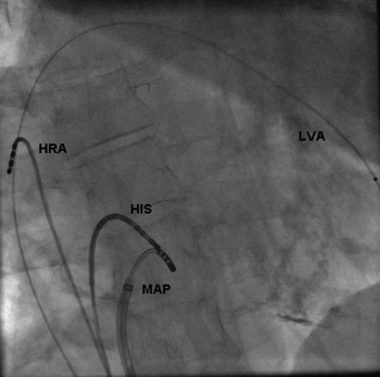
During electrophysiologic study, the tricuspid valve could not be passed with any catheter. Therefore, a ventricular catheter was inserted into the left ventricle through a persisting atrial septal defect. A diagnostic catheter was introduced into the right atrium. Owing to anatomic malformations, a coronary sinus-catheter could not be positioned. The HIS-catheter had to be placed in a more superior and less septal position to record an adequate HIS-signal (Fig 1).
Catheter positions. HIS=HIS-bundle; HRA=high right atrium; LVA=left ventricular apex; MAP=ablation catheter in slow pathway region.

During programmed atrial stimulation, an A-H-jump of about 70 ms was reproducibly induced. Programmed ventricular stimulation demonstrated decremental V-A-conduction. Finally, supraventricular tachycardia with a stable cycle length of 290 ms was reproducibly induced by programmed atrial stimulation after intravenous application of orciprenaline. The earliest atrial signal was recorded in the HIS-region and additional ventricular entrainment led to a characteristic “VAV”-sequence, which confirmed the diagnosis of AV-nodal re-entrant tachycardia and excluded focal atrial tachycardia. Tachycardia was terminated by ventricular pacing.
After mapping of the HIS-region with the ablation catheter, four radiofrequency impulses were delivered. During radiofrequency impulses, junctional beats were regularly observed. An adequate distance between HIS-catheter and ablation catheter ensured safety of the procedure (Fig 1). Subsequently, neither AV-nodal re-entrant tachycardia nor any other supraventricular or ventricular tachycardia were inducible after subsequent application of orciprenaline and intensive programmed supraventricular and ventricular stimulation. The patient was free of symptoms during a clinical follow-up at 6 months.
Conclusion
Patients with tricuspid atresia present an atypical AV-node anatomy. Nonetheless, frequently occurring arrhythmias can be found in these patients. Despite the described anatomic malformation, AV-nodal re-entrant tachycardia may occur and can be safely treated by radiofrequency ablation at the shown atypical slow pathway region.
Financial Support
This research received no specific grant from any funding agency, commercial or not-for-profit sectors.
Conflicts of Interest
None.