Real-time three-dimensional echocardiography is emerging as a valuable clinical tool in the setting of congenital cardiac disease, as it gives new insight into the dynamic changes of intra-cardiac structures during the cardiac cycle.Reference Sufeng, Weinert, Thiele and Lang1–Reference Li and Sanders3 It provides the clinician with immediate three-dimensional images, which can be manipulated and cut in different planes to allow best visualisation of the area of interest. Three-dimensional data sets take seconds to acquire, and can be processed away from the bedside. Two-dimensional images can be reconstructed from the three-dimensional images in multiple planes, independent of the site of acquisition of the data set.
The mitral valve is a complex structure, the true shape and function of which are difficult to visualise accurately in only two dimensions. In adults, the mitral valvar annulus has been demonstrated to decrease in size during systole, and also to change its shape in order to best perform its function without impinging on the left ventricular outflow tract.Reference Kaplan, Basheim and Sheehan4–Reference Glasson, Komeda and Daughters8 Located at the base of the heart, the mitral valve cannot remain planar throughout the cardiac cycle. The base of the left ventricle decreases its circumference during systole, but since the attachments of the leaflets of the valve do not decrease in size, the shape of the valve must change in order to accommodate the ventricular contraction.Reference Ormiston, Shah, Tei and Wong9 These observations have not been reproduced in children. Contrarily, recently it has been suggested that the mitral valve behaves rather differently in children, and decreases its area in diastole.Reference Nii, Roman, Macgowan and Smallhorn10 The inter-relation between mitral annular function and left ventricular function in both health and pathological states remains poorly understood.
Given the complex geometry of the mitral valve, and its dynamic nature, real-time three-dimensional echocardiography is the ideal tool with which to investigate the alterations in valvar dimensions during the cardiac cycle, and to assess mitral regurgitation.Reference Schwartz, Cao, Azevedo and Pandian11–Reference Breburda, Griffin, Pu, Rodriguez, Cosgrove and Thomas13 Using real-time three-dimensional echocardiography, we studied the motion of the mitral valvar annulus in systole and diastole in normal children, and analysed the effect of varying degrees of regurgitation on the annular function.
Materials and methods
A commercial real-time three-dimensional imaging system Philips Sonos 7500 or IE33 (Philips Co, Netherlands) with a 3–5 MHZ matrix phased array transducer was used to acquire real-time three-dimensional echocardiographic data. All patients underwent a standard cross-sectional echocardiographic assessment in addition, using the same system.
Cross-sectional and three-dimensional echocardiography data was acquired for 11 subjects with mitral regurgitation with normal left ventricular function, and 20 age-matched controls. All recordings were taken with the patients breathing normally. Full volume loops of the mitral valve were acquired in the views in which there was clear valvar definition.
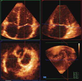
Stored full volume data sets were assessed off-line by two investigators blinded to clinical and cross-sectional echocardiographic findings, using Q lab software version 4.1. In the multiplanar review mode of the software, the mitral valvar annulus was transected in three planes, specifically 2 long-axis planes perpendicular to each other, and one short axis plane (Fig. 1). These three planes were used accurately to define the hinge points of the annulus, choosing a frame in mid-systole and another frame in late diastole. The frame corresponding to the phases of the cardiac cycles were chosen with reference to the electrocardiogram and the motion of the cardiac structures. The mid-systolic frame was at the end of the T wave with the mitral valve closed, and the late-diastolic frame was just prior to the QRS complex with the mitral valve open, before atrial systole.
The 3-D data set (bottom right panel) is opened in the multiplanar review mode of the Q lab software. The mitral valvar annulus is transected in three planes: two long-axis planes perpendicular to each other (green and red boxes), and one short axis plane (blue box).

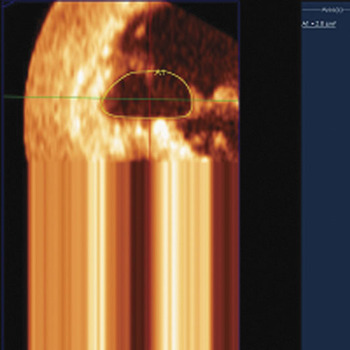
The annular area was then traced around, and the area calculated by the software (Fig. 2). When the hinge point was not clearly definable, the plane of interrogation was moved along the presumed border to ascertain the exact point of attachment of the valve using the multiplanar review mode.
Tracing of mitral valvar annulus area in diastole. The full volume loop has been opened in the multiplanar review mode of Q lab, and the plane displayed is the short axis view of the annulus. The annulus is traced around manually, and the software calculates the area.

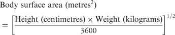
We measured the descent of the annulus first by establishing the level of the annulus at the hinge point in the diastolic frame. This was marked, followed by marking the hinge point in the endsystolic frame. The distance between the two was measured using the measuring tools provided, and was taken as the annular descent. For the purpose of this study, the diastolic length of the left ventricle was considered to be the distance from the mid-point of the annulus to the endocardial apex. All valves were assessed by 2 blinded observers, with 10 randomly selected valves being used to check intra-observer variability. The values obtained were indexed to body surface area in both groups, calculating body surface area using the Mosteller formula:Reference Mosteller14
Patients were divided into sub-groups of mild, moderate or severe mitral regurgitation, depending on the degree of incompetence, which was assessed semi-quantitatively using a clinical system of scoring severity that included the cross-sectional echocardiographic findings, symptomatology, need for surgery, and radiographic findings by clinical symptoms the patients were scored as mild, moderate, and severe, with values from 1 to 3. If surgery was required for mitral regurgitation, a score of 3 was assigned. The cross-sectional echocardiogram was visually assessed by an individual observer and graded as mild, moderate, and severe, and scored in comparable fashion. The chest radiograph was reviewed by an independent observer and again graded from 1 to 3 for mild, moderate, and severe findings based on the degree of pulmonary venous hypertension. We summed these scores, providing categories of mild regurgitation for those scoring zero to 3, moderate regurgitation for 4 to 6, and severe regurgitation for those scoring more than 7. Additionally, we used the indexed orificial area of regurgitation, which is an index measured on real-time three-dimensional echocardiography, developed in our echocardiography laboratory. This index is derived by measuring the regurgitant orificial area by visualisation and measurement of the area of the vena contracta of the regurgitant jet on multiplanar review of three-dimensional colour Doppler full volume data sets. This area is then indexed to the body surface area, and the resultant value can be used as a clinical tool to assess the severity of mitral regurgitation. We have found this to be a measurement with extremely good reproducibility, even in inexperienced hands, and one which correlates well with clinical symptoms.
Our study received approval from the Local Research Ethics Committee, and complied with the Declaration of Helsinki.
Statistical calculations
Statistical calculations were performed using SPSS version 14. Continuous variables were described as the mean plus or minus one standard deviation, with 95% confidence intervals. Inter-group comparisons were carried out using the Mann Whitney U test for nonparametric variables, and Cronbach’s alpha test for inter and intra-observer reliability.
Results
Change in mitral valvar annular area in normal children
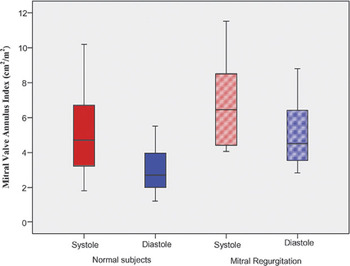
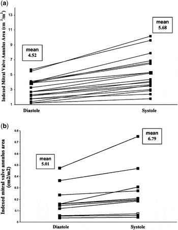
There were 20 normal subjects, 7 female, with a mean age 8.48 plus or minus 5.7 years. Mean body surface area was 1.036 plus or minus 0.50. The mitral valvar annular area indexed to body surface area for each patient increased in systole and decreased in diastole, with a systolic index of 5.28 plus or minus 1.68 centimetres2/metres2, and a diastolic index of 3.05 plus or minus 0.90 centimetres2/metres2 (p is less than 0.0001). The index changed from systole to diastole in normal hearts by a mean of 41.13 plus or minus 9% of the systolic area (Table 1, Fig. 3).
Mitral Valve Annulus Areas.

Sub-Group Analysis*: To detect the effect of severity of MR on the change in annular dimension, only patients with moderate or severe mitral regurgitation were included in a group, and compared to a group comprising of the normal subjects and the patients with mild mitral regurgitation
MVA: mitral valve annulus area; BSA: body surface area. Mitral valve excursion is expressed as percentage of diastolic length of left ventricle
Significant p values are given in bold type
Mitral valvar annular area indexed to body surface area is shown in systole (red) and diastole (blue) for normal subjects (solid colour), and patients with mitral regurgitation (hatched).

Change in area in patients with valvar regurgitation
We assessed 11 patients, all with mitral valvar regurgitation and normal left ventricular function as judged by an experienced clinician. The mean age was 6.52 plus or minus 5.3 years, and mean body surface area 0.92 plus or minus 0.48. The patients were well matched with their controls, with no significant difference between the age or body surface area. The degree of regurgitation was severe in 3, moderate in 4, and mild in 4 patients. This group included 1 patient with rheumatic heart disease, 2 patients with mitral valvar prolapse, and 3 patients with complex congenital heart disease. The other 5 patients had isolated mitral regurgitation.
The indexed annular area in systole was larger in those with mitral regurgitation when compared to normal subjects, with a mean of 6.79 plus or minus 2.55 centimetres2/metres2 versus 5.28 plus or minus 1.68 centimetres2/metres2, but this did not reach statistical significance (p equals 0.072). The indexed valvar area in diastole was significantly larger in patients with regurgitation, with a mean of 5.01 plus or minus 1.78 cm2/m2 as opposed to 3.05 plus or minus 0.90 cm2/m2 in normal subjects (p equals 0.003). The difference between systolic and diastolic areas was expressed as a percentage of the systolic area. This proportional change of indexed area from systole to diastole showed a trend towards being smaller in patients with regurgitation, at 24.8% plus or minus 11.6%, but this did not reach statistical significance (p equals 0.249, Fig. 4).
Mitral valvar annular area indexed to body surface area is shown in systole and diastole for normal subjects (a) and patients with mitral regurgitation (b).

Valvar descent
In normal hearts, the descent in systole was 8.7 plus or minus 2.4 millimetres, compared to 7.3 plus or minus 3.2 mm millimetres in patients with regurgitation. Expressed as a percentage of left ventricular diastolic length, this is 17.04 plus or minus 3.16% for normal hearts, and 13.95 plus or minus 4.53% for patients with valvar regurgitation, which is not significantly different (p equals 0.70).
Analysis of subgroups
To detect the effect of severity of regurgitation on the change in annular dimension, only patients with moderate or severe regurgitation were included in a group, and compared to another group comprising of the normal subjects and those with mild regurgitation. The systolic mean indexed annular area for the normal subjects and those with mild regurgitation was 5.29 plus or minus 2.36 centimetres2/metres2, as opposed to 7.29 plus or minus 2.45 centimetres2/metres2 for those with moderate and severe regurgitation (p = 0.043). The mean indexed diastolic annular area for the normal subjects and those with mild regurgitation was 3.34 plus or minus 1.54 centimetres2/metres2, compared to 5.16 plus or minus 1.99 centimetres2/metres2 for those with moderate and severe regurgitation (p equals 0.014). The percentage change in indexed annular area from systole to diastole, therefore, is blunted, being 36.52 plus or minus 12.22 centimetres2/metres2 in the normal subjects and those with mild regurgitation as compared to 29.60 plus or minus 9.88 centimetres2/metres2 in those with moderate and severe regurgitation (p equals 0.96).
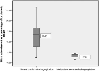
Between these subgroups, the difference in valvar excursion was significant, at 15.84 plus or minus 4.23% versus 12.78 plus or minus 5.10% for patients with moderate and severe regurgitation (p equals 0.012, Fig. 5).
Mitral valve descent is expressed as a percentage of left ventricular diastolic length, and shown for normal subjects and all patients with mitral regurgitation.

Inter and intra-observer reliability
There was very good reliability between the two observers, and for the same observer after a delay of 2 months. Inter-observer reliability was 0.989 for measurement of annular area in systole, and 0.977 in diastole, measured on the same data set. Intra-observer reliability was 0.998 for annular area in systole, and 0.994 in diastole, again measured on the same data set.
Discussion
Our study aids understanding of mitral valvar annular dynamics in children. Studies using cross-sectional transthoracic echocardiography, as well as three-dimensional techniques, have demonstrated the mitral annulus to decrease in systole in adults.Reference Levine, Handschumacher and Sanfilippo7, Reference Ormiston, Shah, Tei and Wong9 It is clear, however, that the geometric changes of the mitral annulus during the cardiac cycle are not straightforward, and are still under discussion. Using a three-dimensional echocardiographic technique, Pai and colleaguesReference Pai, Tanimoto, Jintapakorn, Azevedo, Pandian and Shah15 reported that the mitral annulus in normal adults had its largest area at end-systole, and was smallest at end-diastole. In contrast to the bulk of the available data in adults,Reference Nii, Roman, Macgowan and Smallhorn10 our study corroborates the finding that the mitral annular area, in children, decreases in diastole and increases in systole.
Regurgitation through the mitral valve causes annular dilation, and depresses dynamic annular function. Its effects on the cardiac cycle and on annular motion are unclear. Our study gives further insight into the complexity of mitral valve annular motion in the presence of volume loading of the left ventricle. The dynamic function of the mitral valve is complex and heterogeneous.Reference Salgo, Gorman and Gorman16 The annulus is “saddle-shaped”, and hence is not a simple ellipse. In adults, annular motion has been shown to be non-uniform across the whole of the valve during the cardiac cycle, with not all segments contracting equally. The largest contribution to reduction of the annulus in diastole is from the contraction of its inferior part, supporting the mural leaflet to the valve, with less contribution from the superior component. Part of the antero-superior annulus, that in continuity with the leaflets of the mitral valve, increases in size in systole.Reference Tsakiris, Von Bernuty, Rastelli, Bourgeois, Titus and Wood17, Reference Toumanidis, Sideris, Papamichael and Moulopoulos18 This has a sound physiological reasoning, as it allows unrestricted forward flow from a contracting ventricle through the outflow tract. Differential contraction of regions of the annulus may be the cause of the difference in changes of the valvar area seen between adults and children.
Mitral regurgitation also causes dilation of the left atrium, concomitant with dilation of the annulus. This distortion of the annulus affects the three-dimensional shape of the valve, and also the forces applied to the valve by the papillary muscles. The alteration in dynamic valvar function will be the net result of these interactions. Our results suggest that descent of the annulus is decreased in patients with mitral regurgitation. Previous studies have shown a relationship between mitral annular motion and left ventricular function.Reference Simonson and Schiller19 It can be seen that mitral regurgitation causing abnormal mitral annular motion affects the geometry of the left ventricle and thereby its function. The leaflets of the aortic and mitral valves are in fibrous continuity, and so aortic valvar dynamic function must also be considered to have an effect on mitral annuar function,Reference Timek, Green and Tibayan20 albeit that this effect has not, as far as we are aware, been studied in children. Measurements of left ventricular function, such as estimations of fractional shortening, are observed to increase in mitral regurgitation, but we have found that annular descent is decreased in these patients. This may be a true reflection of occult left ventricular dysfunction in those with mitral regurgitation. Previous studies in adults demonstrate the decreased alteration in size of the mitral annulus during the cardiac cycle in patients with mitral regurgitation,Reference Kaplan, Basheim and Sheehan4, Reference Glasson, Komeda and Daughters8, Reference Gorman, McCaughan and Ratcliffe21, Reference Otsuji, Handschumacher and Schwammenthal22 although in these studies, the smallest annular area is reported to be seen during systole. Kaplan and colleaguesReference Kaplan, Basheim and Sheehan4 demonstrated this lack of systolic reduction in area to be due to decreased accentuation of the saddle shape adopted by the valve. Our preliminary results suggest the degree of blunting of the change in annular area to be related to the degree of mitral regurgitation. Our cohort of patients, however, was too small to allow meaningful analysis in those with mild, moderate, and severe regurgitation.
Why does the annulus of the mitral valve reduce in size in diastole in children? The mitral valve lies at the crux of the heart, and therefore its function and shape are affected by all the surrounding structures, in particular the left atrium and left ventricle, but also the right heart and the tricuspid valve.Reference Nii, Roman, Macgowan and Smallhorn10 Hearts are more compliant in children than adults, and it may be this that produces the different behaviour of the annulus.
Additional variability arises in paediatric practice from the high proportion of patients with concomitant lesions, as mitral regurgitation is rarely an isolated problem. The presence of concurrent abnormalities, which also have haemodynamic and anatomical effects, interacts with the alterations in geometry of the mitral valve, and the quantification of mitral regurgitation.
In this study, we have only looked at the mitral valvar annulus in systole and diastole, and not at changes in the intervening phases of the cardiac cycle. More detailed analysis of the whole cardiac cycle may provide better understanding of the changes occurring. In addition, we have looked at the annulus only as a two-dimensional area, failing to take into account the three-dimensional configuration of its saddle shape. Our method of study, nonetheless, using multiplanar review, allows us very accurately to define the valvar hingepoints. The measured area, therefore, is an accurate depiction of annular area. Our study was done in patients with normal ventricular function, as assessed by cross-sectional echocardiography. Patients with depressed ventricular function, including those with regional abnormalities of wall motion, should also be studied by this method, as this may provide further insights into the forces acting on the mitral valve. Further investigation needs to be done in children into the geometry of the valve and its supporting structures to elucidate the cause of the differences in behaviour from valves in adults, and real-time three-dimensional echocardiography will be a valuable tool in this quest. Further study should include examination of the area of the valvar annulus at each point throughout the cardiac cycle, and segmental analysis of the valve to determine which regions contribute to the changes in area. Our study is a small pilot investigation, and we have not been able to divide our cohort into sub-groups according to age or body size. A larger study involving greater numbers will permit elucidation of the transition from the pattern of change seen during childhood to that observed in adults, and the age or body size at which this occurs.
Measurement of area using three-dimensional images has been shown to have less variability between observers than measurement using cross-sectional echocardiography.Reference Binder, Rosehhek, Porenta, Maurer and Baumgartner23 This probably reflects the greater accuracy of the measurements achieved on three-dimensional echocardiography. Measurements are quick and relatively simple to perform, and can be done off-line, which keeps the time required for examination short, making real-time three-dimensional echocardiography ideal in paediatric practice.
Conclusion
Real-time three-dimensional echocardiography provides a simple and accurate method of assessing the mitral valve in children in both normal and pathological states. The area of the annulus in children reduces in size in diastole, and therefore does not behave in the same manner as the adult mitral valve. Mitral regurgitation blunts the dynamic function of the annulus.