INTRODUCTION
Haemonchus contortus is a parasitic nematode which infects the abomasum of small ruminants, notably sheep and goats. The parasite is a voracious blood feeder and the blood loss associated with infection can cause severe anaemia, loss of condition and death of the host in severe cases (Symons and Steel, 1978). Currently, the strategic use of anthelmintic drugs, together with pasture management practices, is the main method of control available, but the emergence of anthelmintic-resistant populations of this and other species of parasite means that these strategies may not be sustainable in the future (Waller et al. 1996; van Wyk et al. 1997). As an alternative to chemotherapy, the development of effective vaccines against helminth infections is being pursued. A major area of focus for vaccine development has been the parasite gut, targeting so-called hidden or covert antigens, that is antigens to which the host does not normally mount an immune response over the course of a natural infection, but which are accessible to antibodies ingested by the parasite, (reviewed by Newton and Munn, 1999). One of the most promising groups of antigens of this type is the Haemonchus galactose-containing glycoprotein complex (H-gal-GP), localized to the luminal surface of the gut of the parasite (Smith et al. 1994, 1999).
Vaccination with native H-gal-GP confers substantial protection against challenge infection with, typically, a 70% reduction in worm burden and a >90% reduction in the number of nematode eggs excreted in faeces, when compared with control animals (Smith et al. 1999), making this one of the most efficacious anti-nematode antigens reported for any host to date.
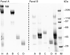
The H-gal-GP complex has a characteristic profile on non-reducing SDS-PAGE (Fig. 1) with 4 main protein zones designated A, B, C and D of 233, 172, 40 and 31 kDa, respectively (Smith et al. 1999). The complex contains at least 2 types of proteolytic enzyme, namely 2 pepsin-like aspartyl proteases associated with the 40 kDa zone C (Longbottom et al. 1997; Smith et al. 2003b) and a family of at least 4 putative zinc metalloproteases associated only with the 233 and 172 kDa zones, A and B (Redmond et al. 1997; Smith et al. 1999). Although these MEPs cannot be separated from the H-gal-GP complex under native conditions, various combinations of them have been evaluated in immunization/challenge trials in sheep. A combination of all 4, separated from the rest of the complex by gel filtration in 8 M urea, significantly reduced H. contortus egg counts by 45–50% (Smith et al. 2003a), whereas, MEP3 alone or MEPs 1, 2 and 4 in combination, electro-eluted from the complex following SDS gel electrophoresis, each reduced egg counts by some 33% (Smith et al. 2003a). Thus the MEPs are clearly important vaccine candidates and a greater understanding of their structure and function will be invaluable in the development of recombinant vaccine antigens. The cloning and characterization of H. contortus MEP1 has been reported previously (Redmond et al. 1997). In the present study we describe MEPs 2, 3 and 4 and compare the structures of the individual members of this family, both to each other and to similar enzymes from other nematode species.

Fig. 1. Separation of H-gal-GP complex by 2-stage 1D SDS-PAGE, Published previously in Smith et al. (1999); figure reproduced with permission. H-gal-GP (Panel A, lane H) was separated under non-reducing conditions into 4 zones designated A–D. Each zone was excised, electro-eluted and re-electrophoresed separately. Lanes A–D in Panel A show these zones under non-reducing conditions; lanes A–D in Panel B show the same zones under reducing conditions. In Panel B lane H contained reduced H-gal-GP, while lane M contained molecular weight marker proteins.
MATERIALS AND METHODS
Construction and immunoscreening of a cDNA library
A cDNA library from 11-day H. contortus was constructed, as described previously (Skuce et al. 1999), in UniZAP-XR (Stratagene, UK) according to the manufacturer's instructions. An aliquot of the amplified library was screened with a pooled sheep antiserum raised against H-gal-GP (Smith et al. 1994). Immuno-positive clones were plaque-purified and subsequently re-screened with sheep antiserum raised against the 172 or 233 kDa zone components of H-gal-GP (Smith et al. 1999).
Characterization of clones
Clones that were immunopositive with both the anti-172 and anti-233 kDa zone sera were subjected to in vivo excision using ExAssist® helper phage (Stratagene), according to the manufacturer's instructions. Plasmid DNA was subsequently isolated using a Wizard™ DNA purification kit (Promega). To characterize the immunopositive clones, insert DNA was PCR-amplified using the T3 and T7 pBluescript vector primers. The PCR products were fractionated on a 0·8% (w/v) agarose gel and transferred to a Hybond N+™ nylon membrane (Amersham) under alkaline conditions, in accordance with the manufacturer's instructions. Blots were probed with an MEP1 gene-specific probe, prepared as described previously (Redmond et al. 1997). Hybridization was performed at 42 °C overnight in DIG Easy Hyb buffer® (Boehringer Mannheim). Membranes were subsequently washed in conditions of moderate (0·5×SSC, 0·1% SDS, 42 °C) or high (0·1×SSC, 0·1% SDS, 68 °C) stringency. Blots were developed using a DIG nucleic acid detection kit (Boehringer Mannheim), used according to the manufacturer's instructions. Clones of interest were sequenced on an Applied Biosystems 377 automated sequence analyser (The Advanced Biotechnology Centre, Imperial College School of Medicine, London, UK).
Analysis of transcripts by RT-PCR in different developmental stages
cDNA (10 ng) from different life-cycle stages of H. contortus, prepared as described previously (Redmond et al. 1997), was used as a template in the reverse transcription polymerase chain reactions (RT-PCR) using gene-specific primers. The PCR conditions employed were: initial denaturation for 5 min at 94 °C, followed by 30 cycles of 94 °C for 1 min, 55 °C for 1 min, 72 °C for 3 min, with a final 7 min extension at 72 °C. The integrity of the cDNA transcripts was assessed by PCR amplification (same conditions) of the constitutively expressed H. contortus extracellular superoxide dismutase (SODe) gene (Liddell and Knox, 1998).
PCR amplification of mep2 and mep4
Oligonucleotide primers were designed based on the partial sequences for mep2 and mep4 held in public databases (NCBI Accession numbers AF080117.1 and AF132519.1, respectively) to amplify the 5′- and 3′-ends of each gene when used in conjunction with vector-specific primers and using an aliquot of the H. contortus Day 11 mixed sex adult cDNA library as a template.
Full-length cDNAs representing mep2 and mep4 were amplified by reverse transcription from total RNA extracted from Day 22 adult H. contortus using oligonucleotide primers designed to the putative 5′- ends of the sequences and to the region flanking the putative termination codons in their open reading frames (ORFs). Reverse transcription and cDNA amplification were performed using the SuperScript™ One-Step RT-PCR System (Invitrogen), in accordance with the manufacturer's instructions. The cDNAs representing mep2 and mep4 reverse transcription products generated in this way were ligated into pGEM-T Easy™ vector (Promega), transformed into JM109 competent cells and the resultant plasmids sequenced. Sequence was generated from the entire length of sense and antisense strands of each of 3 independent clones from each transformation, using vector- and gene-specific primers.
All sequences representing each mep were aligned using the CAP EST assembler (maintained at Istituto FIRC di Oncologia Molecolare, Milan http://bio.ifom firc.it/ASSEMBLY/assemble.html), employing a minimum sequence overlap cut-off of 30 bases and an identity cut-off of 98%. Contiguous sequences generated in this way were compared with those in public databases, including the GenBank™ non-redundant database using the Basic Local Alignment Search Tool (BLASTn and BLASTx) program from the National Center for Biotechnology Information. Details regarding all primers used may be obtained from the corresponding author.
Purification of native H-gal-GP, gel electrophoresis and mass-spectrometry analysis
H-gal-GP was prepared from Triton X-100 extracts of H. contortus gut membranes by lectin affinity chromatography on peanut lectin-agarose, as described previously (Smith et al. 1994, 2000). For one-dimensional SDS-PAGE, 10 μg of H-gal-GP was separated on 4–15% gradient gels (BioRad). For the 2-stage one-dimensional procedure, non-reduced H-gal-GP was first fractionated by SDS-PAGE with Coomassie Blue in the cathode buffer. Subsequently, gel slices containing each band were excised and electro-eluted, according to the methods of von Jagow and Shägger (1994). The electro-eluted proteins were then resolved by SDS-PAGE under reducing conditions. For two-dimensional PAGE, samples (~400 μg) were treated using the 2D Clean Up™ kit (Amersham Biosciences) and re-solubilized in a buffer containing 7 M urea, 2 M thiourea, 2% (w/v) ASB14 detergent before loading on 11 cm immobilized pH gradient (IPG) strips (pI 3–10 linear, Amersham Biosciences) to perform the isoelectric focusing. The strips were focused for ~36000 Vh and subsequently, washed for 15 min in SDS equilibration buffer (50 mM Tris-HCl, pH 8·8, 6 M urea, 30% (w/v) glycerol, 2% (w/v) SDS, bromophenol blue). Electrophoresis in the second dimension was carried out in 12·5% pre-cast gels (Amersham Biosciences) for 90 min at 600 V.
The mass spectrometry analysis was performed as reported previously (Jensen et al. 1998). In short, protein spots were ‘in-gel digested’ using trypsin, subsequently purified and analysed by MALDI-TOF mass spectrometry (PerSeptive Biosystems, Voyager DE PRO). The Mascot™ search engine (MatrixScience.com) was used to analyse the mass spectrometry data and to assign protein identifications to the spots.
Molecular weight determination of H-gal-GP components
The molecular weights and relative abundance of the components of H-gal-GP were determined by analysing scanned gel images with Phoretics 1D™ software version 2003.02 (Non-Linear Dynamics Ltd). Four individual 1D SDS-PAGE gels, each containing a separate preparation of H-gal-GP were run under non-reducing conditions. After staining with Coomassie blue, the gels were dried on to 3 MM paper (Whatman) and recorded using an Epson Perfection 1670™ desktop scanner (Seiko Epson Corporation), at a resolution of 300 dots per inch (dpi). Molecular weights were estimated by comparison of the relative mobility of the components of interest with a series of wide-range molecular weight standards (Sigma).
Prediction of trans-membrane domains, signal peptides and post-translational modifications
The presence of putative trans-membrane domains or signal peptides within the H. contortus MEP sequences was determined using the Phobius algorithm (Kall et al. 2004), which discriminates between proteins with trans-membrane topology and those with signal peptides. Inferred amino acid sequences were also analysed with the NetNGlyc and NetOGlyc software programs (www.expasy.org) to identify putative N-linked and O-linked glycosylation sites, respectively.
N-terminal amino acid sequencing
Amino acid sequencing was carried using standard Edman degradation protocols run on a PE Applied Biosystems 494 LC Protein Sequencer.
MEP activity assay
The endopeptidase activity of protein preparations containing putative MEPs was measured in a two-stage assay utilizing the fluorogenic substrate Glu-Ala-Ala-Phe-4 methoxy-2-naphthylamide (4-MNA, Sigma) (Erdos et al. 1989). Metalloendopeptidase activity in the sample cleaves the substrate on the amino side of the hydrophobic phenylalanine residue producing Phe-4-MNA. After an initial incubation (2 h at 37 °C), of the test sample with substrate, the MEP activity was stopped by the addition of 3 μmol phosphoramidon, a potent MEP inhibitor. The second stage of the assay involved the addition of 72 mU of leucine aminopeptidase (Sigma) and incubation for a further 1 h at 37 °C. Leucine aminopeptidase cleaves the Phe-4-MNA bond, liberating 4-MNA, which was detected on a Perkin Elmer LS50B Luminescence spectrometer, with an excitation wavelength of 340 nm and an emission wavelength of 425 nm. Samples were assayed for the pH optimum over the pH range 5·2–9·0. The reactions at pH 5·2–7·2 were buffered with 100 mM morpholinoethanesulphonic acid (MES) and those from pH 7·2–9·0 were buffered with 100 mM Tris/HCl. MEP activity was expressed as pmol 4-MNA/μg protein/h, calculated from a standard curve prepared from 4-MNA with a concentration range of 1 to 32 μM, in 100 mM MES, pH 7·0.
MEP inhibitor sensitivity
To further characterize the proteolytic activity of H-gal-GP, the assay for MEP activity was carried out, in triplicate, at pH 7·2 in 100 mM MES, as described above, in the presence of the following protease inhibitors; 1 mM 4-(2-aminoethyl) benzenesulfonyl fluoride (AEBSF), 10 μMtrans-epoxysuccinyl-L-leucylamido-(4-guanidino)butane (E64), 2 mM EDTA, 1,10 phenanthroline, 2 mM phosphoramidon or 100 μM pepstatin. Samples were pre-incubated with inhibitor for 15 min at room temperature prior to assay. The results were expressed as the percentage residual activity remaining compared with an uninhibited control sample. All chemicals were purchased from Sigma, Poole, Dorset, UK, unless otherwise stated.
RESULTS
Immunoscreening and sequencing
Immuno-screening the H. contortus cDNA library with anti-H-gal-GP sera and subsequent re-screening of plaque-pure immuno-positives with antisera raised against zones A or B of the H-gal-GP complex identified 31 clones which gave a positive result with each of these sera. When insert DNA from these clones, amplified by PCR, was Southern blotted and probed with DIG-labelled mep1 (Redmond et al. 1997), under conditions of moderate stringency, 30 of 31 clones gave positive hybridization signals (data not shown). One of these positive clones hybridized to the probe when the blot was re-probed at high stringency. Subsequent DNA sequencing confirmed that this single clone was identical to the sequence for mep1. None of the clones hybridized to a probe prepared from a fragment of mep2 (Redmond et al. 1997), even at low stringency. The other 29 mep1-like clones were positive in cross-hybridization studies, at high stringency, with a probe prepared from one of the largest inserts (from clone #13), indicating that they represented the same gene product. Sequencing of clone #13 revealed that it encoded another member of the metalloendopeptidase family, which was designated mep3. The single clone (#21) which was negative on Southern blots, even at moderate stringency, using the mep1 probe, was identified by sequencing as a fourth, distinct metalloendopeptidase, and thus designated MEP4.
Sequence data for H. contortus MEPs
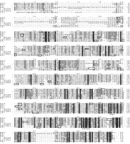
Full length sequence data were obtained for mep2, -3 and -4 and deduced amino acid sequences were aligned, in Fig. 2, with the sequence of MEP1b, a splice variant of mep1 (Redmond et al. 1997), containing an additional 31 residues at the N terminus (NCBI Accession number AF047416). These H. contortus MEPs were also aligned with an MEP from the dog hookworm, Ancylostoma caninum (NCBI Accession number AF273084) and with an MEP from the free-living nematode Caenorhabditis elegans (NCBI Accession number AAC48223). Human neprilysin (NCBI Accession number P08473) was also included as a typical neutral endopeptidase, together with human endothelin-converting enzyme 1 (ece1; NCBI Accession number CAI20192). The sequence data identified these metalloproteases as belonging to the M13 zinc metalloendopeptidase (MEP) family (EC 3.4.24.11), also known as neutral endopeptidases or neprilysins. The M13 peptidases are members of clan MA(E) in the MEROPS classification system (Rawlings et al. 2004).

Fig. 2. Multiple sequence alignment of Haemonchus contortus metalloendopeptidases, MEPs 1–4, compared with MEPs from Ancylostoma caninum, Caenorhabditis elegans and with human neprilysin and endothelin-converting enzyme 1 (ece1). The conserved zinc-binding sites HExxH and ENxADxGG are indicated with *. The fully conserved cysteines are denoted with a closed square ([squf ]). The cysteines that are conserved within the nematode MEPs are marked with an open square (□) and the potentially unpaired cysteine in MEP3 (C211, MEP3 numbering) is marked with an open circle (○). Potential N-linked glycosylation sites in each sequence in the alignment are boxed.
The alignment in Fig. 2 revealed a number of highly conserved features in all of these enzymes. Each of the H. contortus MEPs has a hydrophobic region, serving as either a putative N-terminal transmembrane domain, or as a signal peptide. This observation is consistent with the integral membrane location of the M13 neprilysins and with the finding that detergent is needed to solubilize H-gal-GP from H. contortus gut membranes.
The larger size of MEP4 is accounted for by an amino acid sequence insertion of ~100 residues immediately following the transmembrane domain/signal peptide, which is not present in any of the other proteins (Fig. 2). This segment of sequence is rich in proline and threonine residues, and a BLAST search with this part of the MEP4 sequence indicated that it has some homology with human intestinal mucin polypeptide (42% identity over 150 amino acids) (Gum et al. 1994).
As members of the M13, integral membrane, zinc metallopeptidase family, each protein had the typical HExxH (residues H641, E642, H645, MEP 1b numbering) zinc-binding domain, with the exception of MEP2, where the first histidine was substituted with an arginine. Typically, this class of metallopeptidase also has a secondary zinc-binding domain (ENxADxGG) which relies on the presence of a glutamic acid residue (E714, MEP 1b numbering) to provide the necessary charge to co-ordinate the zinc atom. Again, MEP2 differs from the other proteins in its alignment in that it has a glutamine (Q) rather than a glutamate (E) at this position. All of the sequences analysed have 8 fully conserved cysteines; C116, C124, C178, C456, C687, C763, C803 and C817 (MEP1b numbering).
Endothelin-converting enzyme is expressed as a covalently linked homo-dimer, bound by a disulphide bridge between the C428 residues (Fig. 2, human ECE 1 numbering) on each monomer. There is no equivalent cysteine residue in any of the H. contortus MEPs, the other nematode MEPs or indeed human neprilysin. There are, however, 6 other cysteines which are conserved amongst the nematode sequences (C86, C670, C696, C697, C706, C785, MEP1b numbering) which do not have equivalents in the human enzyme sequences. MEP3 also has another, potentially unpaired cysteine (C211, MEP3 numbering).
All 4 H. contortus MEPs have potential N-linked glycosylation sites, consistent with their lectin-binding properties and thus with their identification as glycoproteins. However, when the sequences were aligned, together with the other nematode and human MEPs there was no clear pattern to the distribution of these sites (Fig. 2). MEP1b had 6 potential sites, MEP2 had 5, MEP3 had 4 and MEP4 had 8 sites. The proline/threonine rich region near the N-terminus of MEP4 had 56 putative O-linked glycosylation sites. There were no predicted O-linked glycosylation sites identified in any of the other H. contortus MEPs or in the human neprilysin or ECE1 sequences. Only the C. elegans MEP sequence was predicted to have O-linked glycosylation, and that was confined to a single site, also near the N-terminus.
Molecular weight of the major components of H-gal-GP
The main components of H-gal-GP have been described previously as zones A, B, C and D (Smith et al. 1999) because of their migration pattern on non-reducing SDS-PAGE gels. Analysis of the molecular weights of these components yielded slightly higher molecular weights than those published previously (Smith et al. 1999). The peak of staining intensity for zone A indicated a molecular weight of 251 kDa, whereas zone B was ~190 kDa, zone C ~49 kDa and zone D ~34 kDa (data not shown).
Membrane topology of the MEP family
Analysis of the MEP sequences with the Phobius algorithm showed that MEPs1b and 3 were predicted to have membrane topology typical of the neprilysin family of metalloendopeptidases. In contrast, MEPs 2 and 4 were predicted to have signal peptides, a feature of secreted proteins. When this algorithm was used to analyse the sequences of human neprilysin, human ECE1, A. caninum MEP and C. elegans MEP, each of these sequences was found to have typical neprilysin-like membrane topology.
Identification of MEP fragments in H-gal-GP
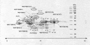
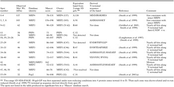
After H-gal-GP was resolved on two-dimensional electrophoresis gels (Fig. 3) 65 prominent spots were excised for tryptic digestion and subjected to mass spectrometry. Of these, 46 were identified as being derived from either the 4 MEPs or the 2 aspartyl proteases known to be present in H-gal-GP (Table 1; (Smith et al. 1999, 2003b). Searches with the peptide mass data generated from the remaining 19 spots, did not identify any proteins in the database. Isoforms, that is spots with very similar mass spectrometry fingerprints, were grouped into the 12 clusters shown in Fig. 3. Detailed examination of the peptide masses revealed that within 7 of the 10 MEP clusters almost all of the polypeptides identified were confined to either the N or C terminal halves of the complete MEP sequences (Table 1). In these cases, the observed molecular weights of the MEP spots were also too small to represent intact MEPs, indicating that they were fragments of the mature proteins (Table 1).

Fig. 3. Two-dimensional SDS-PAGE gel of Haemonchus contortus H-gal-GP. H-gal-GP (400 μg) was separated by isoelectric focusing on a linear immobilized pH gradient from pH 3 to pH 10, followed by second dimension electrophoresis on a 12·5% gel. Spots excised for tryptic digestion and subsequent mass spectrometry are numbered individually. Isoforms, identified as having very similar fingerprints, were grouped into clusters named as follows: protease of origin, then, in parenthesis, domain of origin (N=N terminal; C=C terminal; blank=whole molecule) then molecular weight in kDa.

Previously, H-gal-GP was separated by a 2-stage 1D SDS-PAGE procedure (Smith et al. 1999), in which the complex was first resolved under non-reducing conditions into the 4 zones, A, B, C or D (Fig. 1A). Each zone was subsequently excised and re-electrophoresed separately under reducing conditions into bands named after the zone of origin and the molecular weight of the band in kDa (e.g. A91, B41, etc. Fig. 1B; Smith et al. 1999).
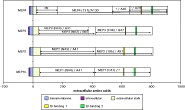
N-terminal amino acid data had been derived earlier for 6 of these bands (Smith et al. 1999) and were also acquired for spot 29, a prominent component within the MEP1 (C47) cluster (Table 1 and Fig. 3). These sequence and molecular weight data were used to match these bands to their equivalent spot cluster from 2D electrophoresis (Table 1). Similarly, by matching these derived sequences with the predicted sequences in Fig. 2, it was also possible to establish several cleavage sites within the MEPs (Table 1). The extracellular domain of each MEP was cleaved between amino acids 22 and 49 from its N-terminus to create the ‘mature’ enzyme (Fig. 4). Each ‘mature’ protease was cut again into 2 almost equal fragments, although in the case of MEP4, the presence of the mucin-like domain made the N terminal fragment considerably larger. Further clipping of the C-terminal fragment of MEP4 yielded a ~29 kDa band, detected by the two-stage 1D gels; there was evidence of further processing of the N-terminal domain of MEP3 in that at least 3 spots of different sizes were identified by 2D electrophoresis and mass spectrometry (Fig. 3, Table 1). However, these fragments must have remained linked by disulphide bonds, because they were not detected on non-reducing gels (Fig. 1).

Fig. 4. Deduced linear structure of the 4 H-gal-GP metalloproteases. The relative sizes of the intracellular, transmembrane and extracellular domains are shown together with the zinc binding-site positions within the extracellular domain. The position of the cleavage site which separates each mature enzyme from the cell membrane are marked, and the fragments resulting from further internal processing of each mature enzyme are identified using the nomenclature employed in Fig. 3.
Analysis of transcripts by RT-PCR in different developmental stages
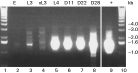
A representative MEP, in this case MEP3, was assessed by PCR using gene-specific primers. MEP3 mRNA was present at high levels from the 4th larval stage through to the adult. There was no evidence of expression in either eggs or exsheathed 3rd-stage larvae and only a trace of expression in the ensheathed L3 (Fig. 5). The integrity of the cDNA templates was assessed by carrying out a PCR with gene-specific primers for the constitutively expressed extracellular SODe (Liddell and Knox, 1998). This assay showed SODe mRNA expression in all stages of the life-cycle (data not shown).

Fig. 5. cDNA preparations from various life-cycle stages of Haemonchus contortus were analysed for expression of MEP3. The gel shows RT-PCR products from reactions which used cDNA from the following life-cycle stages as a template: lane 2, eggs (E), lane 3, 3rd-stage larvae (L3), lane 4, exsheathed 3rd-stage larvae (xL3), lane 5, 4th-stage larvae (L4), lane 6, Day 11 adult H. contortus (D11), lane 7, Day 22 adult H. contortus (D22) or lane 8, Day 28 adult H. contortus (D28) as a template. A positive control (+), which used mep3 plasmid cDNA as a template, lane 9, was also included. Lanes 1 and 10 contain DNA markers.
MEP activity assay
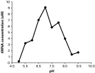
The output of the 2-stage fluorescent assay was measured in arbitrary fluorescence units, and specific enzyme activity was calculated from a standard curve prepared from 4MNA, which showed a linear relationship between 4MNA concentration and arbitrary fluorescent units detected at 425 nm (R2=0·99). The assay revealed that H-gal-GP had a broad activity curve with a maximum activity at pH 7·2 (Fig. 6), producing 9 μM of 4-MNA/μg/h.

Fig. 6. Graph showing the metalloendopeptidase activity of the H-gal-GP complex against the fluorogenic substrate Glu-Ala-Ala-Phe-4 methoxy-2-naphthylamide, over the pH range 5·2 to 9·0.
H-gal-GP inhibitor sensitivity
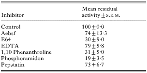
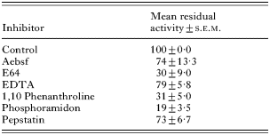
The MEP activity in H-gal-GP was markedly decreased after the inclusion of phosphoramidon, a potent inhibitor of this class of protease, in the incubation mixture (Table 2). As would be expected, 1, 10 phenanthroline, an efficient chelator of zinc ions, also had a marked effect on MEP activity. However, there was only a minimal effect when EDTA was included in the assay (Table 2). There was a marked reduction in enzyme activity with the cysteine protease inhibitor E64.
DISCUSSION
This paper describes the molecular cloning and partial characterization of a family of 4 metalloendopeptidases isolated from the intestine of the parasitic nematode H. contortus. This peptidase family is the major component of the H-gal-GP complex, which is a highly protective antigen (Smith et al. 1994). These are, typically, Type II integral membrane proteins that possess the zinc-binding domain HExxH with an additional glutamic acid serving as an extra zinc ligand (HExxH+E).
Neprilysin was first identified in the brush border membranes of rabbit kidney through its ability to cleave the insulin B chain (Kerr and Kenny, 1974). Subsequently, it was recognized as an enzyme capable of cleaving, and therefore inactivating a range of bioactive peptides, including enkephalin and substance P (Malfroy et al. 1978; Matsas et al. 1984). The substrate specificity of the neutral endopeptidases is relatively broad, with oligopeptides being cleaved preferentially on the N-terminal side of a hydrophobic amino acid residue. In addition to neprilysin, the M13 family also includes the related peptidases, endothelin converting enzymes (ECE-1 and ECE-2), the KELL blood group antigen (Lee et al. 2000) and PEX, an enzyme thought to be involved in the regulation of bone metabolism (Francis et al. 1995). Most of the biochemical studies of neprilysin and related peptidases have been carried out on mammalian enzymes, and their function appears to be the regulation of bioactive peptides. However, similar peptidases have been identified in invertebrates, such as insects and nematodes (Isaac, 1988; Sajid and Isaac, 1995). The genome sequencing projects for Drosophila melanogaster and C. elegans (Chen et al. 2005; Drysdale and Crosby, 2005) have discovered extensive families of neprilysin-like genes in both of these organisms, with D. melanogaster having 24 members and C. elegans having 22. This is not surprising considering that more than 200 bioactive peptides have been identified in various insect species (Gade, 1997; Isaac et al. 2000). Similarly, in C. elegans the FMRFamide-related peptides (FaRPs) are the most studied nematode neuropeptides, with genes coding for >50 FaRPs identified to date (Li et al. 1999).
In H. contortus, the expression of at least 2 members of the MEP family, MEPs-1 and -3, coincides with the development of the gut for blood-feeding (Redmond et al. 1997), indicating that the enzymes may be involved in digesting the bloodmeal. This argument is strengthened by the finding that the MEPs are localized to the luminal surface of the gut of the parasite, as part of the H-gal-GP complex, which also contains at least 2 pepsin-like aspartyl proteases (Longbottom et al. 1997; Smith et al. 2003b). The aspartyl protease activity of H-gal-GP will degrade haemoglobin and a variety of other proteins in in vitro assays (Longbottom et al. 1997).
Studies of proteolytic enzymes from the gut of the canine hookworm, A. caninum, have identified homologues of the major classes of peptidase found in H. contortus and shown that they act in a semi-ordered pathway to degrade haemoglobin (Williamson et al. 2004). It was found that, while a recombinant hookworm MEP was capable of degrading gelatin in a zymogram, it could not hydrolyse intact haemoglobin but that it acted downstream of aspartyl and cysteine proteases to further process the peptides produced by the digestion of haemoglobin by those enzymes (Williamson et al. 2004). The similarities in the blood-feeding habit and gut protease repertoire between A. caninum and H. contortus also strongly suggest that a similar pathway for haemoglobin digestion exists in H. contortus.
It was not possible to compare the enzymic activity of each of the H. contortus MEPs in isolation because they cannot be separated unequivocally from the H-gal-GP complex without denaturing them irreversibly. However, this complex exhibits activity over a broad pH range against the specific MEP substrate Glu-Ala-Ala-Phe-4MNA (pH 5·5–9·0), with a maximum activity at pH 7·2 (Fig. 6). The activity was markedly inhibited by the specific MEP inhibitor phosphoramidon and by the chelating agent 1,10 phenanthroline, which is consistent with the identification of MEP-like activity. The marked inhibition with the cysteine protease inhibitor E64 could indicate that cysteine proteases are required to activate the MEPs. While the current study did not identify any cysteine proteases among the 19 spots which were not MEP or aspartyl proteases, previous studies have reported N-terminal sequence data, associated with the 35 kDa zone of H-gal-GP which was consistent with that of a cysteine protease (Smith et al. 1999). This suggests that the inconsistent appearance of cysteine proteases in H-gal-GP preparations is possibly there as a contaminant which co-purifies with the complex rather than being an integral part of the complex.
The sequences of H. contortus MEPs-1b and -3 indicate that these enzymes are Type II integral membrane proteins, consistent with the integral membrane location of the M13 neprilysins and with the finding that detergent is needed to solubilize H-gal-GP from H. contortus intestinal cell membranes (Smith et al. 1999). In contrast, MEPs-2 and -4 appear to have cleavable signal peptides, suggesting that they may be secreted. Analysis of the 2D electrophoresis and mass spectrometry data indicates that all 4 MEPs in H-gal-GP are cleaved within the extracellular domain close to the membrane, as would be expected for secreted proteins. Thus, it is unclear how the complex is anchored in the cell membrane. Perhaps cleavage is not always complete and/or it is possible that the numerous S-S linked MEP fragments interact with other proteins, which are still membrane bound. It is also possible that the cleavage of the MEPs is an artefact resulting from the concomitant solubilization of the H-gal-GP complex and other membrane-associated proteases, notably the cysteine proteases, also thought to be involved in digesting the blood meal (Knox et al. 1999). However, it is noteworthy that the pattern of bands observed in reducing gels is very consistent between different batches of H-gal-GP (Smith et al. 1999).
Soluble fragments of MEPs-1 and -2, similar if not identical to those reported here have been detected in excretory/secretory products from adult H. contortus (Yatsuda et al. 2003). Excretory/secretory products are obtained by incubating worms harvested from donor animals in simple media, usually for 24–48 h. During this time some parasites may die, releasing gut membrane proteins into the medium in a non-physiological manner. For example, microsomal aminopeptidases were also detected by Yatsuda et al. (2003) in their H. contortus products, even though these are known to be integral membrane proteins localized to gut microvilli (Smith et al. 1997). This would possibly offer an explanation for the apparent discrepancy between their results and ours.
Under non-reducing conditions, MEP3 had an observed mass of ~190 kDa, yet the predicted molecular weight of the core polypeptide was only 87·9 kDa. A likely explanation for this discrepancy is that the mature enzyme exists as an S-S linked dimer, which would have a core polypeptide mass of 175·8 kDa and that the remainder of the observed mass would be made up of the carbohydrate moiety. With 8 potential N-linked glycosylation sites in such a dimer this would give an average mass of ~2 kDa per glycan unit. This is consistent with the glycan masses detected from another H. contortus intestinal glycoprotein, H11 (Haslam et al. 1996).
MEPs 1, 2 and 4 migrated together as Zone A, under non-reducing conditions, with a molecular weight of 251 kDa, yet the predicted masses for each core polypeptide were 85·6, 87·4 and 103·3 kDa, respectively. Perhaps Zone A consists of MEP4 disulphide linked to either MEP1 or MEP2 or a mixture of them both. However, such hetero-dimers would only have core polypeptide masses of ~190 kDa and would have 13 or 14 potential N-linked glycosylation sites which would mean that, if the individual N-linked glycan units were of similar mass to those on the MEP3 homodimer, the total mass would only be ~218 kDa. However, MEP4 has numerous O-linked glycosylation sites in the mucin-like domain near its N-terminal, which could account for the additional observed mass. Steric interactions between the glycan and peptide moieties within such a domain would induce the peptide core to adopt a stiff and extended structure (Jentoft, 1990), which would effectively hold the protein, and perhaps the whole complex, away from the cell membrane. This putative structure of a protein held away from the cell membrane by a stiff, mucin-like domain is somewhat at variance with the prediction of a cleavable signal peptide. Thus, it may be that, in this instance, the putative signal peptide remains un-cleaved, anchoring the protein in the cell membrane.
The MEPs of H. contortus have previously been identified as major candidates in the development of recombinant vaccines against this globally important parasite of sheep and goats. Increased understanding of the structure and function of this interesting family of proteases should contribute toward the development of recombinant gut antigens from H. contortus as vaccines, and may also aid progress in developing vaccines against other blood-feeders, such as the hookworms.
We thank K. Hall for running the 2D gels and K. McLean and L. Patterson for MALDI-TOF analysis and N-terminal amino acid sequencing, respectively. This work was funded by the Scottish Executive Environment and Rural Affairs Department. We also acknowledge Blackwell Publishing for permission to reproduce Fig. 1.