Introduction
Maternally inherited diabetes and deafness syndrome is characterised by progressive sensorineural hearing loss (SNHL) and diabetes mellitus. A mutation in mitochondrial deoxyribonucleic acid (DNA) at locus 3243 (A>G, where A = adenine and G = guanine) is responsible for this syndrome. This mutation also causes a more severe phenotype comprising mitochondrial encephalomyopathy, lactic acidosis and stroke-like episodes.
We present the case of a patient with maternally inherited diabetes and deafness syndrome who was successfully rehabilitated with cochlear implantation. We also discuss the implications of a genotype-phenotype correlation with regards to the rate of SNHL progression.
Case report
A 33-year-old woman was referred to the audiological medicine department in September 2000 for assessment of her progressive, severe to profound, symmetrical, bilateral SNHL. She was using bilateral analogue hearing aids, required modifications to her telephone and doorbell at home, and used a loop system to watch television. Her hearing loss had been first diagnosed at the age of 22 years. She denied any vertigo or tinnitus. At 23 years of age, she had been diagnosed with type two diabetes mellitus.
The patient's mother had developed progressive, bilateral SNHL at 25 years of age, and had been diagnosed with type two diabetes mellitus at 34 years of age.
In view of this family history, aetiological investigations were carried out to exclude a mitochondrial disorder. Magnetic resonance imaging (MRI) scans of the brain and internal auditory meati were found to be normal. A peripheral blood sample was sent for leucocyte DNA analysis, identifying the mitochondrial DNA mutation 3243 A>G. The level of heteroplasmy (i.e. the ratio between mutant and wild-type (‘normal’) DNA) in the sample was reported as intermediate (31–55 per cent).
Over the next 41 months, the patient's hearing loss, as assessed by pure tone audiometry, progressed at an average rate of 11.5 dBHL per year in the right ear and 6.45 dBHL per year in the left ear (averaging pure tone thresholds at 0.5, 1, 2 and 4 kHz). Because of this progressive hearing loss, the patient no longer received any benefit from conventional amplification, and she was referred for a cochlear implant assessment at 35 years of age. She was subsequently implanted with a Nucleus CI 24 in her left (better-hearing) ear (Cochlear, Melbourne, Victoria state, Australia).
One week after implant activation, assessment revealed free-field thresholds of between 35 and 45 dBA from 250–4000 Hz. At six weeks, the patient was able to use both a standard and mobile telephone, and was communicating well with her husband. Her confidence had markedly improved, to the point that she was undertaking interviews for a job. By nine months, she had started a course at a local college, could converse while travelling in the car using a lapel microphone, was able to hear the television with a loop system, and could use an MP3 player with a Walkman-style cable for the implant.
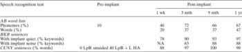
Over the first year of follow up, the patient's scores for the Arthur Boothroyd word lists test and for both the Bamford–Kowal–Bench and the City University of New York sentences tests showed a consistent improvement, as shown in Table I.
Speech recognition scores pre- and post-implantation

Wk = week; mth = months; yr = year; AB = Arthur Boothroyd; BKB = Bamford–Kowal–Bench; NA = not assessed; CUNY = City University of New York; LpR = lip-reading; L HA = left hearing aid
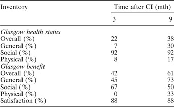
As part of her three- and nine-month reviews, the patient completed the Glasgow health status inventory and the Glasgow benefit inventory questionnaires.Reference Robinson, Gatehouse and Browning1 Her results are shown in Table II, and, again, indicated improvement between three and nine months post-implantation, illustrating the therapeutic effectiveness of cochlear implantation in this patient.
Glasgow health status inventory and Glasgow benefit inventory results after cochlear implantation

CI = cochlear implantation; mth = months
Discussion
Maternally inherited diabetes and deafness syndrome was first described in 1992.Reference Ballinger, Shoffner, Hedaya, Trounce, Polak and Koontz2 It is caused by a point mutation at nucleotide 3243 in the mitochondrial transfer ribonucleic acid (RNA) (leu)-UUR gene, substituting guanine for adenine (3243 A>G). Epidemiological studies estimate that the prevalence of the mutation within the general adult population is ≥16.3/100 000.Reference Majamaa, Moilanen, Uimonen, Remes, Salmela and Karppa3 In the Japanese population, the prevalence of this mutation in patients with SNHL ranges from 0.3 to 1.7 per cent.Reference Usami, Abe, Akita, Namba, Shinkawa and Ishii4, Reference Nagata, Kunahara, Tomemori, Arimoto, Isoyama and Yoshida5
The other syndrome associated with the 3243 A>G mutation is a more severe phenotype comprising mitochondrial encephalomyopathy, lactic acidosis and stroke-like episodes.Reference Goto, Nonaka and Horai6 The discrepancy between maternally inherited diabetes and deafness syndrome and the more severe phenotype may be a result of variability in the degree of heteroplasmy. It has been reported that the skeletal muscle of individuals with the more severe phenotype contains 80 per cent mutant mitochondrial DNA, as compared with 40 per cent in the muscle of a patient with maternally inherited diabetes and deafness syndrome.Reference Van den Ouweland, Lemkes, Ruitenbeek, Sandkujil, de Vijlder and Struyvenberg7
In view of this possible genotype-phenotype correlation, the literature was reviewed to determine whether the rate of SNHL progression in maternally inherited diabetes and deafness syndrome correlated with the degree of mutant heteroplasmy. The severity of hearing impairment has been shown to correlate with the degree of heteroplasmy in muscle, but the rate of progression shows no such correlation.Reference Uimonen, Moilanen, Sorri, Hassinen and Majamaa8 One study showed correlation between the heteroplasmy levels in biopsied muscle and the rate of SNHL progression, but no such correlation for levels in peripheral leucocytes was mentioned.Reference Majamaa-Voltti, Winqvist, Remes, Tolonen, Pyhtinen and Uimonen9 Patients in both these studies were diagnosed with the 3243 A>G mutation but represented a range of phenotypes in addition to maternally inherited diabetes and deafness syndrome.
In population-based cohort studies of cases with the 3243 A>G mutation, SNHL progression was found to occur at a rate of between 2.9 and 4.4 dBHL/year.Reference Uimonen, Moilanen, Sorri, Hassinen and Majamaa8, Reference Majamaa-Voltti, Winqvist, Remes, Tolonen, Pyhtinen and Uimonen9 Yamasoba et al. Reference Yamasoba, Oka, Tsukuda, Nakamura and Kaga10 analysed the rate of hearing loss progression compared with the proportion of mutant heteroplasmy in peripheral leucocytes. The rate of progression ranged from 1.5 to 7.9 dBHL/year (mean 3.2 dBHL/year). These patients were all found to have low levels of heteroplasmy in their leucocyte mitochondrial DNA (30 per cent or less). A higher rate of progression was observed in one patient (11.5 dBHL/year in the right ear, 6.45 dBHL/year in the left ear, mean 8.98 dBHL/year), and this may reflect the higher level of heteroplasmy (intermediate; 31–55 per cent) found in the leucocytes of this patient.
• Progressive sensorineural hearing loss can form part of a syndrome resulting from the mitochondrial deoxyribonucleic acid (DNA) mutation 3243 A>G
• Cochlear implantation is a successful method of rehabilitation in patients with maternally inherited diabetes and deafness syndrome
• The level of DNA mutant heteroplasmy may influence the rate of SNHL progression and may hence be useful in patient counselling
• Current tissue sampling methods provide only an indirect estimate of the level of heteroplasmy in the inner ear. In future, comparison of peripheral and cochlear samples will further our understanding of genotype-phenotype correlation
In patients with maternally inherited diabetes and deafness syndrome, the characteristic audiometric profile comprises an initial bilateral, symmetrical, high frequency loss, progressing across all frequencies.Reference Yamasoba, Oka, Tsukuda, Nakamura and Kaga10, Reference Oshima, Ueda, Ikeda, Abe and Takasaka11 The hearing loss is cochlear in origin, as demonstrated in clinical and histopathological studies. A study of patients with maternally inherited diabetes and deafness syndrome showed absent otoacoustic emissions (i.e. abnormal outer hair cell function).Reference Yamasoba, Oka, Tsukuda, Nakamura and Kaga10 Auditory brainstem responses in these patients showed no central abnormality. In a temporal bone study of a patient with maternally inherited diabetes and deafness syndrome and profound SNHL,Reference Yamasoba, Tsukada, Oka, Kobayashi and Kaga12 examination of the cochlea showed severe degeneration of the stria vascularis and outer hair cells, and reduced numbers of spiral ganglion cells. These areas of the cochlea are highly metabolically active and do not divide; hence, reduced levels of adenosine triphosphate (ATP) associated with mitochondrial dysfunction results in dysfunction of the Na+-K+ ATP pumps, leading to ion imbalance and subsequent cell injury and death.Reference Cortopassi and Hutchin13
There is no evidence in the literature of a relationship between diabetes mellitus and the nature of the SNHL.Reference Oshima, Ueda, Ikeda, Abe and Takasaka11 In a study of 54 patients with the 3243 A>G mutation and maternally inherited diabetes and deafness syndrome, SNHL preceded onset of diabetes in 23 patients, and was diagnosed simultaneously with diabetes in seven patients.Reference Guillausseau, Massin, Dubois-LaForgue, Timsit, Virally and Gin14 There was no difference in the rate of progression of hearing loss between diabetic and non-diabetic patients with the 3243 A>G mutation.Reference Majamaa-Voltti, Winqvist, Remes, Tolonen, Pyhtinen and Uimonen9
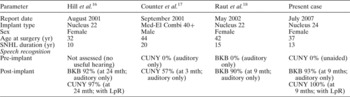
This case report represents the fourth published case with maternally inherited diabetes and deafness syndrome to receive a cochlear implant. As shown in Table III, these patients were affected during their young adult life, and progressed over a period of 10 to 20 years to the point that they required an implant. Serial audiometry is crucial in determining the rate of progression and the optimum rehabilitation strategy. Levels of heteroplasmy vary between tissue types.Reference Remes, Majamaa, Herva and Hassinen15 Current methods of measuring heteroplasmy provide an indirect measure of heteroplasmy within the cochlea. A direct analysis of the level of heteroplasmy in cochlear tissue, e.g. from patients undergoing implantation, would further our understanding of the genotype-phenotype correlation in the 3243 A>G mutation.
Comparison of present case with previously reported MIDD patients with cochlear implantation

MIDD = maternally inherited diabetes and deafness syndrome; yr = years; SNHL = sensorineural hearing loss; CUNY = City University of New York sentences; BKB = Bamford–Kowal–Bench sentences; mth = months; LpR = lip-reading
Conclusion
The case presented here confirms the fact that cochlear implantation is a successful method of rehabilitation in patients with maternally inherited diabetes and deafness syndrome. In our maternally inherited diabetes and deafness syndrome patient, the rate of SNHL progression was faster than previously reported. This may be attributable to the higher levels of mutant mitochondrial DNA present. The degree of heteroplasmy may have implications when counselling patients regarding the rate of progression of their hearing loss. Future analysis of the level of mutant heteroplasmy within cochlear tissue, from patients undergoing cochlear implantation, may add significantly to our current understanding of the condition and its prognosis.