Pompe disease is an autosomal recessive type II glycogen storage disease, characterised by deficiency of acid alpha-glucosidase.Reference Chen, Zhang and Quan1,Reference Kishnani, Hwu, Mandel, Nicolino, Yong and Corzo2 Clinical presentation includes cardiomegaly due to hypertrophic cardiomyopathy, respiratory distress and failure to thrive. Enzyme replacement therapy which slows the progression of the disease as well as reverses the ventricular hypertrophy.Reference Chen, Zhang and Quan1,Reference Bali, Goldstein and Banugaria3 The clinical outcomes are variable due to the severity of the disease and the delay in therapy. The first child’s course was complicated by respiratory decompensation, ventilator dependence and death. The second child had early initiation of therapy resulting in an eventual improvement in the cardiac hypertrophy.
Case 1
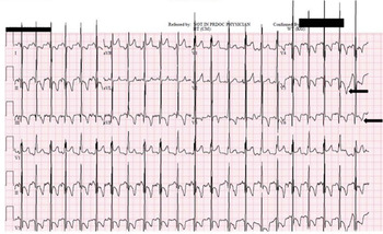
A full-term girl was born with prenatal diagnosis significant for biventricular hypertrophy and no maternal diabetes (Fig 1). Initial evaluation at the referring hospital included an electrocardiogram that demonstrated normal sinus rhythm, depolarisation abnormalities consistent with high voltages, prominent R wave in precordial leads, as well as ST abnormality and T wave inversion in inferolateral leads, indicating severe biventricular hypertrophy. Postnatal echocardiogram confirmed the biventricular hypertrophy with normal anatomy; normal function with no left ventricular outflow obstruction metabolic workup was within normal limits with elevated NT-Pro BNP at 32,798 ng/L (normal < 450 ng/L). At 5 months of age, she developed a significant upper respiratory illness with parainfluenza and rhinovirus. Her respiratory status deteriorated, and she required intubation and mechanical ventilation. CT showed atelectasis of the entire left lung with compression of the left mainstem bronchus due to the significant cardiac hypertrophy. Testing confirmed the diagnosis of Pompe disease. Patient underwent tracheostomy and was discharged home on low ventilator settings. She had two splice site mutations: c.1327-2A>G in Intron 8 and c.1438-1G>C in Intron 9. This mutation was predicted to be cross-reactive immunological material status positive, indicating a possible good response to enzyme therapy and she was started on weekly therapy. However, there was no improvement in the left ventricular hypertrophy. At 20 months of age, she presented to an outside hospital in cardiac arrest due to acute hypoxic respiratory failure. She had significant post arrest hypoxic encephalopathy and family decided to withdraw care.
Electrocardiogram demonstrating severe biventricular hypertrophy with normal sinus rhythm, depolarisation abnormalities consistent with high voltages, prominent R wave in precordial leads, as well as ST abnormality and T wave inversion in inferolateral leads.

Case 2
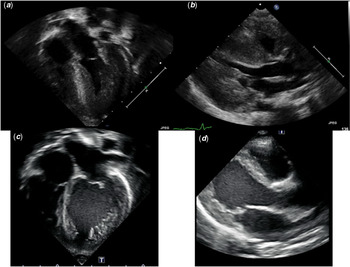
A full-term boy with fetal arrhythmias on prenatal ultrasound had moderate biventricular hypertrophy with mildly depressed biventricular function on postnatal echocardiogram (Fig 2a and b). The intracardiac anatomy was otherwise normal. Creatine phosphokinase enzyme level was elevated to 1439 U/L (normal range 35–350 U/L), and liver enzymes were elevated. Evaluation for Pompe disease was initiated and was found to be abnormal with the GAA enzyme activity of 0.40 nmol/h/mg (normal > 10 nmol/h/mg). The findings were consistent with classic infantile Pompe disease. Molecular testing revealed two pathogenic variants in the GAA gene; based on his molecular testing, he is predicted to be CRIM positive. The patient was heterozygous for a single nucleotide change (c.953 T > C; p. Met318Thr) in exon 5; he was also found to be heterozygous for a single nucleotide change (c.2560C>T; p. Arg854*) in exon 18. Biweekly enzyme replacement therapy was initiated on 11th day of life. At 17 months of age, he has normal cardiac function with complete resolution of the biventricular hypertrophy (Fig 2c and d).
2D-echocardiogram images ((a) apical view and (b) parasternal view) demonstrating moderate biventricular hypertrophy. LVIDd 2 cm (z score 1.2), LVPWd 0.48 cm (z score 2.9), IVSd 0.56 cm (z score 2.1), ejection fraction 49%. Follow-up images ((c) apical view and (d) parasternal view) at 17 months of age with biweekly enzyme replacement therapy demonstrating resolution of biventricular hypertrophy. LVIDd 3.1 cm (z score 1.7), LVPWd 0.39 cm (z score 0.35), IVSd 0.43 cm (z score 0.00), ejection fraction 64%. LVIDd = left ventricular internal dimension at end-diastole; LVPWd = left ventricular posterior wall thickness at end diastole; IVSd = interventricular septum thickness at end diastole; EF = ejection fraction. The above images were captured during diastole.

Discussion
The presentation of Pompe disease may vary from the classic infantile form to a late onset juvenile and adult form, with no cardiac manifestations.Reference Chen, Zhang and Quan1 The classic infantile form usually presented during the first few months of life and is characterised by hypertrophic cardiomyopathy.Reference Chen, Zhang and Quan1,Reference Kishnani, Hwu, Mandel, Nicolino, Yong and Corzo2 The infantile form is the most severe form and can be fatal within the first year of life if not treated early.Reference Bali, Goldstein and Banugaria3
Enzyme replacement therapy is the only treatment effective in managing patients with the infantile form of Pompe disease.Reference Chen, Zhang and Quan1 The Food and Drug Administration approved it in 2006.Reference Chen, Zhang and Quan1,Reference Kishnani, Hwu, Mandel, Nicolino, Yong and Corzo2 Prior to this, supportive and conservative measures were the only treatment available. Early initiation of enzyme therapy is essential in delaying disease progression and improving outcomes. Biweekly infusions have demonstrated prolonged and invasive-ventilation-free survival.Reference Nicolino, Byrne and Wraith4 The greater number of receptors on cardiac muscle cell surface makes the cardiac muscles more responsive to enzyme therapy compared to skeletal muscle.Reference Levine, Kishnani, Chen, Herlong and Li5,Reference Raben, Jatkar and Lee6
The clinical response to enzyme replacement therapy (ERT) depends on several factors including age of initiation, extent of pre-existing damage to the muscle as well as cross-reactive immunological material status.Reference Bali, Goldstein and Banugaria3 Individuals with negative status are unable to create any deficient protein and hence recognise the enzyme as a “foreign protein”. Therefore, these individuals respond poorly to the therapy due to the presence of high titers of neutralising anti-bodies to the recombinant enzyme.Reference Bali, Goldstein and Banugaria3 In contrast, individuals with positive status typically have some residual non-functional protein resulting in low antibody titers and therefore have a better clinical response. Determining the status is therefore crucial in order to gauge clinical response and dictates whether the patient requires immunomodulatory therapy prior to ERT.Reference Bali, Goldstein and Banugaria3 Both our patients were of positive status and thus expected to respond to the enzyme therapy. There are reports of a higher dose of weekly infusions resulting in better outcomes in patients with positive status.Reference van Gelder, Poelman and Plug7 While enzyme therapy continues to be the mainstay of treatment, studies to explore gene therapy as a definitive treatment for Pompe disease are ongoing.Reference Hordeaux, Dubreil and Robveille8
Beneficial effects of enzyme therapy are unpredictable when started after the age of 5 months.Reference Chen, Chen and Chiu9 Cases with severe hypertrophy have been reported to be non-responsive to therapy.Reference Raben, Jatkar and Lee6 Thus, we believe that the delay in the diagnosis and treatment with ERT in our first case contributed to poor outcome. She developed a viral respiratory illness with complete left lung atelectasis, due to left bronchus compression secondary to cardiomegaly, resulting in decompensation, intubation and chronic ventilator dependence. Despite the initiation of weekly therapy, her cardiac status did not improve. In contrast, our second patient had a prompt diagnosis at birth with initiation of enzyme therapy at 11 days of life and a significant improvement in her cardiac status. These contrasting cases demonstrate that early diagnosis and early initiation of enzyme replacement therapy are imperative for improvement in cardiac status and increasing the likelihood of a good outcome.
Although Pompe disease is rare with an incidence of 1 in 138,000 children,Reference Chen, Zhang and Quan1 it should be considered as an important differential diagnosis in newborn infants with significant biventricular hypertrophy. Since the early initiation of therapy can significantly decrease the morbidity and mortality, every attempt should be made to make an early diagnosis.
Acknowledgements
None.
Financial Support
This research received no specific grant from any funding agency, commercial or not-for-profit sectors.
Conflicts of Interest
The authors have no conflicts of interest relevant to this article to disclose.
Ethical Standards
Not applicable.