Introduction
The family Heteroderidae of plant-parasitic nematodes contains cyst-forming (or cyst) genera and non-cyst-forming (or cystoid) genera. In China, cyst-forming genera, including Heterodera Schmidt, 1871, Globodera Skarbilovich, 1959 and Cactodera Krall & Krall, 1978, have been reported (Chen et al., Reference Chen, Zheng and Ma1994; Pan et al., Reference Pan, Lin and Xue1997; Zhuo et al., Reference Zhuo, Wang, Ye, Peng and Liao2013), but not non-cyst-forming genera. Cryphodera Colbran, Reference Colbran1966, a non-cyst-forming genus, was established in 1966. This genus currently comprises six species (Karssen & Van Aelst, Reference Karssen and Van Aelst1999). During a survey of cyst nematodes in China's tropical and subtropical regions in May 2012, a Cryphodera species was recovered from ramie (Boehmeria nivea (Linn.) Gaudich) in Yiyang, a city in Hunan Province in the subtropics. Comparative morphological, morphometric and molecular studies of the nematode revealed that it differed from all other nominal species in the genus and it is herein described as a new species, Cryphodera sinensis n. sp. Phylogenetic analyses on small subunit (SSU), D2D3 expansion domains of large subunit (LSU D2D3) and internal transcribed spacer (ITS) rRNA sequences were performed to investigate the relationship of C. sinensis n. sp. with other available DNA sequences of cystoid and cyst nematodes.
Materials and methods
Collection and examination of nematodes
Cryphodera sinensis n. sp. was extracted from ramie in Yiyang City, Hunan Province, China. Females were isolated from soil samples around ramie roots by the sieving technique (Subbotin et al., Reference Subbotin, Mundo-Ocampo, Baldwin, Hunt and Perry2010a) or dissected from roots. Males and second-stage juveniles (J2) were extracted from soil samples by the Baermann funnel method. J2s were also released from crushed females.
Females were transferred to 4% formalin and processed according to the method by Bajaj et al. (Reference Bajaj, Walia, Dabur and Bhatti1989). Perineal patterns of mature females were prepared by the procedures for Meloidogyne (Hartman & Sasser, Reference Hartman, Sasser, Barker, Carter and Sasser1985). Males and J2s were relaxed by gentle heat, fixed in 4% formalin and processed by the glycerin-ethanol method (Feng, Reference Feng2001). Nematodes were observed, illustrated, measured and photographed with the aid of a Nikon ECLIPSE Ni microscope equipped with a Nikon Digital Sight Camera and exclusive NIS-Elements BR software (Nikon, Japan).
Molecular analysis
For molecular analysis, the DNA samples of single J2s and males were prepared according to the method by Mundo-Ocampo et al. (Reference Mundo-Ocampo, Troccoli, Subbotin, Cid del Prado, Baldwin and Inserra2008). Three rRNA fragments, i.e. SSU, LSU D2D3 and ITS, were amplified. SSU was amplified as two partially overlapping fragments using two pairs of primers. The first primer pair was 988F (5′-CTCAAAGATTAAGCCATGC-3′) and 1912R (5′-TTTACGGTCAGAACTAGGG-3′) and, the second primer pair was 1813F (5′-CTGCGTGAGAGGTGAAAT-3′) and 2646R (5′-GCTACCTTGTTACGACTTTT-3′) (Holterman et al., Reference Holterman, Karssen, van den Elsen, van Megen, Bakker and Helder2009). Detailed protocols of polymerase chain reaction (PCR) amplification were as described by Holterman et al. (Reference Holterman, Karssen, van den Elsen, van Megen, Bakker and Helder2009). Two partial SSU fragments were integrated as a near-full-length SSU sequence using DNAStar software (Burland, 2000). Primers for LSU D2D3 amplification were D2A (5′-ACAAGTACCGTGAGGGAAAGTTG-3′) and D3B (5′-TCGGAAGGAACCAGCTACTA-3′) (Subbotin et al., Reference Subbotin, Sturhan, Chizhov, Vovlas and Baldwin2006). Primers for ITS amplification were TW81 (5′-GTTTCCGTAGGTGAACCTGC-3′) and AB28 (5′-ATATGCTTAAGTTCAGCGGGT-3′) (Subbotin et al., Reference Subbotin, Waeyenberge and Moens2000). Detailed protocols of PCR amplification were as described by Tanha Maafi et al. (Reference Tanha Maafi, Subbotin and Moens2003). The sequencing program was the same as described in Zhuo et al. (Reference Zhuo, Cui, Ye, Luo, Wang, Hu and Liao2010). The newly obtained sequences were deposited in the GenBank database.
The sequences of C. sinensis n. sp. were compared with GenBank accessions from other nematode species using the BLAST homology search program (http://blast.ncbi.nlm.nih.gov/). The closest sequences were selected in phylogenetic analyses. Outgroup taxa for each dataset were chosen according to previous molecular phylogenetic analysis for cyst nematodes (Subbotin et al., Reference Subbotin, Vierstraete, De Ley, Rowe, Waeyenberge, Moens and Vanfleteren2001; Mundo-Ocampo et al., Reference Mundo-Ocampo, Troccoli, Subbotin, Cid del Prado, Baldwin and Inserra2008; Bernard et al., Reference Bernard, Handoo, Powers, Donald and Heinz2010). DNA sequences were aligned by ClustalW (http://workbench.sdsc.edu, Bioinformatics and Computational Biology group, Department of Bioengineering, UC San Diego, California, USA), using default parameters. The alignment quality was examined by eye and optimized. The model of base substitution was evaluated using MODELTEST (Posada & Crandall, Reference Posada and Crandall1998; Huelsenbeck & Ronquist, Reference Huelsenbeck and Ronquist2001). The Akaike-supported model, the base frequencies, the proportion of invariable sites, and the gamma distribution shape parameters and substitution rates were used in phylogenetic analyses. Bayesian analysis was performed to confirm the tree topology for each gene separately using MrBayes 3.1.0 (Huelsenbeck & Ronquist, Reference Huelsenbeck and Ronquist2001) running the chain for 1 × 10⁶ generations and setting the ‘burn-in’ at 1000. The MCMC (Markov Chain Monte Carlo) method was used within a Bayesian framework to estimate the posterior probabilities of the phylogenetic trees (Larget & Simon, Reference Larget and Simon1999) using the 50% majority-rule.
Results
Description of Cryphodera sinensis n. sp
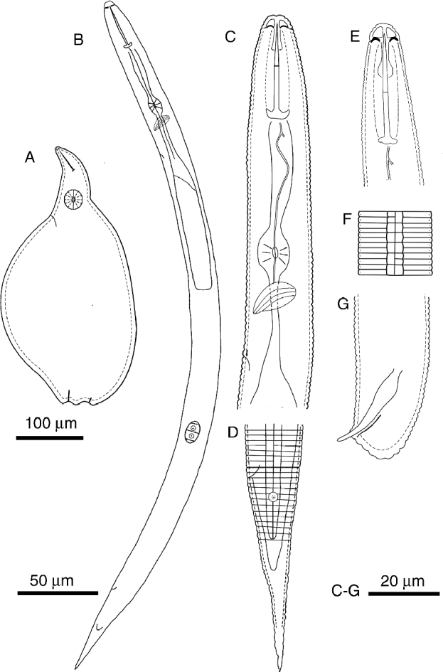
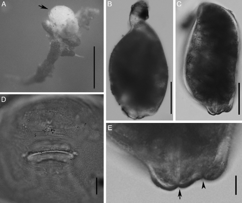
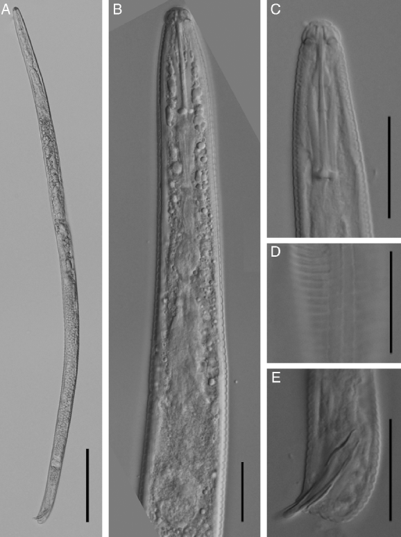
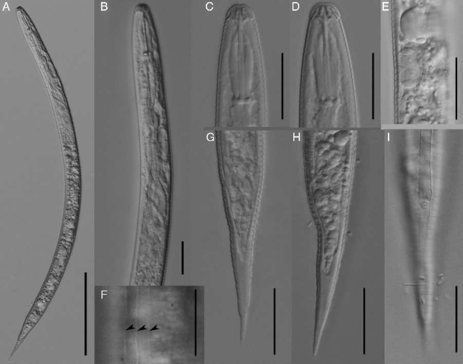
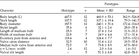
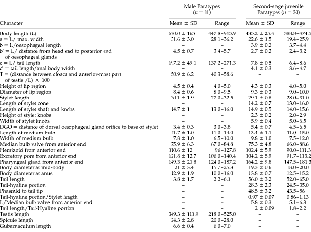
In the morphometrics of females, males and second-stage juveniles (figs 1–4, tables 1 and 2), abbreviations follow those of Siddiqi (Reference Siddiqi2000).
Cryphodera sinensis n. sp.: (A) female; (B) second-stage juvenile (J2); (C) anterior region of J2; (D) lateral field and tail of J2; (E) head region of male; (F) lateral field of male; (G) tail of male.

Females of Cryphodera sinensis n. sp. under the light microscrope: (A) a female (arrow) parasitizing a root of ramie; (B, C) entire body; (D) perineal pattern; (E) vulva–anus region (arrow = vulva; arrowhead = anus). Scale bars: 1000 μm (A); 100 μm (B, C); 20 μm (D, E).
Males of Cryphodera sinensis n. sp. under the light microscope: (A) entire body; (B) pharyngeal region; (C) anterior region; (D) lateral lines in the middle body; (E) tail. Scale bars: 100 μm (A); 20 μm (B–E).
Second-stage juveniles of Cryphodera sinensis n. sp. under the light microscope: (A) entire body; (B) pharyngeal region; (C, D) anterior region; (E) genital promordium; (F) lateral lines (arrowed); (G, H) tail; (I) phasmids. Scale bars: 100 μm (A); 20 μm (B–E, G–I); 10 μm (F).
Morphometrics in μm of females of Cryphodera sinensis n. sp.
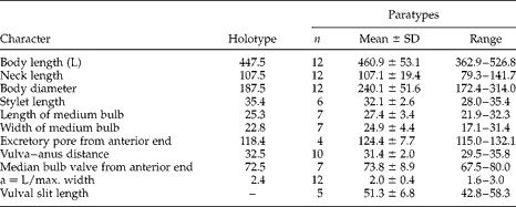
n, number of specimens observed; SD, standard deviation.
Morphometrics in μm of males and second-stage juveniles of Cryphodera sinensis n. sp.
n, number of specimens observed; SD, standard deviation.
Female
Body oval without vulval cone, completely annulated, surrounded by a distinct subcrystalline layer. Neck distinct, usually bent laterally. Labial region not offset, labial disc distinct. Except for the stylet and median bulb, other internal structures difficult to observe. Stylet long, with rounded knobs sloping posteriorly. Median bulb nearly rounded to oval, with distinct valve plates situated centrally. Excretory pore located posterior to median bulb. Vulva terminal, lips pronounced and protruding. Anus subterminal, dorsal, near the vulva. Area between anus and vulva distinctly concave. Perineal pattern ovoid with fine, smooth and continuous striae. Striae surround anus and vulva respectively. Phasmids indistinct. Eggs and juveniles (55 ± 19 (30–75)) retained inside the body. No cyst formed.
Male
Body slender, ventrally curved when heat-relaxed. Lateral fields with three incisures, areolated. Annulus width at mid-body 1.7–2.1 μm. Labial region slightly offset, with a prominent labial disc and two post-labial annuli, basal annulus broader and longitudinal striations present. Stylet long, with rounded knobs sloping slightly posteriorly. Median bulb oval, occupying c. 50% of corresponding body diameter, with 3–4 μm long valve plates situated centrally. Pharyngeal glands developed, ventrally overlapping the intestine. Hemizonid distinct, posterior to median bulb, c. 4 μm long, 3–6 annuli anterior to excretory pore. Testis single, anteriorly outstretched. Spicules slender, arcuate with blunt rounded tip. Gubernaculum thin and almost straight. Tail very short, hemispherical, anal lips forming a short tube. Phasmids indistinct.
Second-stage juveniles (J2)
Body slender, tapering posteriorly, ventrally curved when heat-relaxed. Labial region slightly offset, hemispherical, with one weakly offset labial disc and three distinct post-labial annuli. Stylet long, cone straight, shaft cylindrical. Stylet knobs well developed with distinct anterior projection. Median bulb oval, occupying c. 50% of corresponding body diameter, with 3–4 μm long valve plates situated centrally. Pharyngeal glands filling body cavity. Hemizonid distinct, immediately anterior to excretory pore. Lateral fields with three incisures, areolated. Annulus width at mid-body 1.7–2 μm. Genital primordium oval, 12.5 (10–14) μm long, 7.9 (6–10) μm wide, 159 (145–174) μm anterior to tail tip. Tail conoid, gradually tapering to a point terminus with mucro-like tip, hyaline region relatively long, occupying 44.8–54.7% of tail length, posterior half not annulated. Phasmids large, lens-like, 2–6 annuli posterior to anus.
Type host, material and etymology
Type host. Roots and rhizosphere of ramie (Boehmeria nivea) from Yiyang City (latitude 28°55.977′N, longitude 112°25.818′E), Hunan Province, China. Material collected in May 2011. The soil type is clay and the climate is subtropical.
Deposition of type specimens. Holotype female, two paratype males and six paratype J2s are deposited on slides in the USDA Nematode Collection, Beltsville, Massachusetts, USA; one paratype female, two paratype males and five paratype J2s deposited on slides in the Department of Nematology, University of California, Riverside, California, USA. Other voucher specimens are available at the Plant Nematode Research Laboratory, College of Natural Resources and Environment, South China Agricultural University, Guangzhou, China.
Etymology. Specific epithet refers to the geographical location (adj. sinensis = Chinese, from China).
Diagnosis and relationships
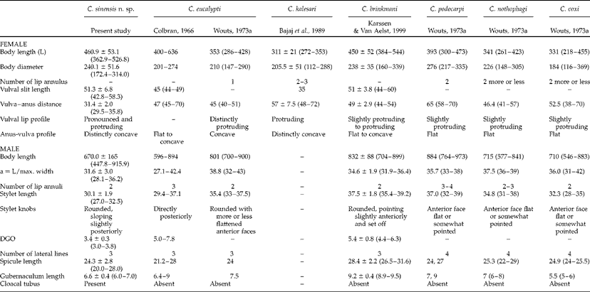
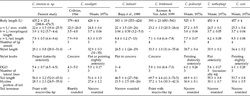
Cryphodera sinensis n. sp. has the general characteristics of the genus Cryphodera. Diagnostic characters of the new species and all species comparisons in the genus Cryphodera are compiled in tables 3 and 4, and a key to all species within Cryphodera is also provided. The new species, on the basis of pronounced and protruding vulval lips, concave anus–vulva profile, three lateral lines in males and J2s, three post-labial annuli and anteriorly concave stylet knobs in J2s, appears morphologically most similar to C. eucalypti Colbran, Reference Colbran1966; C. kalesari Bajaj, Walia, Dabur & Bhatti, Reference Bajaj, Walia, Dabur and Bhatti1989 and C. brinkmani Karssen & Van Aelst, Reference Karssen and Van Aelst1999.
Comparative morphometrics (in μm) of females and males in species of Cryphodera with ranges in brackets.
DGO, distance of dorsal oesophageal gland orifice to base of stylet.
Data referred to Colbran (Reference Colbran1966), Wouts (Reference Wouts1973a), Bajaj et al. (Reference Bajaj, Walia, Dabur and Bhatti1989), Karssen & Van Aelst (Reference Karssen and Van Aelst1999), Siddiqi (Reference Siddiqi2000) and Sturhan (Reference Sturhan2010). –, not available.
Comparative morphometrics (in μm) of second-stage juveniles (J2) in species of Cryphodera with ranges in brackets.
DGO, distance of dorsal oesophageal gland orifice to base of stylet.
Data referred to Colbran (Reference Colbran1966), Wouts (Reference Wouts1973a), Bajaj et al. (Reference Bajaj, Walia, Dabur and Bhatti1989), Karssen & Van Aelst (Reference Karssen and Van Aelst1999), Siddiqi (Reference Siddiqi2000) and Sturhan (Reference Sturhan2010). –, not available.
† Colbran (Reference Colbran1966) reported stylet length for J2 of C. eucalypti was 26–35.9 μm. Wouts (Reference Wouts1973a) mentioned that the minimum 26 μm belonged to J2 of Heterodera. Bajaj et al. (Reference Bajaj, Walia, Dabur and Bhatti1989) and Karssen & Van Aelst (Reference Karssen and Van Aelst1999) accepted the point of Wouts (Reference Wouts1973a). Thus, the value of C. eucalypti described by Colbran (Reference Colbran1966) is omitted here.
‡ Wouts (Reference Wouts1973a) described four incisures in the field of C. nothophagi J2. Sturhan (Reference Sturhan2010) re-examined paratype J2 of C. nothophagi, which revealed three incisures in the field, with the inner incisure occasionally diverging into two. Thus, the point of Sturhan (Reference Sturhan2010) is accepted here.
Cryphodera sinensis n. sp. differs from C. eucalypti by the shorter vulva–anus distance (29.5–35.8 v. 40–70 μm), the presence of cloacal tubus, the tail terminus shape of the J2 (point with mucro-like tip v. bluntly rounded), the shorter J2 stylet (28–31 v. 31–35 μm), the stylet knob shape of J2 (distinctly anteriorly projected v. anterior faces flat or slightly pointed anteriorly) and the longer J2 tail (56.0 ± 3.2 v. 51.6 ± 1.3 μm). It differs from C. kalesari by the larger female (362.9–526.8 v. 272–353 μm in length; 172.4–314.0 v. 112–288 μm in width), the shorter female stylet (28.0–35.4 v. 37–42 μm), the shorter vulva–anus distance (29.5–35.8 v. 48–72 μm), the longer J2 stylet (28.0–31.0 v. 24–29 μm), the tail terminus shape of the J2 (point with mucro-like tip v. narrow rounded), the longer J2 tail and hyaline portion of tail (52.0–65.0 v. 27–54 μm; 24.5–35 v. 18–26 μm) and the presence of males in C. sinensis n. sp. but no males found in C. kalesari. From C. brinkmani, the new species differs by the shorter female stylet (28.0–35.4 v. 39.2–42.3 μm), shorter vulva–anus distance (29.5–35.8 v. 44–54 μm), shorter male stylet (27.0–32.5 v. 35.4–39.2 μm), fewer J2 lip annuli (3 v. 4), shorter J2 stylet (28.0–31.0 v. 31.6–35.4 μm) and shorter J2 tail (52.0–65.0 v. 61.3–76.5 μm).
In addition, C. sinensis n. sp. can be easily distinguished from the remaining three species of Cryphodera, namely C. podocarpi (Wouts, 1973) Luc, Taylor & Cadet, Reference Luc, Taylor and Cadet1978, C. nothophagi (Wouts, 1973) Luc, Taylor & Cadet, Reference Luc, Taylor and Cadet1978 and C. coxi (Wouts, 1973) Luc, Taylor & Cadet, Reference Luc, Taylor and Cadet1978, by the shape of the vulval lips (pronounced and protruding v. protruding slightly out of body contour), anus–vulva profile (concave v. flat), the shorter vulva–anus distance (29.5–35.8 v. at least 38 μm), fewer incisures in the male lateral field (3 v. 4) and fewer lip annuli in J2 (3 v. 4 in C. nothophagi and C. coxi, and 5 in C. podocarpi).
Molecular profiles and phylogenetic relationships
The 1740-bp near-full-length SSU sequence was based on DNA template of a single J2. The GenBank accession number of the sequence is JX566453. A BLASTN search of C. sinensis n. sp. revealed a high match with the only sequenced Cryphodera species in this gene, i.e. C. brinkmani (GenBank accession number JQ965679). The identity of the two sequences was 99.6% (1651/1657) without a gap.
The rDNA sequences of 787-bp LSU D2D3 from a single male (body length up to 900 μm), 788-bp LSU D2D3 from another single male (body length only 500 μm, the same size as a J2) and the same J2 as mentioned above were sequenced, respectively. GenBank accession numbers of the three sequences are JX566454, JX566455 and JX566456. The identities of the two male sequences were 99.5% (783/788) with one insertion/deletion (0.1%). The identities of the juvenile sequence and the two male sequences were 99.4% (782/788) with one insertion/deletion (0.1%) and 99.6% (785/788) without a gap, respectively. Intraspecific D2D3 sequence variation for C. sinensis n. sp. was 3–7 nucleotides (0.4–0.6%). A BLASTN search of C. sinensis n. sp. on the LSU D2D3 revealed a high match with the only sequenced Cryphodera species in this gene, i.e. C. brinkmani (GenBank accession number DQ328705). The identities of the sequence from C. brinkmani and the three sequences from the new species were 93.8% (621/661) with one insertion/deletion (0.1%), 94% (622/661) without a gap, and 93.8% (621/661) without a gap. The interspecific D2D3 sequence variation was 39–40 nucleotides (6%).
The 1037-bp, 1040-bp and 1042-bp ITS-rDNA sequences were sequenced, respectively, based on the same DNA template of males and J2. GenBank accession numbers of the three sequences are JX566457, JX566458 and JX566459. The identities of the two male sequences were 98.8% (1026/1040) with three insertions/deletions (0.2%). The identities of the juvenile sequence and the two male sequences were 98.5% (1022/1042) with five insertions/deletions (0.4%) and 98.8% (1027/1043) with four insertions/deletions (0.3%), respectively. Intraspecific ITS sequence variation for C. sinensis n. sp. was 17–25 nucleotides (1.1–1.5%). A BLASTN search of C. sinensis n. sp. on the ITS revealed high matches with four Cryphodera isolates, including three unidentified isolates and C. brinkmani, the highest being with an unidentified isolate, namely Cryphodera sp. A (GenBank accession number JF894388–JF894396). The identities of the sequences from the new species and Cryphodera sp. A were 97.1–98.9% (631–640/647, 627–640/648 or 632/649) with 0–3 gaps. Comparing with C. brinkmani (AF274418), the three sequences of C. sinensis n. sp. had 212, 219 and 224 variable sites with 82.2, 81.9 and 81.1% identities (777/989, 772/996 and 778/997) and 78, 91 and 90 insertions/deletions (7.9, 9.1 and 9.0%), respectively. The identities of C. sinensis n. sp. (JX566457–JX566459) and Cryphodera sp. B (JF894400–JF894402) ranged from 77.3 to 78.7%, and C. sinensis n. sp. (JX566457–JX566459) and Cryphodera sp. C (JF894397–JF894399), 83.0–83.7%. The interspecific ITS sequence variation within Cryphodera ranged from 15.1 to 22.8%.
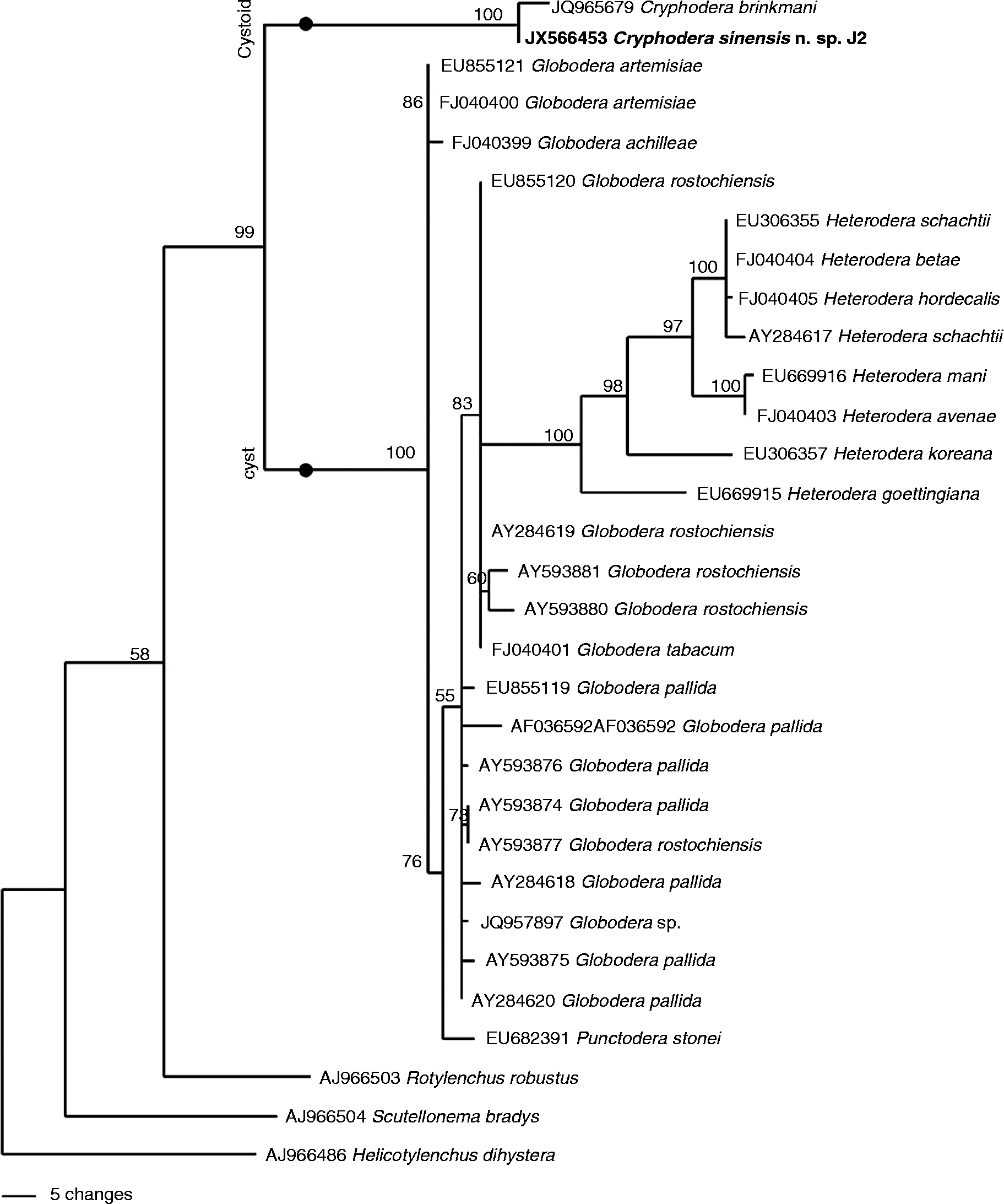
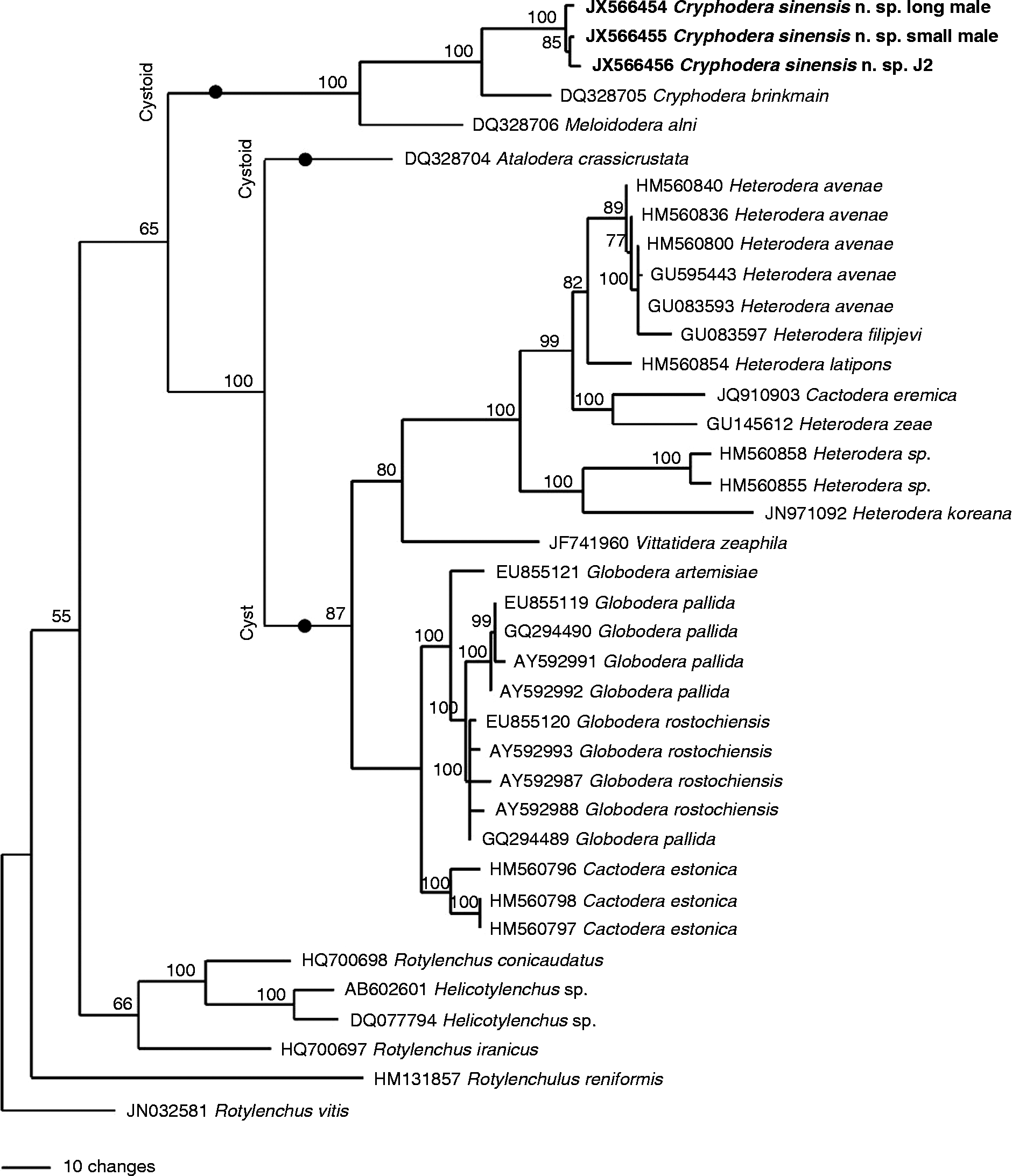
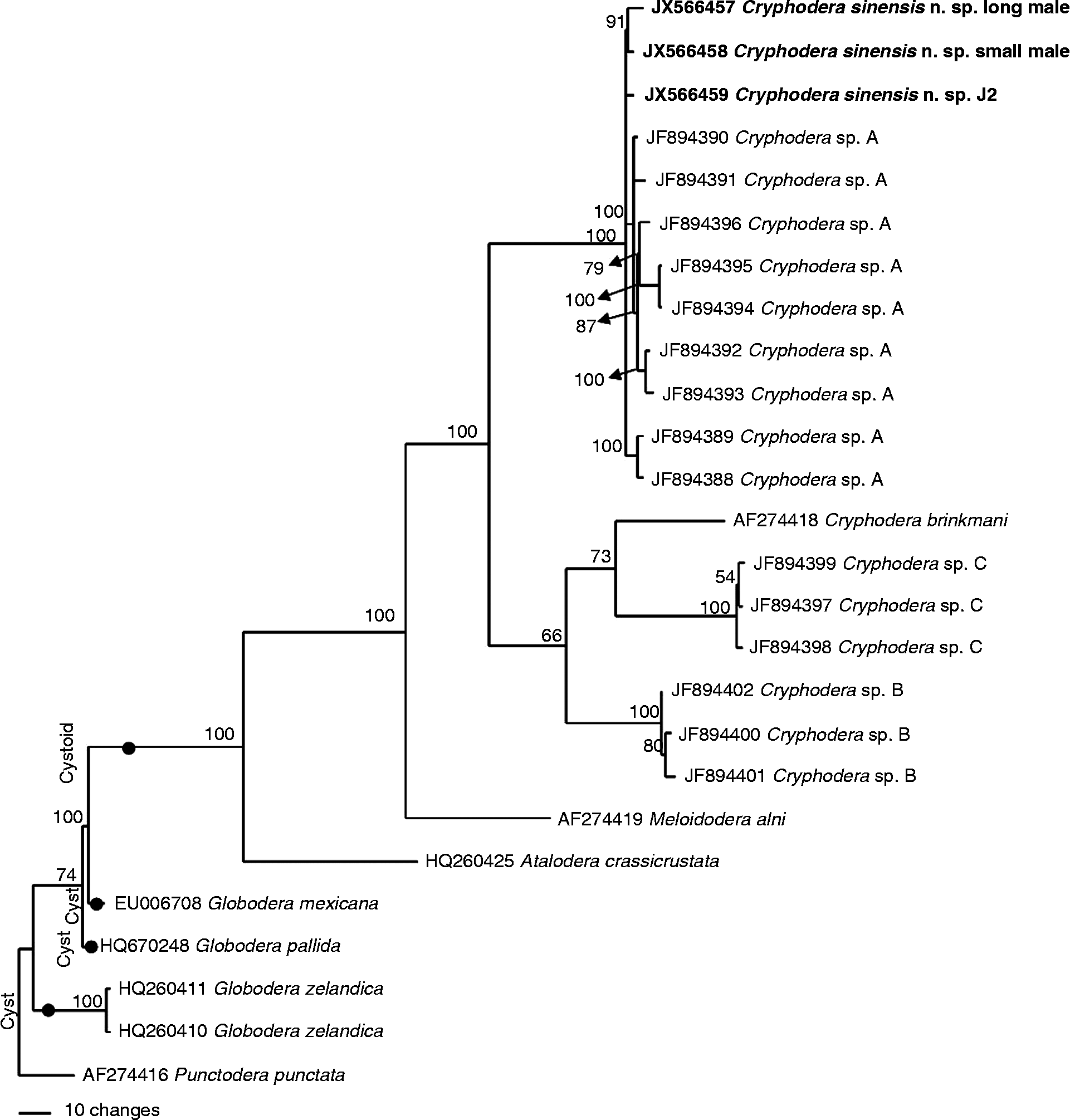
The molecular phylogenetic relationships of the new species are shown in figs 5–7. A phylogenetic tree based on SSU from a multiple alignment of 1753 total characters was constructed (fig. 5). This dataset has 1551 constant characters (88.5%). The average nucleotide composition is as follows: 23.95% A, 23.93% C, 27.49% G and 24.63% T. When using Helicotylenchus dihystera and Scutellonema bradys within the family Hoplolaimidae as outgroup taxa, the molecular phylogeny strongly supported a monophyly of Heteroderidae with 99%. Cryphodera sinensis n. sp. is in a 100%-supported monophyletic clade with C. brinkmani.
The 10,001st Bayesian tree inferred from SSU under TrN+I+G model ( − lnL = 4228.6084; freqA = 0.2395; freqC = 0.2393; freqG = 0.2749; freqT = 0.2463; R(a) = 1; R(b) = 3.3399; R(c) = 1; R(d) = 1; R(e) = 8.6593; R(f) = 1; Pinva = 0.7202; Shape = 0.7907). Posterior probability values exceeding 50% are given on appropriate clades.
The 10,001st Bayesian tree inferred from LSU D2D3 under TVMef+I+G model ( − lnL = 4735.6504; freqA = 0.1976; freqC = 0.2262; freqG = 0.3324; freqT = 0.2438; R(a) = 0.5072; R(b) = 4.9033; R(c) = 1.9702; R(d) = 0.5097; R(e) = 9.0255; R(f) = 1; Pinva = 0.4639; Shape = 0.9702). Posterior probability values exceeding 50% are given on appropriate clades.
The 10,001st Bayesian tree inferred from ITS under GTR+I+G model ( − lnL = 3759.562; freqA = 0.2117; freqC = 0.2186; freqG = 0.2789; freqT = 0.2908; R(a) = 1.0389; R(b) = 6.3099; R(c) = 1.5176; R(d) = 0.1822; R(e) = 6.3099; R(f) = 1; Pinva = 0; Shape = 0.6136). Posterior probability values exceeding 50% are given on appropriate clades.
A phylogenetic tree based on LSU D2D3 was from a multiple alignment of 887 total characters (fig. 6). This dataset has 576 constant characters (64.9%). The average nucleotide composition is as follows: 19.76% A, 22.62% C, 33.24% G and 24.38% T. When using Rotylenchus vitis and Rotylenchulus reniformis within the family Hoplolaimidae as outgroup taxa, the molecular phylogeny barely supported a monophyly of Heteroderidae with 65% support. Cryphodera sinensis n. sp. is positioned in the cluster of the family Heteroderidae and is in a 100%-supported monophyletic clade with C. brinkmani. This clade is sister to Meloidodera alni, another cystoid nematode with 100% support.
A phylogenetic tree based on ITS-rRNA was from a multiple alignment of 932 total characters (fig. 7). This dataset has 586 constant characters (62.9%). The average nucleotide composition is as follows: 21.17% A, 21.86% C, 27.89% G and 29.08% T. Using Punctodera punctata as outgroup taxon, all Cryphodera sequences reside within a well-supported monophyletic clade with 100% support. The three sequences of C. sinensis n. sp. are positioned within a clade with Cryphodera sp. A with 100% support.
Discussion
Since the genus Cryphodera and the type species C. eucalypti were proposed in 1966 (Colbran, Reference Colbran1966), five other species, namely C. podocarpi, C. nothophagi, C. coxi, C. kalesari and C. brinkmani, have been recognized in this genus (Karssen & Van Aelst, Reference Karssen and Van Aelst1999; Siddiqi, Reference Siddiqi2000). All these species were found in Oceania and Asia, e.g. C. eucalypti in Australia (Colbran, Reference Colbran1966), C. podocarpi, C. nothophagi and C. coxi in New Zealand (Wouts, Reference Wouts1973a), C. kalesari in India (Bajaj et al., Reference Bajaj, Walia, Dabur and Bhatti1989) and C. brinkmani from Japan (Karssen & Van Aelst, Reference Karssen and Van Aelst1999). In China, non-cyst-forming genera have never been reported to date. This is the first record of the non-cyst-forming genus Cryphodera in China. In this study, C. sinensis n. sp., a non-cyst-forming nematode, was obtained from Boehmeria nivea in Hunan Province, China. It's worthy to note that two obviously different sizes of males were present in the same population of C. sinensis n. sp. The longest male is about twice the length of the J2 and the smallest male is shorter than J2, but no morphological differences are observed. In order to clarify if these two very different sized males and juveniles are the same, DNA from a single longer or smaller male and J2 were sequenced separately, and LSU D2D3 (JX566454–JX566456) and ITS (JX566457–JX566459) were obtained. The sequence comparison results revealed that D2D3 and ITS sequence variations were 3–7 nucleotides (0.4–0.6%) and 17–25 nucleotides (1.1–1.5%) respectively. In Heterodera orientalis, ITS sequence variation was up to 1.4% (Mundo-Ocampo et al., Reference Mundo-Ocampo, Troccoli, Subbotin, Cid del Prado, Baldwin and Inserra2008). Intraspecific ITS sequence variation for Cryphodera sp. A was 2.5% (Nguyen et al., Reference Nguyen, Sturhan and Subbotin2011). Cryphodera sp. A was found in a survey for Heteroderidae from natural forests in Vietnam. Three Cryphodera species including Cryphodera sp. A, Cryphodera sp. B and Cryphodera sp. C could be distinguished by molecular data (Nguyen et al., Reference Nguyen, Sturhan and Subbotin2011). Interestingly, the identities of the ITS sequences from C. sinensis n. sp. and Cryphodera sp. A are 97–99% and the phylogenetic tree based on ITS-rRNA showed that C. sinensis n. sp. is in the same clade with Cryphodera sp. A with 100% support and short branch differences. Based on these data and morphological observations, molecular variation among the males of different sizes and juveniles is intraspecific, suggesting that C. sinensis n. sp. and Cryphodera sp. A are the same species.
Diagnostic morphological characters of Cryphodera were proposed by Siddiqi (Reference Siddiqi2000). Except for the shorter vulva–anus distance and the presence of cloacal tubus, other morphological characters of C. sinensis n. sp. conform to the diagnostic characters of Cryphodera. In the new species, the range of vulva–anus distance is from 29.5 to 35.8 μm, but the minimum vulva–anus distance is 38 μm in other Cryphodera species. Moreover, males of C. sinensis n. sp. have cloacal tubus v. no cloacal tubus in other Cryphodera species. A similar difference was also reported in Heterodera. Males of only some species of Heterodera have cloacal tubus (Subbotin et al., Reference Subbotin, Mundo-Ocampo, Baldwin, Hunt and Perry2010b). The phylogenetic analysis placed C. sinensis n. sp. in a 100%-supported monophyletic clade with C. brinkmani based on SSU and LSU D2D3 and in a 100%-supported clade of Cryphodera including Cryphodera sp. A, Cryphodera sp. B, Cryphodera sp. C and C. brinkmani based on ITS. Therefore, the presence of a cloacal tube is variation at the species level. In the genus Cryphodera, the perineal pattern was only previously examined for C. brinkmani (Karssen & Van Aelst, Reference Karssen and Van Aelst1999). Striae between the vulva and anus in C. sinensis n. sp. are less than those in C. brinkmani. The perineal pattern in the genus Meloidogyne is an important diagnostic character to distinguish some species. Unfortunately, this feature has not been examined for other Cryphodera species and needs further study. Through this study, C. sinensis n. sp. represents the seventh Cryphodera species. Due to this addition, two morphological characters of the genus Cryphodera need to be updated, i.e. range of the vulva–anus distance could be 29.5 to 72 μm and a cloacal tube could be absent or present.
About nine genera within Heteroderidae are non-cyst-forming genera according to Siddiqi (Reference Siddiqi2000) including Cryphodera, Meloidodera and Atalodera. Different classifications among these three genera were proposed. For instance, Wouts (Reference Wouts1972, Reference Wouts1973a, Reference Woutsb) placed Cryphodera and Meloidodera in the subfamily Meloidoderinae and Atalodera in Ataloderinae; however, Siddiqi (Reference Siddiqi2000) proposed Cryphodera and Atalodera in the subfamily Ataloderinae and Meloidodera in the subfamily Meloidoderinae. In our phylogenetic trees based on LSU D2D3 and ITS, Cryphodera is more closely related to Meloidodera than Atalodera, which is similar to the tree inferred from ITS1 rRNA gene reported by Vovlas et al. (Reference Vovlas, Trisciuzzi, Troccoli, De Luca, Cantalapiedra-Navarrete and Castillo2013). Unfortunately, few DNA sequence data are available for non-cyst-forming genera. As more sequence data become available, the classification of non-cyst-forming nematodes in the Heteroderidae should be re-evaluated.
Key to species within the genus Cryphodera
1 – J2 with three lip annuli2
– J2 with four to five lip annuli4
2–Vulva–anus distance < 40 μm; male present, with cloacal tubus; tail of J2 with point, mucro-like tipC. sinensis n. sp.
–Vulva-anus distance ≥ 40 μm; male absent, if present, lacking cloacal tubus; tail of J2 with rounded terminus3
3–Female with one lip annulus; stylet length of J2 ≥ 31 μmC. eucalypti
– Female with two to three lip annuli; stylet length of J2 < 30 μmC. kalesari
4 – Male with three incisures in lateral field; tail of J2 with point, mucro-like tipC. brinkmani
– Male with four incisures in lateral field; tail of J2 with narrow rounded terminus5
5 – J2 with five lip annuliC. podocarpi
– J2 with four lip annuli6
6 – J2 with conical lip region and distinct flat labial disc; stylet knobs of J2 flat anteriorlyC. nothophagi
– J2 with hemispherical lip region and inconspicuous labial disc; stylet knobs of J2 slightly pointing anteriorlyC. coxi
Acknowledgements
This research was supported by the special fund for agro-scientific research in the public interest of China with grant no. 200903040.