Introduction
Scinax Wagler, 1830 is a genus of anuran amphibians of the family Hylidae, widely distributed across the Americas and occurring from eastern and southern Mexico to Argentina, Uruguay and the islands of St. Lucia and Trinidad as well as Tobago (Frost, Reference Frost2019). The genus comprises about 97 recognized species that occur in tropical and subtropical areas, with a particular diversity in the Atlantic forests of south-eastern Brazil (Cardoso & Pombal, Reference Cardoso and Pombal2010). Scinax gr. ruber (Laurenti, 1768) is a nocturnal species usually found in open biotopes with short vegetation and, more rarely, around temporary ponds in the forests. However, it is often observed in anthropic landscapes such as rural and urban environments (Solís et al., Reference Solís, Ibáñez and Jaramillo2010).
The genus Rhabdias Stiles & Hassal, 1905 includes about 83 species of nematodes parasitic in the lungs of amphibians and reptiles worldwide; 17 recognized species were reported from Neotropical hosts (Kuzmin & Tkach, Reference Kuzmin and Tkach2019).
After the examination of nematodes found in amphibians collected in the Gunma Ecological Park in Santa Bárbara municipality, state of Pará, Brazil, we found some individuals of S. gr. ruber harbouring lung parasites of the genus Rhabdias, which were morphologically distinct from their congeners. Herein, we describe these nematodes as a new species based on morphological data and provide its molecular differentiation using sequences of the partial mitochondrial Cytochrome c Oxidase subunit I (CO1) gene.
Material and methods
Host collection and morphological study
Seven specimens of S. gr. ruber were collected by hand in the Gunma Ecological Park, Santa Bárbara municipality, state of Pará, Brazil, in April 2016. The anurans were anesthetized and euthanized with 2% lidocaine hydrochloride and their internal organs examined under a stereomicroscope at the Laboratory of Cell Biology and Helminthology of the Federal University of Pará. Nematodes were rinsed in saline and fixed in hot 70% alcohol. Two specimens of nematodes were transferred to 100% ethanol and stored in a freezer at −20°C for subsequent molecular analysis.
After fixation, nematodes were cleared in lactophenol and mounted on temporary slides for morphological analysis. Measurements were taken using an Olympus BX41 microscope (Olympus, Tokyo, Japan) equipped with a drawing tube, and photomicrographs were taken under an Olympus BX53 microscope, equipped with differential interference contrast optics and a digital imaging system. The type specimens were deposited in the invertebrate collection of the Museu Paraense Emílio Goeldi (MPEG), Belém, state of Pará, Brazil. For comparative studies, we also examined the type specimens (holotype and two paratypes) of Rhabdias tobagoensis Moravec & Kaiser, Reference Moravec and Kaiser1995 stored in the Helminthological Collection of the Institute of Parasitology, Biology Centre CAS (České Budějovice, Czech Republic). All measurements are presented as the value of the holotype, followed by the mean of the paratypes, followed by the range in parentheses; measurements are given in micrometres, unless otherwise indicated.
Molecular analyses and phylogenetic study
DNA was isolated from one of the specimens using the QIAGEN QIAamp DNA Mini Kit (Hilden, Germany) and following the manufacturer's instructions. Polymerase Chain Reaction (PCR) was conducted to amplify the partial mitochondrial CO1 gene, according to the methodology and primers proposed by Prosser et al. (Reference Prosser, Velarde-Aguilar, León-Règagnon and Hebert2013). After verification by agarose gel electrophoresis, successfully amplified PCR products were purified with the QIAGEN QIAquick PCR Purification Kit, according to the manufacturer's instructions, and cycle-sequenced using the Applied Biosystems™ BigDye™ Terminator v3.1 Cycle Sequencing Kit (Carlsbad, Califórnia, USA). All these steps were conducted in the Laboratory of Biology and Parasitology of Wild Reservoir Mammals at the Oswaldo Cruz Institute, Oswaldo Cruz Foundation (LABPMR/IOC/Fiocruz), Rio de Janeiro, Brazil. Sequences were run on an Applied Biosystems™ 3730 DNA Analyzer at the DNA Sequencing Platform of the Oswaldo Cruz Foundation (RPT01A/PDTIS/FIOCRUZ). Forward and reverse sequences were assembled into a contig and edited for errors and ambiguities with the Geneious 9.1.8 suite (https://www.geneious.com). In addition, a matching CO1 sequence of Rhabdias pseudosphaerocephala Kuzmin, Tkach & Brooks, Reference Kuzmin, Tkach and Brooks2007 was obtained in the University of North Dakota from the same sample as the type series of the species originally described by Kuzmin et al. (Reference Kuzmin, Tkach and Brooks2007), following the protocols published by Machado et al. (Reference Machado, Kuzmin, Tkach, Santos, Gonçalves and Melo2018).
For molecular phylogenetic analyses, we compared our sequence with 27 publicly available CO1 sequences from Rhabdias spp. and the newly obtained sequence of R. pseudosphaerocephala from Rhinella marina (Linnaeus, 1758) (Bufonidae) from Nicaragua. Two CO1 sequences of Serpentirhabdias Tkach, Kuzmin & Snyder, 2014 available in GenBank were used as outgroups. We built another matrix for pairwise nucleotide comparisons with 67 publicly available CO1 sequences of Rhabdias spp. and our new sequences. The sequences were aligned using default parameters of the program MUSCLE (Edgar, Reference Edgar2004) under Geneious 9.1.8 (https://www.geneious.com), and the resulting alignment was trimmed to the length of the shortest sequence. Amino acid translation was used to confirm reading frame positions and find unexpected stop codons.
We carried out phylogenetic reconstructions using the maximum likelihood (ML) algorithm as implemented in PhyML 3.0 (Guindon et al., Reference Guindon, Dufayard, Lefort, Anisimova, Hordijk and Gascuel2010). The Nucleotide evolutionary model was chosen based on the Akaike information criterion (AIC) using the smart model selection algorithm (Lefort et al., Reference Lefort, Longueville and Gascuel2017) within PhyML. Node support was assessed by the approximate likelihood ratio test for branches, non-parametric branch support based on a Shimodaira–Hasegawa-like (aLRT SH-like) procedure (Anisimova and Gascuel, Reference Anisimova and Gascuel2006). The best-fit model chosen by AIC on PhyML was the general time reversible (GTR) model including estimates of the gamma (G) distributed among-site rate variation model of nucleotide substitution.
We also carried out a Bayesian phylogenetic inference (BI) analysis using MrBayes version 3.2.6 (Ronquist et al., Reference Ronquist, Van Der Mark and Teslenko2012) on XSEDE in the CIPRES Science Gateway (Miller et al., Reference Miller, Pfeiffer and Schwartz2010). To account for different evolutionary processes at each of the three codon positions, BI analyses were performed with distinct models per codon position chosen by the Bayesian information criterion (BIC) using the program PAUP* version 4.0a147 (Swofford, Reference Swofford2002). We performed Markov chain Monte Carlo samplings for 10,000,000 generations with four simultaneous chains in two runs. Node support was assessed by Bayesian posterior probabilities (BPP) calculated from tree samples every 100 generations, after the removal of a ‘burn-in’ fraction of 25%. We assessed the robustness of our sampling using the program Tracer v1.7.1 (Rambaut et al., Reference Rambaut, Drummond, Xie, Baele and Suchard2018) to calculate the effective sample sizes (ESS) of parameters. Values above 100 effectively independent samples were considered sufficiently sampled.
Uncorrected pairwise p-distances were calculated using the program PAUP* version 4.0a147 (Swofford, Reference Swofford2002).
Results
Systematics
Family: Rhabdiasidae Railliet, 1915
Rhabdias glaurungi sp. nov.
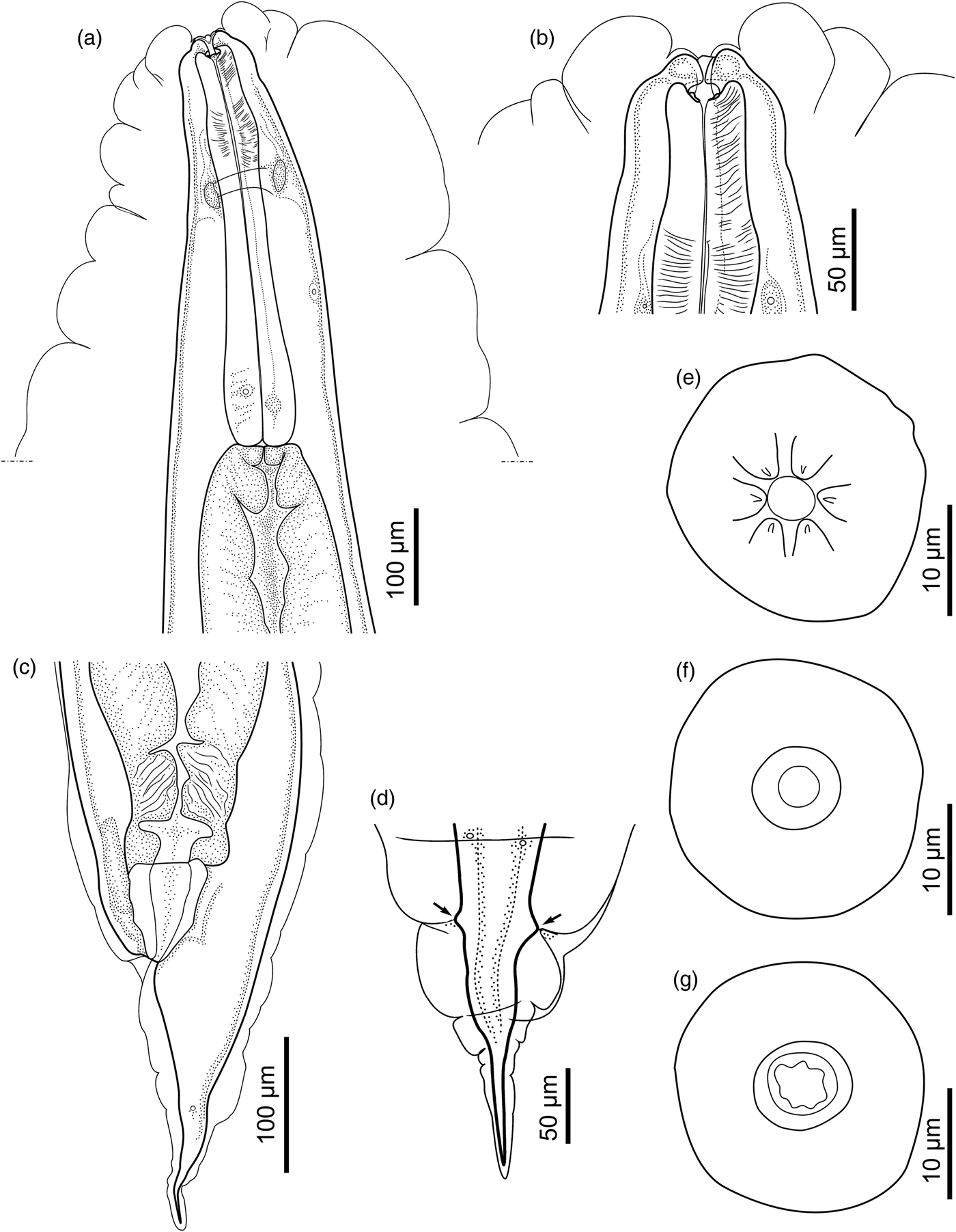
Line drawings of Rhabdias glaurungi sp. nov. (a) Anterior part of body, lateral view; (b) anterior end, lateral view; (c) posterior part of body, lateral view; (d) caudal end, dorso-ventral view, showing phasmids (arrowheads); (e) anterior end of body, en face view; (f) optical section through vestibulum; (g) optical section through posterior part of buccal capsule.

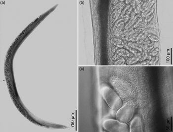
Light microscopical photographs of Rhabdias glaurungi sp. nov. using differential interference contrast optics. (a) General view; (b) region of vulva, lateral view; (c) U-bend of posterior seminal receptacle.

Taxonomic summary
Type host. Scinax gr. ruber (Laurenti, 1768) (Hylidae)
Localization in host. Lungs.
Type locality. Gunma Ecological Park, Santa Bárbara, Pará, Brazil, (1°12′25.08″S, 48°15′02.42″W).
Type material. Holotype (MPEG 00219); Paratypes (MPEG 00220).
Infection parameters. One out of seven infected S. gr. ruber harboured 14 nematodes.
GenBank Accession number. MK820652.
ZooBank registration. The Life Science Identifier (LSID) for R. glaurungi n. sp. is 95B7CA7E-E970-41E1-B452-E89799ED92F0.
Etymology. The new species is named after the fictional character ‘Glaurung’, the first of the Dragons from J.R.R. Tolkien's mythopoeic collection ‘The Silmarillion’.
Description
Based on holotype and seven paratypes, all gravid hermaphrodites. Body 5.04; 4.84 (3.54–6.73) mm long, gradually tapering from mid-region to anterior and posterior extremities (fig. 2a). Anterior end rounded (fig. 1b), posterior end pointed (fig. 1c, d). Body width at vulva 291; 293 (237–344), width at oesophago–intestinal junction 112; 133 (112–187). Cuticle inflated along entire body. Lateral pores and ducts present.
At anterior end, rounded dilatation of body wall present at level of oesophagus anterior end; dilatation separated from narrower part surrounding lips and posterior part of body by slight constrictions (fig. 1b). Oral opening round. Six small lips of identical shape and size present on apical surface, close to edge of oral opening. Each lip bearing prominent papilla on its inner edge (fig. 1e). Amphidial openings located posterior to lateral lips.
Buccal capsule cup-shaped (fig. 1b). Anterior part of buccal capsule circular in shape, with smooth internal wall (fig. 1f), posterior part with irregularly folded internal surface (fig. 1g). Maximum diameter of buccal capsule 14; 13 (10–16), total depth of both parts 7; 7 (6–9); depth-to-width ratio 0:5; 0:55 (0.45–0.66). Oesophagus club-shaped, 314; 310 (292–332) long (6.2; 6.4% (4.9–8.2%) of body length), with distinct dilatation in middle of anterior half and with posterior bulb. Width of oesophagus anterior end 30; 27 (24–30), maximum width of anterior dilatation 39; 37 (34–43), width posterior to dilatation 32; 31 (29–34). In posterior half, oesophagus gradually widening towards bulb 55; 58 (53–66) wide (fig. 1a).
Nerve ring encircling oesophagus just posterior to its anterior dilatation, anterior to its mid-length, at 112; 115 (108–140) from anterior end; this distance corresponding to 36; 37% (36–42%) of oesophagus length. Excretory pore not observed. Intestine thick-walled, its anterior part much wider than oesophageal bulb. Rectum funnel-shaped, wide in anterior part, tapering posteriorly, prominently cuticularized (fig. 1c).
Reproductive system amphidelphic, typical of the genus. Vulva post-equatorial, situated at 2.9; 3 (2.9–3.2) mm from anterior end (56; 53% (50.8–57.3%) of body length). Lips of vulva not salient (fig. 2b). Vagina short, transverse, cuticularized. Uteri joined, thin-walled, containing numerous eggs (>100 in total); eggs near vulva with fully developed embryos. Egg size 99; 101 (96–103) × 50; 50 (49–52) (N = 11, measured in uteri of holotype and paratypes). Both genital tubes U-bent in proximal parts of seminal receptacles (fig. 2c). Distance from anterior end of body to anterior U-bend 747; 708 (553–816), distance from posterior U-bend to tail end 687; 630 (489–687). Testis zones observed in all specimens, in at least one syngonium. Numerous sperm cells usually observed in seminal receptacles. Proximal parts of syngonia overlapping level of vulva.
Tail narrow, gradually tapering. Posterior end pointed (fig. 1c). Slight elevation of body wall present on ventral side posterior to anus. Tail length 211; 185 (151–216), or 4.2%; 3.8% (3.2–4.3%) of body length. Phasmids located posterior to mid-length of tail (fig. 1d).
Remarks
Rhabdias glaurungi sp. nov. is assigned to the genus Rhabdias of Rhabdiasidae based on the presence of the following morphological characters: inflated body cuticle, small buccal capsule, amphidelphic reproductive system with short transverse vagina and joined uteri; and due to its parasitism in the lungs of amphibian hosts.
Based on the limited geographic range of distribution of Rhabdias species, we decided to compare our new species only with those reported for the Neotropics. However, among the 17 species parasitizing amphibians from the Neotropics, the data regarding the hermaphroditic forms of R. mucronata Schuurmans-Stekhoven, Reference Schuurmans-Stekhoven1952 and R. truncata Schuurmans-Stekhoven, Reference Schuurmans-Stekhoven1952 parasitic in amphibians are unavailable, Schuurmans-Stekhoven (Reference Schuurmans-Stekhoven1952) based the descriptions only on juvenile forms found in the body cavity of the host; thus, we did not use this species in our comparisons.
Rhabdias glaurungi sp. nov. differs from R. alabialis Kuzmin, Tkach & Brooks, Reference Kuzmin, Tkach and Brooks2007 parasitic in R. marina in the presence of distinct lips and papillae on the apical surface. Lips and papillae are absent in the latter species, and the oral opening is slit-like (Kuzmin et al., Reference Kuzmin, Tkach and Brooks2007).
Rhabdias androgyna Kloss, Reference Kloss1971 parasitic in Rhinella cf. margaritifera (Laurenti) (Bufonidae) is much larger than R. glaurungi sp. nov. in both length and width (about 7.9–15 mm long and 269–473 µm wide) but similar to the new species considering the relative proportions of the oesophagus, position of the nerve ring and the vulva. Despite that, R. androgyna differs from the new species by a wider but shallower buccal capsule (19–27 µm wide, 7–9 µm deep, 0.29–0.47 depth-to-width ratio) and by the particular shape of the anterior part of the body, which consists in a shoulder-like circular dilatation with an extended cuticular swelling disposed in two layers (Kloss, Reference Kloss1971; Kuzmin et al., Reference Kuzmin, Du Preez and Junker2015).
In comparison to the new species, Rhabdias breviensis Nascimento, Goncalves, Melo, Giese, Furtado & Santos, 2013 described by Nascimento et al. (Reference Nascimento, Gonçalves, Melo, Giese, Furtado and Santos2013) from Leptodactylus petersii (Steindachner, 1864) (Leptodactylidae) has a particular, curved body shape, and smaller but remarkably wider body (about 2.63–3.63 mm long and 370–543 µm wide at vulva level). Also, R. breviensis has a comparatively smaller buccal capsule (7–13 µm wide, 4–9 µm deep, 0.6–0.7 depth-to-width ratio), a smaller distance of the nerve ring from the anterior end (41–84 µm) and a distinctly post-equatorial vulva, with the distance from anterior end to vulva corresponding to 65–71% of the body length.
Rhabdias elegans Gutierrez, 1945, known from various anurans, mostly bufonids, in the southern parts of South America (Gonzáles & Hamann, Reference González and Hamann2009; Kuzmin et al., Reference Kuzmin, Du Preez and Junker2015), differs from R. glaurungi sp. nov. by its larger body size (4.55–9.5 mm long, after Kloss, Reference Kloss1974). Despite oesophagus length being similar in both species, maximum oesophagus width is generally smaller in R. elegans (42–59 µm, after Kloss, Reference Kloss1974) than in the new species (53–66 µm). In addition, the two species differ in buccal capsule size: 7 × 9 µm in R. elegans (after Kloss, Reference Kloss1974) vs. 13 × 7 µm (on average) in R. glaurungi sp. nov.
Rhabdias fuelleborni Travassos, Reference Travassos1926 is parasitic in bufonid hosts. Despite being much larger (up to 10–12 mm or 16.3 mm) than R. glaurungi sp. nov., it resembles the new species considering the relative proportions of the oesophagus and the relative position of the nerve ring and distance of the vulva from the anterior end. Rhabdias fuelleborni also has a slightly narrower portion at the level of the oesophageal apex, similar to that in R. glaurungi sp. nov. However, R. fuelleborni has a cuticular inflation more distinct in the anterior part, a comparatively longer tail (about 313–345 µm corresponding to 4.7–5.4% of body length) and a wider buccal capsule (18–19 µm vs. 10–16 µm) (Travassos, Reference Travassos1926; Kloss, Reference Kloss1971; Kuzmin et al., Reference Kuzmin, Du Preez and Junker2015).
Rhabdias galactonoti Kuzmin, Melo, Filho & Santos, 2016 from Adelphobates galactonotus (Steindachner, 1864) (Dendrobatidae) is slightly larger than R. glaurungi sp. nov., but similar in some metrical characters like the relative dimensions of the oesophagus and the tail, the position of the nerve ring and the position of the vulva. However, in R. galactonoti the inner surface of the anterior part of the buccal capsule is regularly folded (Kuzmin et al., Reference Kuzmin, Melo, Silva Filho and Santos2016), whereas in R. glaurungi sp. nov. it is smooth. The position of the vulva in R. galactonoti is pre-equatorial and its tail is narrower and more elongated when compared to the tail in the new species.
Rhabdias hermaphrodita Kloss, Reference Kloss1971 was described from Rhinella crucifer (Wied-Neuwied, 1821) (syn. Bufo crucifer Wied-Neuwied, 1821) in south-eastern Brazil (Kloss, Reference Kloss1971, Reference Kloss1974). The species is larger than R. glaurungi sp. nov., with a body length of up to 12.9 mm (Kloss, Reference Kloss1971). Though R. hermaphrodita was rather superficially described (no information on apical structures, no measurements of the buccal capsule), it follows from the images presented by Kloss (Reference Kloss1974, Figs 41–49) that the cuticle is inflated mostly in the anterior part of the body and the rounded dilatation of the body wall at the anterior end is absent. In R. glaurungi sp. nov. the cuticular inflation covers the entire body and the anterior dilatation is present.
Rhabdias kuzmini Martinez-Salazar & Leon-Regagnon, 2007 from a bufonid host Incilius occidentalis (syn. Bufo occidentalis) (Camerano, 1879) Camerano (Martínez-Salazar & León-Règagnon, Reference Martínez-Salazar and León-Règagnon2007) differs from R. glaurungi sp. nov. by the much larger body size (about 14.2–19.2 mm long), the presence of two lateral pseudolabia instead of the lateral lips, the larger buccal capsule (35–47 µm wide and 31–70 µm deep), the truncated shape of the anterior end and the cuticular swelling more prominent mostly in the anterior and posterior parts of the body (Martínez-Salazar & León-Règagnon, Reference Martínez-Salazar and León-Règagnon2007).
In comparison to R. glaurungi sp. nov., R. manantlanensis Martinez-Salazar, 2008 from Craugastor occidentalis (Taylor, 1941) (Brachycephalidae) (Martínez-Salazar, Reference Martínez-Salazar2008) has a larger body size (up to 6.48–9.64 mm long), a larger buccal capsule (19–27 µm wide) that is completely surrounded by the anterior part of the oesophagus, a nerve ring that is situated in the mid-region of the oesophagus (the distance from anterior end to nerve ring corresponding to 47.4–49.9% of oesophagus length) and a shorter tail (143–232 µm long, or 1.5–3.3% of body length). Further differences between the two species are the mostly pre-equatorial position of the vulva and the body cuticle that is not or only slightly inflated in R. manantlanensis (Martínez-Salazar, Reference Martínez-Salazar2008).
Rhabdias paraensis Santos, Melo, Nascimento, Nascimento, Giese & Furtado, 2011 from R. marina is geographically close to the new species and possesses a similar cuticular inflation and dilatation of the body wall in the anterior part (Santos et al., Reference Santos, Melo, Nascimento, Nascimento, Giese and Furtado2011). However, the constriction anterior to the dilatation is absent and the lips are less distinct in R. paraensis (Santos et al., Reference Santos, Melo, Nascimento, Nascimento, Giese and Furtado2011; Melo et al., Reference Melo, Nascimento, Macedo, Santos and Kuzmin2016). The buccal capsule is smaller in R. glaurungi sp. nov. (10–16 µm wide, 6–9 µm deep) than in R. paraensis (15–28 µm wide, 4–17 µm deep). The oesophageal bulb is significantly wider in R. paraensis (75–91 µm) than in the new species (53–66 µm).
According to Martínez-Salazar et al. (Reference Martínez-Salazar, Falcón-Ordaz, González-Bernal, Parra-Olea and León2013), R. peninsularis Martinez-Salazar, Falcon-Ordaz, Gonzalez-Bernal, Parra-Olea & Perez-Ponce de Leon, 2013 from a hylid host Pseudacris hypochondriaca Hallowell (1854) has a longer body (5.84–7.28 mm) than R. glaurungi sp. nov. but a shorter oesophagus (334–430 µm long), corresponding to 5.56–6.4% of the body length, and a shorter tail (about 139–213 µm long), representing 2.16–3.39% of the body length. The cuticle in R. peninsularis is inflated mostly at the anterior and posterior ends and comparatively thin and smooth in the mid-region. Besides that, R. peninsularis has an almost globular and larger buccal capsule (15 deep and 15–23 µm wide, 1–1.5 depth-to-width ratio).
Rhabdias pseudosphaerocephala described by Kuzmin et al. (Reference Kuzmin, Tkach and Brooks2007) from R. marina is larger in size (body length 6.17–9.6 mm) but similar to R. glaurungi sp. nov. considering its relative morphometrical dimensions. In contrast to the new species, R. pseudosphaerocephala possesses two lateral pseudolabia and four submedian lips, with prominent grooves on the dorsal and ventral sides of the apical surface (Kuzmin et al., Reference Kuzmin, Tkach and Brooks2007; fig. 3c).
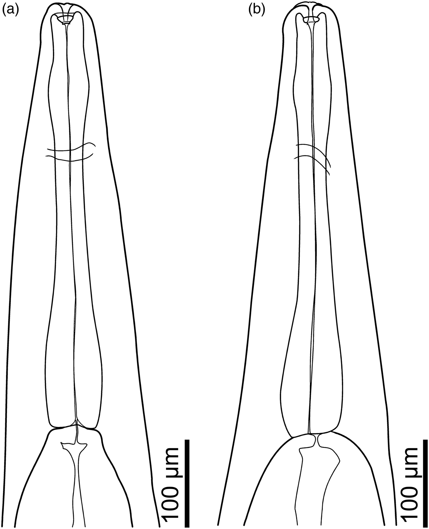
Line drawings of Rhabdias tobagoensis, paratypes. (a) Anterior part of body, dorso-ventral view; (b) anterior part of body, lateral view.

Rhabdias savagei Bursey & Goldberg, Reference Bursey and Goldberg2005 parasitic in a host belonging to the Ranidae (Bursey & Goldberg, Reference Bursey and Goldberg2005) can be easily distinguished from R. glaurungi sp. nov. by the smooth cuticular inflation, the absence of a dilatation in the muscular portion of the oesophagus and the narrower oesophageal bulb (43–52 µm wide vs. 53–66 µm, respectively). The buccal capsule in R. savagei is wider (18–24 µm).
According to Kuzmin et al. (Reference Kuzmin, Melo, Silva Filho and Santos2016), the anterior end of R. stenocephala Kuzmin, Melo, Filho & Santos, 2016 from Leptodactylus spp. (Leptodactylidae) has a particular shape: the narrow anterior part of the body (not considering the cuticular inflation) is separated from the remaining body by a constriction at the level of the anterior part of the oesophagus. Posterior to this constriction, there is an abrupt widening of the body wall. Those characters are not observed in R. glaurungi sp. nov. The buccal capsule in R. stenocephala is similar in size to that in R. glaurungi sp. nov. (15–18 µm wide); however, the inner surface of its anterior part is irregularly folded in apical view, whereas it is smooth in the new species.
Rhabdias tobagoensis parasitic in Pristimantis cf. terraebolivaris (Rivero, 1961) (Craugastoridae) is most similar to R. glaurungi sp. nov. in the shape and dimensions of its body, oesophagus and tail, though the type specimens of R. tobagoensis are somewhat larger (7.34–7.56 mm; after Moravec and Kaiser, Reference Moravec and Kaiser1995) than specimens of the new species. Rhabdias tobagoensis has a wider buccal capsule (18–21 µm), whereas its depth is approximately the same (6–9 µm) as in the new species. The posterior end of the oesophagus in R. tobagoensis is distinctly flattened and even concave, while it is rounded in R. glaurungi sp. nov. The latter feature was not mentioned in the description of R. tobagoensis (Moravec & Kaiser, Reference Moravec and Kaiser1995); however, it was obvious in the type specimens of the species examined in the present study (fig. 3a, b). The oesophagus is longer and more slender in R. tobagoensis (467–485 µm long, 45 µm wide at anterior dilatation and 64–69 µm wide at posterior bulb). Rhabdias tobagoensis lacks the constriction at the anterior end separating the rounded dilatation from the anteriormost part surrounding the lips, as seen in the new species.
An unidentified species of Rhabdias recovered from the hylid frog Scinax staufferi Cope (1865) in Mexico and described by Martínez-Salazar et al. (Reference Martínez-Salazar, Pérez-Ponce de León and Parra Olea2009) is similar to R. glaurungi sp. nov. in body shape and dimensions as well as in the presence of six circumoral lips. It differs from the new species in the more pronounced inflation of the body cuticle at the anterior end, the indistinct dilatation in the muscular part of the oesophagus, a wider buccal capsule (19 µm) and a longer tail (251 µm).
Molecular analyses and phylogenetic study
Alignments of sequences resulted in a matrix comprising 31 taxa and 597 characters, of which 391 were constant and 164 were parsimony-informative variable characters for phylogenetic analyses; and a matrix comprising 69 taxa and 432 characters, of which 274 were constant and 133 were parsimony-informative variable characters for distance analyses.
The evolutionary models selected through BIC for the BI were: TrN + I + G in the first codon positions, HKY + I in the second codon positions and HKY + G in the third codon positions, all models with unlinking state frequencies and parameters. Markov chains provided highly significant estimated ESS values for all parameters.
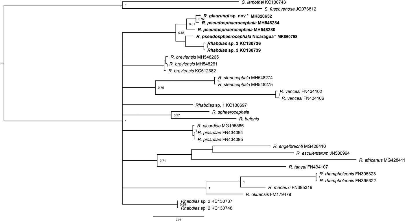
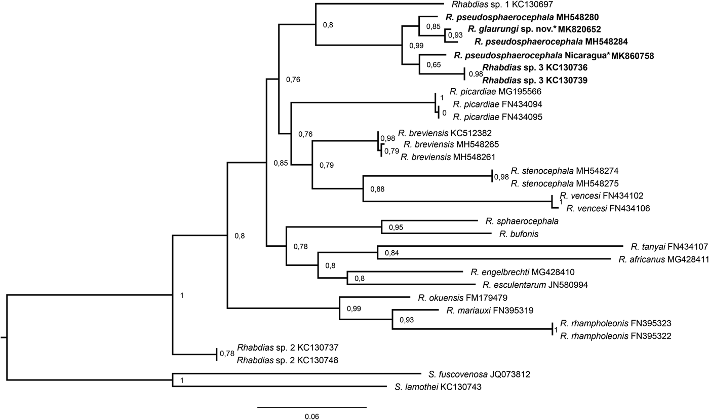
Our phylogenies, based on the CO1 sequences and inferred using two different methods (ML and BI), resulted in generally similar topologies with little variation in nodes and support values (figs 4 and 5).
Phylogenetic relationships among 31 rhabdiasid taxa resulting from maximum likelihood analysis of partial sequences of the mitochondrial CO1 gene. Taxa in bold correspond to the new species and Rhabdias pseudosphaerocephala species complex. Support values (aLRT SH-like) are indicated at the nodes. New sequences obtained in this study are indicated by an asterisk.

Phylogenetic relationships among 31 rhabdiasid taxa resulting from Bayesian inference (10,000,000 generations) of partial sequences of the mitochondrial CO1 gene. Taxa in bold correspond to the new specie and Rhabdias pseudosphaerocephala species complex. Support values (BPP) greater than 70% are indicated at the nodes. New sequences obtained in this study are indicated by an asterisk.

In all topologies, our CO1 sequence of R. glaurungi sp. nov. nested within a clade with sequences of R. pseudosphaerocephala. The sequence of the new species formed a well-supported clade with a sequence of R. pseudosphaerocephala from the north-east of Brazil (aLRT SH-like = 93%, BPP = 97%). Rhabdias glaurungi sp. nov + R. pseudosphaerocephala from the north-east of Brazil formed a moderately well-supported monophyletic group with a sequence of R. pseudosphaerocephala from the south of Brazil (aLRT SH-like = 85%, BPP = 81%). The sequence of R. pseudosphaerocephala from R. marina from Nicaragua formed a weakly supported clade with sequences of Rhabdias sp. 3 from R. marina from Colima, Mexico (aLRT SH-like = 65%, BPP = 52%). This group formed a clade with R. glaurungi sp. nov. + R. pseudosphaerocephala from Brazil with moderate to strong support (aLRT SH-like = 99%, BPP = 85%).
Pairwise p-distance comparisons between CO1sequences of Rhabdias species showed significant differences between the sequence from R. glaurungi sp. nov. and most publicly available CO1 sequences from other species (see supplementary table S1). Of the 67 publicly available CO1 sequences of Rhabdias, 28 sequences differed from R. glaurungi sp. nov. by more than 10%. These sequences are mostly from African hosts, except for R. cf. stenocephala from leptodactylid hosts in Ceará, Brazil and Rhabdias sp. 4 from bufonids in São Paulo, Brazil (see supplementary table S1).
Twenty-two of the remaining sequences showed greater than 7% divergence from R. glaurungi sp. nov. These sequences include nematodes of hosts from Cameroon, Tanzania, Italy and Mexico; Rhabdias sp. 5 from a dactyloid host in Ceará and R. breviensis from leptodactylid, hylid and bufonid hosts in different localities in Brazil (see supplementary table S1).
Lower divergence levels were observed in sequences of Rhabdias sp. 3 from a bufonid host in Mexico, which had about 5% divergence from R. glaurungi sp. nov.
The levels of genetic divergence between R. glaurungi sp. nov. and sequences of R. pseudosphaerocephala were as follows: 3% from a bufonid host in Mexico; 2–3% from bufonid and hylid hosts in the Brazilian Amazon, south and south-east of Brazil; and 1–2% from bufonid hosts in the Brazilian north-east. Intraclade genetic distances ranged from 0 to 1% within the Amazon–south–south-east clade as well as within the north-east clade (see supplementary table S1).
Sequences of R. breviensis split into two clades (figs 4 and 5). The genetic distances between these clades ranged from 1 to 2%, whereas distances within clades were only 0–1%. Rhabdias cf. stenocephala formed two clades as well. Similar to R. breviensis, the genetic distances between clades ranged from 1 to 2% and the distances within clades ranged from 0 to 1%.
Discussion
Rhabdias glaurungi sp. nov. is the 18th nominal species of the genus Rhabdias in the Neotropical region. Species of the genus Rhabdias are characterized by a high level of morphological uniformity. According to Kuzmin et al. (Reference Kuzmin, Tkach and Snyder2003), the number, shape and position of the circumoral lips and pseudolabia are among the essential characters that can be used for specific diagnosis of the genus. Nascimento et al. (Reference Nascimento, Gonçalves, Melo, Giese, Furtado and Santos2013) also emphasized the structure of the lips, the relative position of the vulva and the shape of the tail as distinguishing characters.
From the 17 valid species of the Neotropical realm, R. alabialis, R. elegans and R. paraensis lack the lips, while there is no information on the details of the apical structures of R. hermaphrodita. Despite that, the new species can be distinguished from R. hermaphrodita, which differs from the new species in the cuticular inflation being mostly in the anterior part of the body and the absence of the rounded dilatation of the body wall at the anterior end. Moravec and Sey (Reference Moravec and Sey1990) mentioned the host specificity and geographical distribution of Rhabdias species as auxiliary data for comparisons with its congeners. Thus, R. alabialis, R. elegans, R. paraensis and R. hermaphrodita that parasitize bufonid toads, are both morphologically and biologically distinct from the parasitic nematodes found in S. gr. ruber.
Among the remaining species, R. androgyna, R. fuelleborni, R. kuzmini and R. pseudosphaerocephala are known mostly from bufonid hosts, whereas R. breviensis and R. stenocephala are from the Leptodactylidae, R. manantlanensis and R. tobagoensis are from the Craugastoridae, R. galactonoti was described from the Dendrobatidae and only R. peninsularis is known exclusively from Hylidae. These species seem to be exclusively or mostly associated with one specific amphibian family, except for R. savage, which was reported from the Ranidae, Craugastoridae and Leptodactylidae (Bursey & Goldberg, Reference Bursey and Goldberg2005; Bursey & Brooks, Reference Bursey and Brooks2010). In the latter case, however, there is no molecular evidence of the conspecificity of the forms parasitic in members of different anuran families.
The low number of Rhabdias species known from hylids might be related to their mostly arboreal lifestyle, which makes infection by Rhabdias larvae less likely. However, unlike most hylids, S. gr. ruber usually inhabits open biotopes with short vegetation and breeds in temporary ponds. In addition, the broad distribution, abundance and high adaptability of these frogs to altered environments may facilitate their contact with infective larvae in the soil (Solís et al., Reference Solís, Ibáñez and Jaramillo2010).
The sequence of R. glaurungi sp. nov. showed high genetic divergences compared to most presently available CO1 sequences, as shown in supplementary table S2. The high divergence values observed between our newly generated sequence and sequences from R. mariauxi Lhermitte-Vallarino & Bain, 2009, R. picardiae Junker, Lhermitte-Vallarino & Bain, Reference Junker, Lhermitte-Vallarino, Barbuto, Ineich, Wanji and Bain2010, R. rhampholeonis Lhermitte-Vallarino & Bain, 2009, R. tanyai Junker, Lhermitte-Vallarino & Bain, Reference Junker, Lhermitte-Vallarino, Barbuto, Ineich, Wanji and Bain2010, R. tarichae Kuzmin, Tkach & Snyder, Reference Kuzmin, Tkach and Snyder2003, R. vencesi Junker, Lhermitte-Vallarino & Bain, Reference Junker, Lhermitte-Vallarino, Barbuto, Ineich, Wanji and Bain2010, R. okuensis Lhermitte-Vallarino, Barbuto, Ineich, Wanji, Lebreton, Chrio & Bain, 2008, Rhabdias sp. 1 and Rhabdias sp. 2 could be explained by the geographical distance between these taxa, since these sequences were obtained from species occurring in Africa, the US and Mexico (Kuzmin et al., Reference Kuzmin, Tkach and Snyder2003; Lhermitte-Vallarino et al., Reference Lhermitte-Vallarino, Barbuto, Ineich, Wanji, Lebreton, Chirio and Bain2008; Lhermitte-Vallarino et al., Reference Lhermitte-Vallarino, Barbuto, Junker, Boistel, Ineich, Wanji and Bain2009; Junker et al., Reference Junker, Lhermitte-Vallarino, Barbuto, Ineich, Wanji and Bain2010; Langford & Janovy, Reference Langford and Janovy2013). However, the higher divergence level observed between sequences of the new species and R. cf. stenocephala from Ceará, Brazil is noteworthy and cannot be explained by geographic distance alone. These species represent two distinct phylogenetic lineages of Rhabdias evolved and are undergoing radiation in the Americas, especially in the Neotropics.
However, as stated by Müller et al. (Reference Müller, Morais, Costa-Silva, Aguiar, Ávila and da Silva2018), species of Rhabdias currently known in Brazil may include cryptic species. Although the morphology of the specimens studied by these authors allowed them to identify their nematodes as R. stenocephala, their sequences were obtained not from the type host and the type locality. Hence, the relatively high intraspecific differences (up to 2%) among sequences of R. stenocephala (see supplementary table S1) might potentially reflect the existence of an undescribed, morphologically similar species.
Different divergence levels were observed for the sequences of R. breviensis parasitic in leptodactylid, bufonid, hylid and odontophrynid hosts and collected from several areas in Brazil spanning across different Neotropical biomes. This divergence may not only reflect the geographical distance between collecting sites but may also suggest the existence of cryptic diversity within the R. breviensis species complex, as demonstrated by Müller et al. (Reference Müller, Morais, Costa-Silva, Aguiar, Ávila and da Silva2018).
Sequences from R. pseudosphaerocephala parasitic in bufonid and hylid hosts from different locations in Brazil (Amazonas, Ceará, Paraná, Piauí and São Paulo) were the most similar (genetic distance 1–3%) to the sequence of R. glaurungi sp. nov. Rhabdias pseudosphaerocephala was first described parasitizing R. marina in Nicaragua and represents a species complex according to Müller et al. (Reference Müller, Morais, Costa-Silva, Aguiar, Ávila and da Silva2018). However, R. pseudosphaerocephala differs from the new species in the number of submedian lips. Importantly, the sequence of R. pseudosphaerocephala from the same sample as the type series used in the original description diverged by 3–5% from the Brazilian forms of R. pseudosphaerocephala, by 3% from Rhabdias sp. 3 and by 3% from R. glaurungi sp. nov. Thus, although nested within R. pseudosphaerocephala, both morphological characters and DNA sequence data support the status of R. glaurungi sp. nov. as an independent species, corroborating the conclusion of Müller et al. (Reference Müller, Morais, Costa-Silva, Aguiar, Ávila and da Silva2018) that R. pseudosphaerocephala is a species complex. Therefore, considering the great diversity of amphibians and the comparatively small number of Rhabdias species described so far in the Neotropics, it is likely that a significant number of new species of Neotropical Rhabdias are awaiting discovery.
Author ORCIDs
Y. Willkens, 0000-0002-0904-5200
Supplementary material
To view supplementary material for this article, please visit https://doi.org/10.1017/S0022149X19000476.
Acknowledgements
The authors are grateful to Dr. Arnaldo Maldonado Junior for his assistance with DNA extraction and molecular sequencing, the curators of the Helminthological Collection of the Institute of Parasitology, Biology Centre CAS (České Budějovice, Czech Republic), Dr. Tomáš Scholz and Blanka Škoráková, for the collection material loaned for examination. Dr. Stephen Curran (University of Southern Mississippi, USA) helped to collect R. pseudosphaerocephala during the field trip in Nicaragua.
Financial support
This work was supported by the National Council for Scientific and Technological Development (CNPq) (grant number 431809/2018-6 Universal); Productivity Scholarship Grant (CNPq) to J.N. Santos and F.T.V. Melo. CNPq (process: 304955/2018-3).
Conflicts of interest
None.
Ethical standards
The authors assert that all procedures contributing to this work comply with all applicable institutional, national and international guidelines for the care and use of animals. Specimens were collected under license 0004/06 NUC SUPES PA, project ‘Biodiversity survey of the herpetofauna according to the Research Program on PPBIO eastern Amazonia’ and SISBIO 32660-1, project ‘Amphibian and reptile diversity and associated helminth parasites in the Amazon region’.