Implications
This paper describes an attempt to automate the culture of aquatic organisms for research and aquaculture purposes. Aquatic organisms used in biological research and aquaculture may require large culture and reproduction efforts. We investigated the use a programmable device to reduce the production costs and the needs for personnel and fixed setups. Manual procedures imply critical issues: the need for thermostatic chambers as well as plenty of space and trained operators following repetitive but specific standard operation procedures. The newly developed system allows for a total control of standard operations, simulating the activity of human operators through the robotization of programmed activities, including specific techniques of water changes, disinfection and manipulations. Results indicate that even such demanding species as Hippolyte inermis may be successfully cultured.
Introduction
Various organisms are widely used in biological research in order to understand the functions of life forms (Murthy and Ram, Reference Murthy and Ram2015). Despite the diversity of life forms, cellular and molecular processes, as well as vital functions are sometimes conserved and they can be – at least partially – compared (Griggio et al., Reference Griggio, Voskoboynik, Iannelli, Justy, Tilak, Turon, Xavier, Pesole, Douzery, Mastrototaro and Gissi2014). In addition, several organisms are needed for aquaculture purposes, as live foods to be used only in given phases of the production process, or as main targets of aquaculture practices, during the larval phases (Calado et al., Reference Calado, Figueiredo, Rosa, Nunes and Narciso2003; Buttino et al., Reference Buttino, Ianora, Buono, Vitiello, Malzone, Rico, Langellotti, Sansone, Gennari and Miralto2012). In this case, small programmable devices may dramatically reduce production costs and the need for personnel and fixed setups (Acierno and Zonno, Reference Acierno and Zonno2010; Dixon, Reference Dixon2015). Programmable devices may consistently and cost-effectively repeat standard operations with higher precision, permitting to re-direct the personnel resource to other indispensable tasks.
Common model organisms may not be used for any purpose: ‘choosing the right organism for one’s research is as important as finding the right problems to work on’ (Brenner, Reference Brenner2003). The increasing use of aquatic models is both an opportunity and a challenge for science and aquaculture: studies ranging from diet and culturing density to the management of facility spaces, up to the animal physiology with the aim of optimizing protocols and procedures, make them simpler, cheaper and more efficient as a fundamental step for the success of any model species (Zupo and Maibam, 2011). Although adult specimens, collected in the field, are usually prone to captivity problems (diseases, stress), the management of conditioned organisms implies a wide spectrum of issues, for example, definition of long-term complete diets, set-up of high-density re-circulating systems, reduction of water–volume/organism ratio.
In some cases, model organisms are characterized by remarkable complexity and may be difficult to breed and culture but they are the only targets for particular compounds or for the study of specific mechanisms (Howe et al., Reference Howe, Clark, Torroja, Torrance, Berthelot, Muffato, Collins, Humphray, McLaren, Matthews, McLaren, Sealy, Caccamo, Churcher, Scott, Barrett, Koch and Al2013).
For example, H. inermis (Leach), a marine decapod crustacean living in the seagrass Posidonia oceanica (L. Delile) meadows, is important as a model organism, as it undergoes a peculiar process of protandric sex reversal (Reverberi, Reference Reverberi1950; Zupo et al., Reference Zupo, Messina, Carcaterra, Aflalo and Sagi2008) due to apoptosis of the androgenic gland. Unfortunately, bioassays on its post-larvae, needed to test the effect of experimental apoptogenic compounds (Nappo et al., Reference Nappo, Berkov, Massucco, Di Maria, Bastida, Codina, Avila, Messina, Zupo and Zupo2012), involve complex culture practices (Zupo, Reference Zupo2001). The culture of H. inermis larvae, in fact, involves specific operational work to collect and manage ovigerous females, perform sterile water changes and daily check larvae, avoid contamination by potential pathogens, and distribute alive foods.
Therefore, it is important to develop flexible, programmable and modular culture systems facilitating the automatic production of demanding species, both for scientific and aquaculture purposes. In fact, dedicated culture systems should satisfy the physiological needs of target organisms (temperature, dissolved oxygen, pH, salinity), reduce the abundance of decaying organic matter and the concentration of pollutants (e.g. nitrogen compounds), avoid the introduction of pathogens and reduce the stress that might alter behavioural and physiological patterns.
For this purpose, we devised and registered (Patent No. W02016166696A1) a programmable experimental system for the automatic culture of a range of aquatic organisms characterized by different features, at various developmental stages. The patented culture system is compact and it can be used for research or aquaculture purposes; it is modular and contains a central control unit (CCU), programmable in Ladder language to modify culture protocols and meet the needs of a range of species. We tested the automatic culture system on H. inermis, to verify its productivity and the performances in the culture of larvae of this delicate and demanding species.
Material and methods
General layout
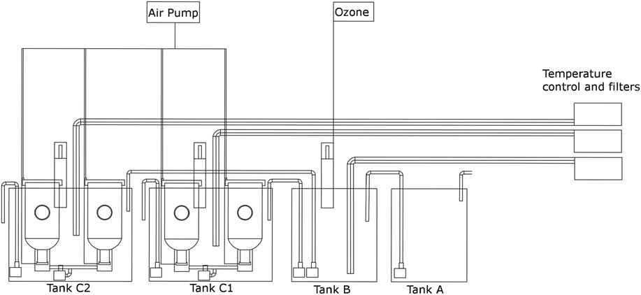
The larval culture equipment consists of four cylindrical units immersed into two tanks (Figure 1; Supplementary Figure S1). Each tank is equipped with a canister filter (Eheim Classic 350) loaded with activated carbon and perlon wool, a chiller (Teco Tr-5 160 w or Teco Tr-10 260 w), a heater (Askoll Tronic 200 w) and a protein skimmer (Ferplast Bluskimmer 550, 350 l/h air flow). An ozone generator (Sander Certizon) is also present in the tank B and it can be operated to assure disinfected water for delicate organisms. Two water-level sensors are located in each tank. The first one reads the maximum level and it is located 200 mm under the tank edge; the second one reads the minimum level and it is located 100 mm under the edge.
Overview of the automated culture equipment. The boxes on the left indicate chillers and filters and they are connected to the corresponding tanks. The first tank on the left (tank A), contains seawater for storage. The pump inside this tank moves water to the tank B, where it is sterilized and filtered. The skimmer inside the tank B is connected to an ozone generator. The two pumps perform the connection between the tank B and the two treatment tanks C1 and C2. Two larval units are disposed in the tank C1 and used for the low-density treatments and two in the tank C2 and used for the high-density treatments. A pump is connected by means of an aspiration tube to the bottom of each couple of cylinders. Air pumps allowed air insufflation in the treatment tanks, cylinders and under the nets located at the bottom of each cylinder.

The first tank (A) is 400×500×500 mm (h); it is a storage that receives continuously pumped water and permits a partial sedimentation of particulate matter. The second tank (B), is 450×500×500 mm (h). Herein, the water received from tank A is sterilized and filtered. Two water pumps (Askoll Biodinamics 4, 620 l/h water flow), located in tank B, move the water to the tanks C1 and C2, when operated. These last tanks are 650×500×500 mm and are equipped with water pumps (Askoll Biodinamics 4), connected to the drainage. Two cylindrical larval rearing units (described below) are submerged in each of the tanks C1 and C2. Only indirect natural light was shed during our experiments. However, in case of need, the system may be served by one or two fluorescent lights located over the tanks and controlled by the CCU.
Larval rearing units
Each larval unit is made of a cylinder, a base, a dripper and a porous stone with its holder. The cylinder is a modification of a standard tronco-conical larval rearing unit commonly used in aquaculture practices, measuring 350 mm in height and 180 mm in diameter, with a total volume of ca. 10 l (Supplementary Figure S2). They are equipped with two holes: the first one, placed laterally, is 36 mm in diameter and it is provided with a 50 µm net; the second, located on the bottom, is 50 mm in diameter and is provided with a 250 µm net. The base and the cylinder holder work synergistically to allow the collection of a large air bubble acting as a stopper, under the 250 µm net. The bubble can be removed by a pump (Sicce Syncra 1.0), located at the base and operated by the CCU. The air, blown into the lower part of the cylinder and inside the cylinder, comes from an air pump (Schego Prima 100l/h), operated as well by the CCU.
The dripper is made of two polyvinyl chloride (PVC) parts, joined to make a single ring and creating an interspace equipped with 16 holes (1-mm diameter) in the outer face. This latter is connected to a water pump (Sicce Syncra 1.5). When activated, it drives water under pressure in the ring cavity and through the holes, along the cylinder walls, creating gentle flows on the cylinder wall, to avoid larvae to remain adherent to the walls during water changes.
Automation
The automation of the larval culture equipment is assured by a CCU, Zelio logic Programmable Logic Controller (model SRC2261BD; Schneider Electric) programmed in FBD language. Level sensors are connected via 0 to 10 volt digital inputs and technical accessories are connected via relay outputs. During the ‘stationary operation’ phase, the system is programmed to activate lights, pumps, skimmers, filters, chillers, heaters and the two aforementioned aerators: the first connected to the porous stones, the second to the bases of the cylinders.
According to the software developed for this equipment, the second phase begins at 0800 h: the water pumps placed at the base of culture cylinders are switched on for 5 h. The pumps connected with the drippers are switched on at 0800 h up to 1200 h, starting to pump water into the cylinders.
Two pumps (Askoll Biodinamics 4), one for each culture tank, are switched on at 1201 h and they pump water to the drainage. Two pumps (Askoll Biodinamics 4) controlled by liquid level floats are subsequently switched on after 30 s, pumping water from tank B to tank C1 and C2, respectively. In order to minimize the mixing of clean water and used water, the drainage pumps are positioned on the left side, whereas clean water inlet pipes are placed on the right side of each tank. For the same reason, inlet pipes are modified in order to create a non-turbulent laminar flow into the tanks C1 and C2.
When tank B is empty, a liquid level float signals to CCU to switch off all the water change pumps. A Boolean logic subroutine activates the refilling and disinfection of tank B with ozone, and then reactivates the ‘stationary operation’ phase. The ozonization process consists of the switching on of the skimmer and the ozonizer in tank B. After 5 h, the ozonizer is turned off, whereas the skimmer is kept working in order to accelerate the removal of residual, potentially toxic, ozone.
Collection of ovigerous females
Individuals of H. inermis were collected in a P. oceanica meadow off Lacco Ameno d’Ischia (Zupo, 1994) and sorted on board the stazione zoologica di napoli (SZN) vessel Phoenicia in May 2013. Samples were examined in the laboratory under a Leica MZ6 stereomicroscope and divided in 1500 ml conical flasks. Larvae produced by ovigerous females were collected, pooled and divided in 16 replicates each cultured in a 800 ml conical flask according to a standard culture procedure described by Zupo and Messina (Reference Zupo and Messina2007). Cultures were repeated in automatic culture system, in two replicates for each of two culture densities, that is, low density (0.1 larvae/ml corresponding to 370 hatched larvae), and high density (0.15 larvae/ml corresponding to 550 hatched larvae). The experiment was repeated twice to obtain four replicates of each culture density.
Larvae were fed on Brachionus plicatilis (5 ind/ml) along with Artemia nauplii (5 ind/ml) for 7 days. Artemia metanauplii enriched with AlgaMac 2000 (BioMarine Inc.) were used in replacement of the previous food from the 8th day onward.
Water analyses and larval density measures
Measurements of chemical, physical and biological parameters were carried out daily in the culture equipment. The main chemical features of the water were measured: redox potential (using a Martini Instruments ORP57WP portable ORP meter), pH (using a Mettler Toledo S62 pH portable tester) and concentration of nitrogen compounds and phosphates (using a Hach DR/2010 spectrophotometer and pre-prepared kits). The temperature was daily measured by means of a digital probe (TFA SDT8A). Larval density in control treatments was daily evaluated by counting all the larvae present in each conical flask after collecting them, individually, by means of a Pasteur pipette. Larval density in the test equipment was evaluated every day in three 200 ml samples of the culture medium, collected in each cylinder after continuous and intense agitation of the medium, to reduce patchiness.
Data treatment
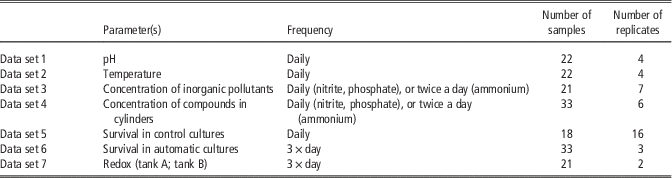
Data collected were organized into data sets, as indicated in Table 1. Data sets were filed into MS Excel 2010 spreadsheets. Survival rates in the automatic larval culture were evaluated by plotting the average number of larvae present each day in each replicate. Before collecting replicate samples and during the collection, the water was strongly agitated in each cylinder by means of a 50 ml plastic stripette. The survival rates were calculated as
 $$\left( {{\rm ANL}{\times}18/{\rm INL}} \right){\times}100$$
where ANL is the average number of larvae contained in 200 ml samples and INL the initial number of larvae/m/.
Experimental plan and data set arrangement

For each measured parameter, the frequency of recordings and the number of samples and replicates considered is given.
Wilcoxon matched-pairs test was applied to evaluate the significance of differences of ammonia, nitrite and phosphate concentrations between tanks C1 and C2 and among different replicates and treatments. Survival rates among low-density cylinders, high-density cylinders and control cultures were analysed by Mann–Whitney test (Supplementary Table S1).
Results
Automatic larval culture
The temperature measured in the tank A (water supply and storage) of the experimental equipment increased from 15.8°C to 22.0°C during 21 days of test (average value of temperature was 17.8 SD 1.8°C), due to seasonal natural variations of the seawater pumped in. Differently, the temperature was stable in tanks B (sterilization tank), C1 and C2 (culture tanks) during the experiment (tank B, 18.5°C, SD 0.1; tank C1, 18.5°C, SD 0.1; tank C2, 18.5°C, SD 0.1) due to the thermoregulation applied. Similarly, the pH was stable during the experiment, as indicated by small standard deviations exhibited in daily measures in all tanks (tank A, pH 8.29, SD 0.06; tank B, pH 8.28, SD 0.05; tank C1, pH 8.28, SD 0.05; tank C2, pH 8.29, SD 0.04). Redox potential was checked three times a day: before, during and after the planned automated ozonation activities, to guarantee the disinfection and the removal of residual ozone.
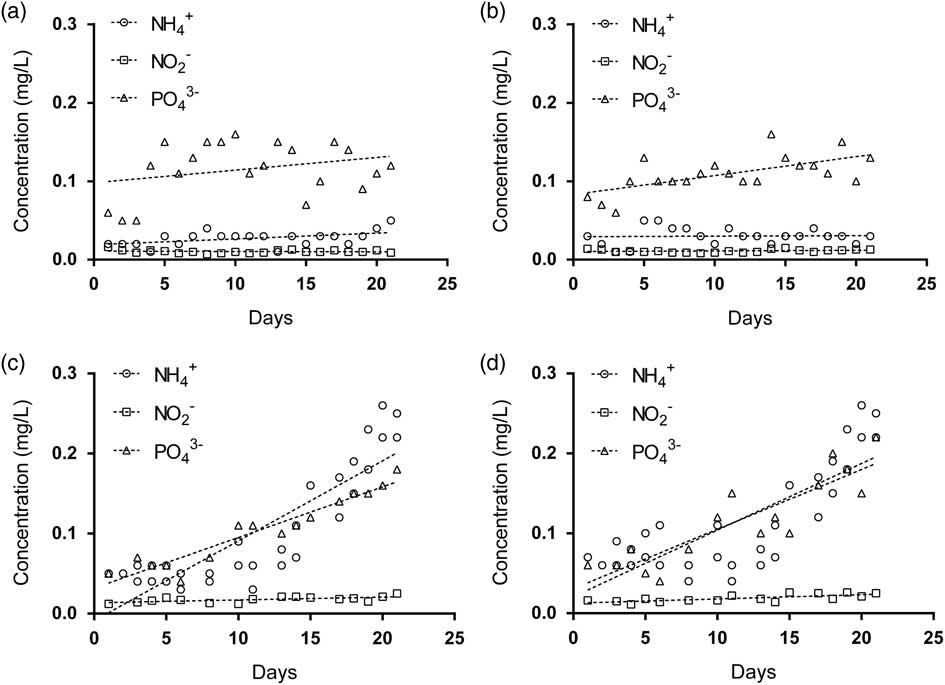
The maximum concentrations of ammonium, nitrites and phosphates in tank A were 0.05, 0.016 and 0.16 mg/l, respectively (Figure 2a). They were 0.05, 0.015 and 0.16 mg/l in tank B, respectively (Figure 2b). Tanks C1 and C2 showed a linear increase of ammonia, nitrite and phosphate concentrations during the experiment. Maximum concentrations of nitrogen compounds in tanks C1 and C2 (Figure 2c and b) were as follows: 0.26 mg/l of ammonia at the 21st experimental day in both tanks; 0.025 mg/l of nitrite at the 21st day in the tank C1 and 0.026 mg/l of nitrite at the 15th and 19th day in the tank C2; 0.18 mg/l. of phosphate in tank C1 and 0.22 mg/l in tank C2 at the 21st day of experiment. Significant differences in the ammonium concentration were observed between the tanks C1 and C2 (Wilcoxon matched-pairs test, P<0.0001). On the contrary no significant differences were observed in nitrite (Wilcoxon matched-pairs test, P>0.05) and phosphate (Wilcoxon matched-pairs test, P>0.05) concentrations in the same tanks.
Concentration and trends of NH4 +, NO2 − and PO4 3− in the tanks A (a), B (b), C1 (c) and C2 (d).

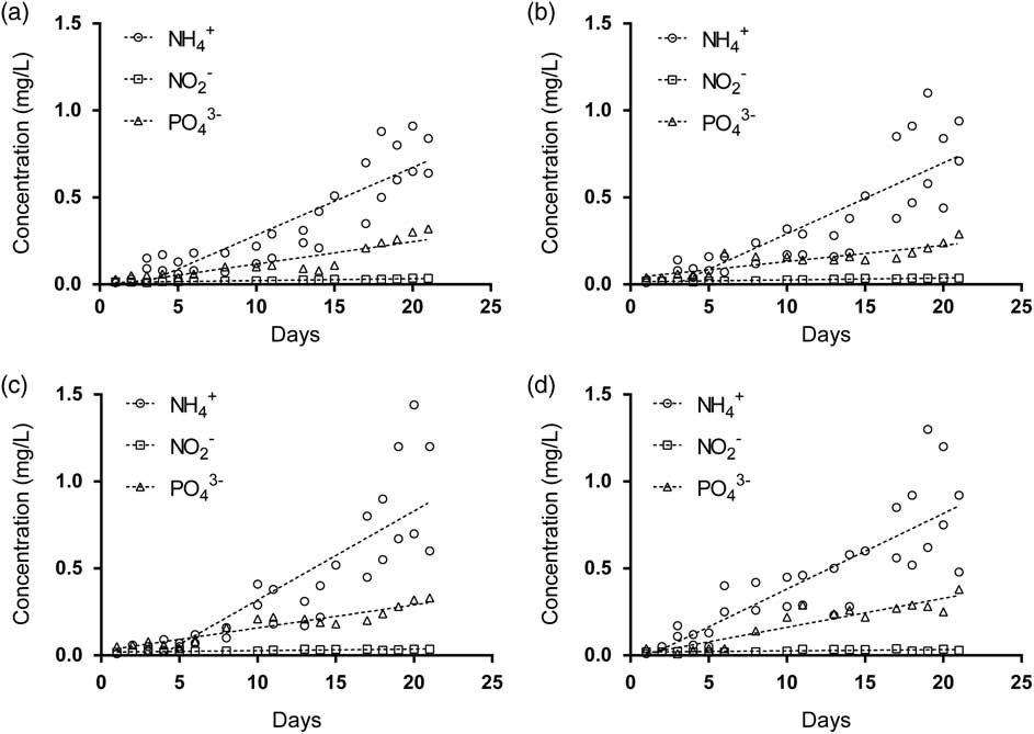
Ammonium concentration (Figure 3) in low-density replicates (Cylinders 1 and 2) reached a maximum of 0.91 mg/l at the 20th day in Cylinder 1 and 1.10 mg/l at the 19th day in Cylinder 2; ammonium concentration reached higher values in replicates at high density (Cylinders 3 and 4), with a maximum concentration of 1.44 mg/l in Cylinder 3 (20th day of experiment) and 1.30 in Cylinder 4 (19th day of experiment).
Concentrations and trends of NH4 +, NO2 − and PO4 3− in low-density Cylinders 1 (a) and 2 (b), and in high-density Cylinders 3 (c) and 4 (d).

The nitrite concentration in low-density replicate cylinders showed the maximum concentration at the 20th day of experiment (0.035 mg/l) in Cylinder 1 and at the 18th day of experiment in Cylinder 2 (0.036 mg/l). High-density replicate cylinders exhibited higher concentrations (0.036 at the 17th and the 20th day in Cylinder 3; 0.038 at the 17th day in the Cylinder 4).
Phosphate concentrations in low-density replicate cylinders reached a maximum value of 0.32 mg/l in Cylinder 1 and 0.29 mg/l in Cylinder 2 at the 21st day of experiment. Phosphate concentrations in high-density replicate cylinders reached a maximum value of 0.33 mg/l in Cylinder 3 and 0.38 mg/l in Cylinder 4 at the 21st day.
Ammonium, nitrite and phosphate concentrations showed linearly increasing trends among low-density and high-density replicates. Ammonium, nitrite and phosphate concentrations exhibited significant differences among low- and high-density replicates (ammonium concentration: Wilcoxon matched-pairs test, P<0.001; nitrite concentration: Wilcoxon matched-pairs test, P<0.01; phosphate concentration: Wilcoxon matched-pairs test, P<0.01).
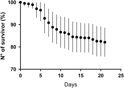
Survival rates showed a linearly decreasing trend in each cylinder, reaching, in both treatments, minimum values at the 16th day (Figure 4). In particular, at the day 16th, the survival in Cylinder 1 was 70.0% (SD 8.7) of the initial larval density; the survival in Cylinder 2 was 65.0% (SD 5.0) of the initial larval density; the survival in Cylinder 3 was 46.7% (SD 12.0) of the initial larval density; the survival in Cylinder 4 was 57.8% (SD 1.9) of the initial larval density. Larval survivorships of each replicate after the 16th day of experiment could not be evaluated because at the end of the larval development, last-stage zoeae started the settlement and they were no more sampled in the water column.
Percent survival rates of Hippolyte inermis larvae cultured 21 days in the automatic system in each culture cylinder (dots) and their standard deviations (vertical bars).

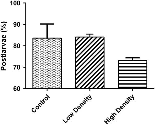
At the end of the first experiment (day 21st) the specimens still present in each replicate cylinder were collected (Figure 5). In total, 318 post-larvae were collected from Cylinder 1 (survival 85.9%; post-larval density 0.086 Pl/ml); 310 post-larvae were collected from Cylinder 2 (survival 83.8%; post-larval density 0.083 Pl/ml); 394 post-larvae were collected from Cylinder 3 (survival 71.6%; post-larval density 0.106 Pl/ml) and 411 post-larvae were collected from Cylinder 4 (survival 74.7%; post-larval density was 0.111 Pl/ml). The remaining, non-metamorphosed larvae were collected as well. In total, 30 larvae were present in Cylinder 1 (8.1%), 17 larvae in Cylinder 2 (4.6%), 43 larvae in Cylinder 3 (7.8%) and 16 larvae in Cylinder 4 (2.9%). At the end of the second experiment, we collected 311 post-larvae from Cylinder 1 (survival 84.1%; post-larval density 0.084 Pl/ml), 306 post-larvae were collected from Cylinder 2 (survival 82.7%; post-larval density 0.083 Pl/ml), 404 post-larvae were collected from Cylinder 3 (survival 73.5%; post-larval density 0.109 Pl/ml) and 399 post-larvae were collected from the Cylinder 4 (survival 72.5%; post-larval density 0.108 Pl/ml). Non-metamorphosed larvae were collected in the Cylinders 1, 2, 3, 4 and they were 5.1%, 5.4%, 8%, 5.5% of the initial stocks, respectively.
Percentage of post-larvae collected in the control culture replicates, low-density replicates (Cylinders 1 and 2) and in the high-density replicates (Cylinders 3 and 4); vertical bars indicate standard deviations.

Average survival in replicates conducted at a low density was 84.13% (SD 1.33), whereas in replicates conducted at a high density was 73.08% (SD 1.33).
No significant differences among low-density and control culture replicates in the survival rates were recorded (Mann–Whitney test, P>0.05). In contrast, significant differences among low-density and high-density replicates were recorded in the survival rates (Mann–Whitney test, P<0.05).
Control larval culture
The control experiment, conducted without the aid of an automated device, according to the standard operation procedures proposed by Zupo (Reference Zupo2000), consisted of 16 replicates of 800 ml conical flasks at a density of 0.1 larvae/ml, and it yielded high survival values (Figure 6). The survival curve showed a sigmoidal shape. Its slope was deepest from day 3 to day 10 and then it became shallower until the end of the experiment. Considerable differences in survival rates among the replicates can be inferred by the high standard deviation in the survival curve from day 4 to the end of the experiment. At the end of the experiment (day 21st), the average survival obtained in the control experiment was 83.6% (SD 6.6) with a density of 0.091 post-larvae/ml (Figure 5). The highest survival rates were observed in the conical flask 5 (93.8%), whereas the lowest survival rates were obtained in the conical flask 10 (70%).
Percent survival rates of Hippolyte inermis larvae cultured 21 days in glass vessels managed by operators (dots) and their standard deviations (vertical bars).

Discussion
Larval culture of H. inermis is a strenuous activity. The survival rates recorded are in line with previous studies (Zupo, Reference Zupo2000), although large differences were observed among replicates. The differences in survival among replicates might be due to the difficult manipulation of fragile zoeae (Luis-Villaseñor et al., 2012), the decay of water quality (Robertson and Austin, Reference Robertson and Austin1998) or bacterial diseases influencing the mortality in individual replicates (Robertson and Austin, Reference Robertson and Austin1998; Luis-Villaseñor et al., 2012). The non-automatized procedures, still assuring a high post-larval production (Zupo, Reference Zupo2000), exhibit some critical issues: a thermostatic chamber is needed, as well as plenty of space and trained operators (Calado et al., Reference Calado, Pimentel, Vitorino, Dionísio and Dinis2008). For this reason, an automatic larval system has been set to work without the intervention of trained biologists and it does not need an intensive daily maintenance. According to Calado (Calado et al., Reference Calado, Figueiredo, Rosa, Nunes and Narciso2003; Calado et al., Reference Calado, Narciso, Morais, Rhyne and Lin2005; Calado et al., Reference Calado, Pimentel, Vitorino, Dionísio and Dinis2008), the automatic culture system has been devised in order to meet the needs of cultured species and reduce research and laboratory resources while optimizing spaces and the use of manpower.
During water changes, both water inflow and outflow were activated simultaneously, so avoiding changes in the water level that could lead to mortality (personal observation). In addition, larvae were not forced to pass through or pressed on the meshes, due to the pressure of the outflowing water (Quinitio et al., Reference Quinitio, Parado-Estepa and Alava1999; Nghia et al., Reference Nghia, Wille, Binh, Thanh, Van Danh and Sorgeloos2007).
As for the chemical properties of the water, temperature and pH are the most important parameters influencing larval survival. Stable temperatures and pH were recorded during the experiment. These parameters can modify larval physiology with consequence on health, residual energy, difficulties to molt and settlement (Palma et al., Reference Palma, Bureau, Correia and Andrade2009; Taylor et al., Reference Taylor, Gilleard, Allen and Deheyn2015). The concentrations of waste compounds were constant during the experiment and, consequently, water analyses in tank B (sterilization tank) yielded constant concentrations, also due to the intensive ozonization and filtration provided.
In spite of inorganic pollutants recorded in tanks A and B, tanks C1 and C2 (culture tanks) were influenced by the presence of larval culture. Tanks C1 and C2, in fact, exhibited very significant differences in the daily concentrations of ammonia. These differences may be correlated to the presence of high-density cultures in the tank C2.
In addition, decomposing brine shrimps may induce higher concentrations of particulate organic matter (POM) and dissolved organic matter (DOM) and, consequently, of nitrogen compounds deriving from their degradation. The accumulation of organic material can lead to a saprophytic, potentially pathogenic, bacterial proliferation and to a consequent decrease of dissolved oxygen (Leonard et al., Reference Leonard, Blancheton and Guiraud2000). In the equipment we tested, bacteria were controlled by the activity of protein skimmers (Brambilla et al., Reference Brambilla, Antonini, Ceccuzzi, Terova and Saroglia2008; Suzuki et al., Reference Suzuki, Hanagasaki, Furukawa and Yoshida2008), as well as the disinfection of incoming water (in tank B) and this reduces the bacterial growth in the filter units.
Ozone addition is effective against a wide range of bacterial, viral, fungal and protozoan pathogens. The effectiveness is concentration dependent and is influenced by the exposure time, pathogen loads and abundance of organic matter (Gonçalves and Gagnon, Reference Gonçalves and Gagnon2011). Although seawater ozonation may generate bromate ions as a by-product (Parrino et al., 2014a and Reference Parrino, Camera-Roda, Loddo, Palmisano and Augugliaro2014b), during our experiment we did not find any evidences of larval mortality induced by the disinfection process.
Density of food and larvae is a key point for the success of any culture. Larvae must be fed ad libitum and, usually, organisms are cultured in a medium containing high food concentration, creating potential problems due to decaying organic matter and possible decrease of dissolved oxygen concentrations. High-density replicates showed significantly higher concentrations of nitrogen compounds and phosphates as compared with low-density replicates. The maximum concentration of nitrite and phosphate ions in high-density replicates was slightly higher then that in the one of low-density replicates. As well, the maximum concentration of ammonia was considerably higher in high-density replicates then that in low-density replicates.
These differences, which increased during the latest days, can be explained by considering that in the semi-closed system, larval units exchanged water only for a few hours each day. During this time the aged, dying or dead feeds were removed. The ammonium concentration was, at least partially, related to the larval density, as the concentration of live feeds is identical in the two treatments.
Nitrogen compounds did not reach lethal concentrations: LC50 (96 h) analysis on decapod crustacean larvae and juveniles showed a species-specific toxicity of nitrogen compounds (Liao et al., Reference Liao, Wang and Lin2011; Romano and Zeng, Reference Romano and Zeng2013). These studies were performed on species that are phylogenetically far from H. inermis. LC50 concentrations measured in above-mentioned studies were generally higher than those recorded in our treatments. However, other processes could influence the mortality in the described equipment, in the case of organisms with a longer period of larval development (Schuenhoff et al., Reference Schuenhoff, Shpigel, Lupatsch, Ashkenazi, Msuya and Neori2003).
The larval survival rates in cylinders showed a linear trend from the day 0 to the day 16; after this time, the number of sampled larvae decreased, due to both behavioural and morphological changes (Zupo and Buttino, Reference Zupo and Buttino2001). In fact latter zoeal stages are characterized by the elongation of pleopods and pereiopods, positive phototaxis and by the tendency to settle to the substrate, as a transition period preceding the benthonic phase, thus decreasing the probability to collect swimming larvae into the culture units. For these reasons, it was impossible to obtain a complete estimation of the larval density from the day 17th to the end of the experiment in the automated culture equipment. Low-density replicates and control culture replicates showed the same post-larval production trends and the differences among them are not significant. In general, the survival rates in the automatic equipment were quite high, with higher survivals in low-density replicates, due probably to lower concentration of pollutants, higher food availability and reduced influence of cannibalism. Despite the statistical differences recorded for the survival rates among low-density and high-density replicates, these last showed lower survival rates but they produced a higher number of healthy larvae per volume unit.
The efficiency of the described automatic culture equipment depends, as demonstrated in other rearing systems (Calado et al., Reference Calado, Figueiredo, Rosa, Nunes and Narciso2003; Calado et al., Reference Calado, Narciso, Morais, Rhyne and Lin2005; Calado et al., Reference Calado, Pimentel, Vitorino, Dionísio and Dinis2008), on various chemical, biological and mechanical influences. The efficiency was improved making use of a filter, frequent water changes, a correct dosage of feeds, the use of ozone and a careful setting of optimal densities. The process was also improved by a fine tuning of the operational software, providing efficient water changes. This improvements conducted to results similar to those produced by manually operated culture system, with lower operational efforts and costs. This automatic equipment largely simplifies the culture of small laboratory organisms and it offers interesting applications for the purposes of aquaculture and scientific or biotechnological research (Nappo et al., Reference Nappo, Berkov, Massucco, Di Maria, Bastida, Codina, Avila, Messina, Zupo and Zupo2012).
Acknowledgements
This research was funded by the Stazione Zoologica Anton Dohrn and it was conducted within a Federico II University Master course, under the supervision of V. Z. The authors acknowledge the Benthic Ecology group (Stazione Zoologica Anton Dohrn) for their assistance during the sampling operations (cpt. V. Rando) and bioassays. Prof. Claudio Agnisola provided suggestions and support to the project development. All animal experiments were carried in accordance with the EU Directive 2010/63/EU for experiments on animals.
Supplementary materials
To view supplementary material for this article, please visit https://doi.org/10.1017/S1751731117000908