Introduction
Adenoidectomy is a commonly performed procedure in the field of otolaryngology. It has traditionally been conducted using the curettage method. This is a relatively ‘blind’ technique which risks nasopharyngeal injury and incomplete adenoid removal; indeed, it has been found to completely remove adenoid vegetations in less than 30 per cent of cases.Reference Bross-Soriano, Schimelmitz-Idi and Arrieta-Gómez1
In 1992, Becker et al. reported the use of endoscopy-assisted adenoidectomy.Reference Eiji, Lawrence, Ananda and Panje2 This technique uses a 0, 30, 70 and 120° rigid nasal endoscope of 2.7 or 4 mm diameter. It has the advantages of improved visualisation and magnification, rigidity, superior haemostasis, reduction of unnecessary trauma, complete removal of adenoid tissue, and improved safety. Endoscopy-assisted adenoidectomy is generally perceived to be more effective in clearing adenoid tissue, compared with the conventional curettage method, but this has not been objectively assessed.
Therefore, we undertook a descriptive, cross-sectional study evaluating the nasopharynx of 41 consecutive paediatric adenoidectomy cases, using a 0, 30 and 70°, rigid, 4 mm endoscope. The study aimed (1) to evaluate the role of such endoscopy in assessing the adenoids before and after traditional curettage adenoidectomy; (2) to assess the effectiveness of curettage adenoidectomy performed alone; and (3) to evaluate the possible role of such endoscopy in improving the results of curettage adenoidectomy.
Materials and methods
We included in the study 41 paediatric patients undergoing adenoidectomy with or without other surgical procedures (e.g. tonsillectomy or ventilation tube insertion), whose parents were willing to give informed consent. We excluded patients with contraindications for adenoidectomy, and those in whom the rigid nasopharyngeal endoscope could not be navigated up to the nasopharynx.
Ethical approval was obtained from our institution's ethical committee.
All patients underwent clinical history-taking, and were assessed pre-operatively using relevant, noninvasive investigations such as pure tone audiometry, impedance audiometry and X-ray (using a soft tissue, lateral neck view with open mouth and neck extension).
The nasal cavity was decongested with cotton soaked in 0.05 per cent oxymetazoline.
The nasopharynx was inspected via the nasal cavity using a 0, 30 and 70° rigid endoscope (Karl Storz, Tuttlingen, Germany) to assess the extent of adenoid tissue (this was the first endoscopy). The operating surgeon was kept unaware of the findings.
Surgery was performed under general anaesthesia with orotracheal intubation, with the patient placed in Rose's position. A Boyle–Davis mouth gag of appropriate size was inserted. A small French rubber catheter was inserted through the nostril and brought out through the mouth, and the ends were clinched for palatal retraction.Reference Gabalski, Mattucci, Setzen and Moleski3 The operating surgeon assessed the size and extent of the adenoid with the index finger of the dominant hand. The adenoids were removed by conventional curettage. Small tags of lymphoid tissue retained after curettage were removed with punch forceps. Pressure haemostasis was achieved by packing the area with sterile cotton gauze packs soaked in adrenaline (1:100 000), for three minutes unless contraindicated. If adrenaline was contraindicated, sterile gauze packs soaked with saline only were used.Reference Wang, Bernheim, Kaufman and Clement4
Thereafter, the patient's nasal cavity and nasopharynx were again examined endoscopically, by the same endoscopist as previously, to determine the completeness of adenoid tissue removal at different sites (this was the second endoscopy). If remnant adenoid tissue was seen, it was removed under endoscopic control, and the final result again assessed endoscopically (this was the third endoscopy).
Following curettage and endoscopy-assisted adenoidectomy, the volume of adenoid tissue removed was measured using the displacement method (utilising a 25 ml measuring cylinder).
Results
Patients' mean age ± standard deviation (SD) was 8.83 ± 2.77 years; most were aged seven to 12 years. The male to female ratio was 1.56:1. The most common symptom was snoring (97.6 per cent), followed by nasal obstruction. Mouth-breathing was seen in 85.5 per cent cases, and a sore throat in 80.5 per cent (Table I).
Patient's clinical features

All the patients had enlarged adenoids on X-ray (soft tissue, lateral neck view).
The most common surgical procedure conducted was adenotonsillectomy (n = 32), followed by adenotonsillectomy with ventilation tube insertion (n = 6). Isolated adenoidectomy was performed in only one case.
In seven cases, both choanae were completely blocked by adenoid tissue, preventing passage of the 4 mm rigid endoscope. In the remaining cases (n = 34), the nasopharynx could be easily accessed with a 0, 30 and 70°, 4 mm, rigid endoscope, and the adenoid tissue extent at various nasopharyngeal sites could be studied satisfactorily.
Before curettage, adenoid tissue was found to be present in all cases in the nasopharyngeal roof and superomedial choanae. Adenoid tissue was also present in the anterior vault (in 91.6 per cent of patients), other parts of the choanae (84.15 per cent), the fossa of Rosenmuller (77.95 per cent) and the eustachian tube opening (76.45 per cent).
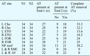
The second endoscopy could be easily performed in all cases. Excellent haemostasis was achieved in all patients. In all cases, the curettage technique failed to completely remove adenoid tissue from the superomedial choanae and the anterior vault. Other sites of incomplete removal were (in descending order of frequency) other parts of the choanae (in 67.2 per cent of patients), the eustachian tube opening (63 per cent), the nasopharyngeal roof (61.78 per cent) and the fossa of Rosenmuller (61 per cent) (Figure 1). The curettage method was most successful in removing adenoid tissue from the nasopharyngeal roof; even so, 38.2 per cent of patients had incomplete removal at this site. Thus, further, endoscopy-guided clearance was necessary in all cases to ensure complete removal of adenoid tissue. Data on the success of curettage adenoidectomy are presented in Table II.
Site of residual adenoid tissue (AT) following conventional curettage. L = left; R = right; Cho = other parts of choana; ETO = eustachian tube opening; FOR = fossa of Rosenmuller; SMC = superomedial choana; AV = anterior vault

Patient results for curettage adenoidectomy

AT = adenoid tissue; N1 = first endoscopy; N2 = second endoscopy; End-1 = pre-operative endoscopy; End-2 = post-curettage endoscopy; L = left; R = right; Cho = other parts of choanae; ETO = eustachian tube opening; FOR = fossa of Rosenmuller; NP = nasopharyngeal; SMC = superomedial choanae; AV = anterior vault
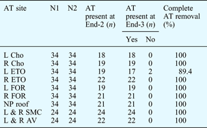
Endoscopy-assisted adenoidectomy successfully removed the residual adenoid tissue from all nasopharyngeal sites, except for the eustachian tube opening in two cases (Figure 2). Thus, the success rate for complete adenoid remnant removal from the eustachian tube opening, under endoscopic guidance, was 89.4 per cent. For all other nasopharyngeal sites, a 100 per cent success rate was achieved. Data on the success of endoscopy-assisted adenoidectomy are presented in Table III.
Site of residual adenoid tissue (AT) following endoscopy-assisted adenoidectomy. L = left; R = right; Cho = other parts of choana; ETO = eustachian tube opening; FOR = fossa of Rosenmuller; SMC = superomedial choana; AV = anterior vault

Patient results for endoscopy-assisted adenoidectomy

AT = adenoid tissue; N1 = first endoscopy; N2 = second endoscopy; End-2 = post-curettage endoscopy; End-3 = post endoscopy-assisted adenoidectomy endoscopy; Cho = other parts of choanae; ETO = eustachian tube opening; FOR = fossa of Rosenmuller; NP = nasopharyngeal; SMC = superomedial choanae; AV = anterior vault
The mean volume ± SD of adenoid tissue removed using conventional curettage was 1.74 ± 0.77 ml (range 0.50–3). In addition, endoscopy-assisted adenoidectomy removed a mean volume ± SD of 0.91 ± 0.34 ml (range 0.5–2). Thus, if endoscopy-assisted adenoidectomy had not been performed, 34.3 per cent of the total adenoid tissue volume would have been retained (this equates to 52.3 per cent of the adenoid tissue volume removed by curettage).
The mean time ± SD required to perform the first, pre-operative endoscopy was 3.56 ± 1.58 minutes (range 1–10), while that for the second, post-curettage endoscopy was 8.82 ± 2.15 minutes (range 2–12).
Intra-operative complications were noted in two patients, in the form of transient tachycardia while packing with adrenaline-soaked gauze, immediately after curettage adenoidectomy. This resolved upon removal of the pack.
One patient developed a reactionary haemorrhage from the tonsillar bed six hours post-operatively; this was managed successfully with bipolar cautery under general anaesthesia.
The patients' mean ± SD hospital stay was 3.76 ± 0.487 days (range 3–5).
Discussion
The objective of adenoidectomy is to remove the hypertrophic adenoid tissue that causes nasal airway obstruction and pathological restriction of nasal airflow. Dissatisfaction with the safety and adequacy of clearance of conventional curettage adenoidectomy has led to the development of alternative techniques, made possible by developments in fibre-optics and endoscopic instrumentation.Reference Parsons5–Reference Yanagisawa and Weaver7
The main disadvantage of curettage is that it is a relatively blind technique that may lacerate the choanae and torus tubarius and graze the nasopharyngeal mucosa; it may also merely skim the adenoid bulk, leaving behind obstructing tissue particularly at the eustachian tube openings and intranasal protrusions and high in the nasopharynx.Reference Koltai, Kalathia, Stanislaw and Heras8 A popular alternative to conventional curettage is endoscopy-assisted adenoidectomy – the second method employed in our study.
Our study was designed to assess the success of conventional curettage adenoidectomy versus endoscopy-assisted adenoidectomy in removing adenoid tissue from different nasopharyngeal sites. We also aimed to assess the possible role of rigid endoscopic evaluation in improving the success of conventional curettage adenoidectomy.
The first, pre-operative endoscopy could not be conducted in two patients due to bilateral inferior turbinate hypertrophy; these patients were thus excluded from the study. In the remaining cases (n = 41), the adenoid tissue was easily assessable prior to curettage, using a 0, 30 and 70°, 4 mm, Hopkin's rod rigid endoscope. The anterior vault and the superomedial portion of the choanae were assessed with a 70° Hopkin's rod rigid endoscope and a posterior rhinoscopy mirror.
Pre-operatively, adenoid tissue was invariably found at the nasopharyngeal roof, followed by the fossa of Rosenmuller (on the left in 73.5 per cent of patients and on the right in 82.4 per cent) and the eustachian tube opening (64.7 per cent on the left and 79.4 per cent on the right). We searched the literature but could not locate any similar studies performed to assess the nasopharyngeal extent of adenoid tissue, prior to performing curettage. In our study, the adenoid tissue observed in the eustachian tube openings and nasopharyngeal vault correlated well with our patients' symptoms of ear ache and snoring, respectively. We expect that the anatomical extent of adenoid hypertrophy would affect its clinical symptomatology.
Following curettage adenoidectomy, we used cotton gauze packs soaked in 1:100 000 adrenaline to pack the nasopharynx for three minutes, to achieve haemostasis. Adrenaline packs were not contraindicated in any of our cases. Patients were closely monitored for adrenaline side effects. We encountered transient tachycardia in two cases, which settled after removing the nasopharyngeal pack.
Excellent peri-operative haemostasis was achieved in all patients. We did not encounter any immediate post-operative haemorrhage; this is consistent with the results of Teppo et al. Reference Paradise, Bluestone, Stool, Alper, Arjmand, Casselbrant, Dohar and Yellon9 However, one patient (2.4 per cent) developed a reactionary haemorrhage from the right tonsillar bed six hours post-operatively; this was managed successfully with bipolar cautery under general anaesthesia. A prospective study has shown that the incidence of reactionary haemorrhage is 2–8 per cent.Reference Drake-Lee and Stokes10 Return to theatre for haemostasis was required in 0.5% and 2%.Reference Guida and Muttucci11–Reference Marshall, Sheppard and Narula13 Thus, our complication rate was within acceptable limits.
In the present study, a second nasopharyngeal endoscopy was conducted following curettage adenoidectomy, and we noted that the extent of superomedial choanae and anterior vault adenoid tissue was unchanged from its pre-operative state (as viewed at the first endoscopy). Other sites harbouring retained adenoid tissue included (in descending order of frequency) other parts of the choanae (in 67.2 per cent of patients), the eustachian tube opening (63 per cent), the nasopharyngeal roof (61.78 per cent) and the fossa of Rosenmuller (61 per cent). Thus, conventional curettage had not cleared the adenoid tissue completely in even a single case. These observations would have not been possible had we not conducted a second endoscopy after the curettage procedure. Hence, the second objective of our study was fulfilled.
Although we assessed all cases with a 70° rigid endoscope and a posterior mirror, we found the superomedial choanae and anterior vault could only be evaluated with a 0 and 30° endoscope, with regards to the extent of adenoid tissue. In addition, these two areas were the most inaccessible sites for conventional curettage removal of adenoid tissue.
In the present study, conventional curettage adenoidectomy successfully removed adenoid tissue from other parts of the choanae, the eustachian tube opening and the fossa of Rosenmuller areas in 33, 16 and 24.5 per cent of patients, respectively. By comparison, Bross-Soriano et al. studied 150 patients with an absolute indication for adenoidectomy, in order to evaluate the efficacy of conventional adenoidectomy, using intra-operative endoscopic inspection of the nasopharynx, and to evaluate the need for endoscopy-guided revision surgery.Reference Bross-Soriano, Schimelmitz-Idi and Arrieta-Gómez1 They found residual adenoid tissue in 107 cases, 45.3 per cent of which involved the pharyngeal part of the eustachian tubes. Our study findings indicate a higher prevalence of residual adenoid tissue across a greater range of nasopharyngeal sites, following curettage adenoidectomy.
Although not used in the present study, we believe that adenoid tissue in the superomedial choanae and anterior vault can also be effectively dealt with using a microdebrider or suction diathermy.
In the present study, endoscopy-assisted clearance achieved complete adenoid removal in all but two cases. In these two patients, a small tag of adenoid tissue could not be removed from the eustachian tube opening; this would have required very tightly curved, small Blakesley or Takahashi forceps, which were unavailable.
Huang et al. treated 15 patients with symptomatic adenoid hypertrophy, using combined conventional and endoscopic adenoidectomy (any residual adenoid tissue was completely removed during an endoscopic revision procedure).Reference Huang, Chao, Chen and Hsiao6 They concluded that (1) this procedure could completely remove large amounts of adenoid tissue without prolonging the operative time, and (2) the endoscope provided a clear, direct view that enabled the surgeon to remove adenoid tissue accurately, to evaluate and stop bleeding effectively, and to avoid unnecessary trauma. Thus, these authors believed that a combined approach employing both conventional and endoscopic adenoidectomy was an effective and safe method for managing enlarged adenoids.
Kulak conducted a similar study of 125 adenoidectomy cases, in which complete removal of adenoid tissue was achieved using an endoscopy-assisted method.Reference Uçar14
In the present study, the first, pre-operative endoscopy took 3.56 minutes on average. Curettage adenoidectomy took an average of 9.2 minutes, while the second, post-curettage endoscopy took an average of 8.82 minutes. In comparison, in Cannon and colleagues' series of 130 endoscopy-assisted adenoidectomy cases, revision endoscopy was performed to clear residual adenoid tissue following curettage and took less than 5 minutes.Reference Cannon, Replogle and Schenk15
In the present study, the mean volume ± SD of adenoid tissue removed via conventional curettage was 1.74 ± 0.77 ml (range 0.50–3), while that removed via endoscopy-assisted adenoidectomy was 0.91 ± 0.34 ml (range 0.5–2). Thus, failure to undertake endoscopy-assisted adenoidectomy would have resulted in retention of 34.3 per cent of the total pre-operative adenoid tissue volume (equating to 52.3 per cent of the adenoid volume removed by curettage).
• The use of an endoscope during adenoidectomy gives the surgeon a clear, direct view, facilitating precise and complete adenoid tissue removal, effective haemostasis, and avoidance of unnecessary trauma
• Endoscopy-assisted adenoidectomy allows better adenoid tissue clearance, compared with conventional curettage adenoidectomy, by enabling localisation of residual adenoid tissue especially in the superomedial choanae and anterior vault
• Conventional curettage adenoidectomy may achieve the desired clinical results in patients with adenoid hypertrophy; however, endoscopic evaluation indicates that it fails to completely remove adenoid tissue
All our patients were discharged on the third post-operative day, giving a mean hospital stay ±SD of 3.76 ± 0.487 days (range 3–5).
The slightly older age of our patient group could be attributed to parents waiting longer before seeking health care for their child, due to differing attitudes to health care compared with other parts of the world.
Conclusion
The use of an endoscope gives surgeons a clear, direct view of the nasopharynx, enabling them to remove adenoid tissue precisely and completely, to control bleeding effectively, and to avoid unnecessary trauma.
Endoscopy-assisted adenoidectomy improves the results of conventional curettage adenoidectomy by enabling accurate localisation of residual adenoid tissue, especially in the superomedial choanae and anterior vault areas.
Conventional curettage adenoidectomy may achieve the desired result in patients with adenoid hypertrophy; however, endoscopic evaluation reveals that it fails to achieve complete adenoid removal and hence is less satisfactory than endoscopy-assisted adenoidectomy. Further research is needed to establish whether anatomically complete adenoid removal leads to real clinical benefit for patients.