INTRODUCTION
Trypanosoma evansi is mainly a parasite of camelids, equids and carnivores, but it is also pathogenic in a number of wild and other domestic host species (Gill, Reference Gill1977). In Africa and Latin America, cattle seem to have low susceptibility to T. evansi, but in Asia clinical signs with a significant impact are regularly described in buffaloes and cattle (Luckins, Reference Luckins1988; Reid, Reference Reid2002; Dargantes et al. Reference Dargantes, Mercado, Dobson and Reid2009).
During the course of T. evansi infection in cattle, parasitaemia is frequently low, but it can occasionally be as high as in other trypanosome infections (Gill, Reference Gill1977; Chobjit et al. Reference Chobjit, Kheowsri, Rodtian, Jongchansittoe and Nuangmek2006). With recurrent trypanocide treatments in the field, especially at subcurative doses, the immune control of parasitaemia increases, which lowers parasitaemia to undetectable levels (Desquesnes, Reference Desquesnes2004); under such conditions, it is sometimes difficult to determine with certainty whether treated animals have actually been cured (complete elimination of the parasite), or whether they have become healthy carriers and could constitute a potential reservoir. Testing a trypanocide in splenectomized animals, hence with reduced natural resistance, would demonstrate the efficacy of the chemical itself, despite the artificial immunosuppression.
T. evansi infection can be cured using trypanocides such as isometamidium chloride and diminazene aceturate, but products such as quinapyramine and melarsomine hydrochloride are more suitable. Melarsomine hydrochloride (Mel Cy) (Cymelarsan®) is the only trypanocide specifically recommended for controlling T. evansi in camels, at a dose of 0·25 mg/kg by deep intramuscular injection. However, the manufacturer (Merial®) has never defined a melarsomine hydrochloride dose for other host species. Studies conducted in various situations have shown infection relapses when using 0·25 mg/kg in cattle and buffaloes; higher doses were then suggested for these Bovinae (Lun et al. Reference Lun, Min, Huang, Liang, Yang and Huang1991; Payne et al. Reference Payne, Sukanto, Partoutomo and Jones1994a; Dia and Desquesnes, Reference Dia and Desquesnes2007).
In Thailand, although isometamidium chloride has occasionally been used and quinapyramine was used in the past (Indrakamhang, Reference Indrakamhang1998), diminazene aceturate is the only trypanocide regularly available, which is used extensively to control surra in horses, buffaloes, cattle, pigs, deer, elephants and dogs (Tuntasuvan and Luckins, Reference Tuntasuvan and Luckins1998). After more than 40 years of regular use, treatment failures, most probably due to chemoresistance to diminazene aceturate, are regularly reported from the field (Tuntasuvan et al. Reference Tuntasuvan, Jarabrum, Viseshakul, Mohkaew, Borisutsuvan, Theeraphan and Kongkanjana2003a,Reference Tuntasuvan, Trongwanichnam, Sukruen, Borisutsuvan, Mohkaew and Chompoochanb; Hin-On et al. Reference Hin-On, Rodtian, Nuangmek, Uthaiwan and Muangyai2004). In some cases, relapses have been observed in cattle from Chiang Mai province, despite treatments at a dose of 10 mg/kg (Rodtian P, unpublished observation).
In our study, we assessed the efficacy of the intramuscular injection of 0·5 mg/kg of melarsomine hydrochloride as a parasitological cure in normal and splenectomized cattle experimentally infected with a Thai strain of T. evansi.
MATERIALS AND METHODS
Animals
Six 10-month-old dairy cattle, 82·75% Holstein Friesian-type breed (crossed with a local breed), from a dairy farm free of trypanosomosis, and testing negative in parasitological (Haematocrit Centrifuge Technique, HCT) (Woo, Reference Woo1970) and serological tests (card agglutination test for trypanosomes (CATT/T. evansi) and ELISA-T. evansi), were randomly selected from a batch of 30 animals. They were kept in a fly-proof stable in Pathumthani province.
To demonstrate the efficacy of the treatment itself, and to ensure that the parasites would not be inhibited or killed solely by a combination of drug effects and host immune responses, half of the experimental animals were immunosuppressed by splenectomy. Thus, 45 days prior to the experiment, 3 of the 6 cattle were randomly selected and splenectomized. For this minor surgery (less than 30 min) an intravenous injection of 2 ml of 2% xylazine solution (Rompun®) was given, as recommended by the manufacturer (Bayer®).
Parasites
A Thai strain of T. evansi previously isolated from highly parasitaemic cattle in Bangkok, and characterized by PCR with specific primers (Masiga et al. Reference Masiga, Smyth, Hayes, Bromidge and Gibson1992; Panyim et al. Reference Panyim, Viseshakul, Luxananil, Wuyts and Chokesajjawatee1993), passed once through a mouse and cryopreserved in liquid nitrogen, was recovered by intraperitoneal injection of 3 mice. At the peak of parasitaemia (days 3–5), the mice were anaesthetized with a combination of zolazepam and tiletamine (Zoletil®) and bled to death; blood was collected in a citrated tube (Fushino® Na citrate, 3·2%) and diluted 1:2 in phosphate saline glucose (PSG). Parasites were counted as previously described (Desquesnes and Tresse, Reference Desquesnes and Tresse1996). After optimized dilution in PSG, approximately 105 trypanosomes were injected into the jugular vein of each of the cattle on Day 0 of the experiment.
Melarsomine hydrochloride treatment
On Day 50, the cattle were weighed and were given a deep intramuscular injection of 0·5 mg/kg of melarsomine hydrochloride (Cymelarsan®, batch D01601A, Merial).
Cattle sampling
Ten ml of blood were regularly collected from the jugular vein of the animals at intervals of 2–7 days from Day 0 to Day 151; 2·5 ml was kept in EDTA tubes for HCT, mice inoculation and preparation of the buffy-coat for PCR. The rest was left at room temperature for 24 h to allow clotting. Sera were collected and kept at −20°C until further use. On Day 99, 2 ml of cerebrospinal fluid (CSF) were collected by puncture at the sacrococcygeal vertebral junction for the CSF mouse inoculation test (CSFMIT).
In the event of death during the experiment, an autopsy was carried out to collect blood, aqueous humour of the eyes, joint fluid from the scapulo-humeral, hip and/or femoro‑tibial joints, CSF from the occipital joint, and brain samples (cerebrum and cerebellum). The blood was used for HCT and BMIT (see below). The CSF was used for the mouse inoculation test (CSFMIT). The CSF, joint fluid and aqueous humour of the eyes were used for fluid pellet microscopic observation and PCR (see below).
Parasitological examinations
Direct microscopic examination was conducted in the mice by observing 10 μl of blood collected from the tip of the tail and placed between a slide and cover-slip (22×22 mm), under dark ground conditions at ×400 magnification; the number of parasites was estimated as previously described (Desquesnes and Tresse, Reference Desquesnes and Tresse1996). The parasitaemia 95% confidence interval (CI) was calculated as CI=mean+/−1·96 S/√n, and the mean parasitaemias were compared using the Student's t-test (Ancelle, Reference Ancelle2002). The haematocrit centrifuge technique (HCT) was used on the cattle after centrifugation of 70 μl of blood in a capillary tube, at 10 000 g for 5 min, and by direct microscopic observation of the buffy coat (Woo, Reference Woo1970).
Fluid-pellet microscopic examination
Duplicate samples of 0·5 ml of cattle aqueous humour of the eye, or joint fluid diluted 1:2 in PSG, or CSF were centrifuged at 10 000 g for 10 min; the supernatant was discarded. One of the pellets was preserved at −20°C for PCR, and the other was re‑suspended in the remaining supernatant and deposited between the slide and cover-slip for direct microscopic examination under dark ground conditions.
Blood-mouse inoculation tests (BMIT) or cerebrospinal fluid-mouse inoculation tests (CSFMIT) were carried out by intraperitoneal injection of 0·25 ml of cattle blood or CSF, respectively, into 1 mouse each. The mice were monitored 3 times a week for 3 weeks by direct microscopic examination of blood from the tail. The animals were considered negative when parasites were still not found 3 weeks after inoculation.
PCR diagnosis
A sample of 100 μl of buffy coat collected from the centrifugation of 1 ml of blood, or pellets from the centrifugation of 0·5 ml of fluids (CSF, joint fluid, aqueous humour of the eyes) underwent DNA purification using the phenol/chloroform method (Sambrook and Russell, Reference Sambrook and Russell2001). PCR was carried out in 10 μl volumes of master mix with 1 μl of sample, using TBR primers according to a technique already described (Masiga et al. Reference Masiga, Smyth, Hayes, Bromidge and Gibson1992). PCR products, including positive and negative controls, were electrophoresed for 1 h at 120 V in 2% agarose gels, together with Generuler® 100-bp DNA ladder plus (Fermentas, Invitrogen Corporation, Van Allen Way, Carlsbad, CA, USA), stained with ethidium bromide and visualized under UV light.
Serological examination
(1) The card agglutination test for trypanosomes (CATT/T. evansi) (antigen lot 10A1B2) was carried out on sera diluted 1:4 as described by the manufacturer (Prince Leopold Institute of Tropical Medicine, Belgium) (Bajyana Songa and Hamers, Reference Bajyana Songa and Hamers1988). (2) The ELISA procedure has been described previously (Desquesnes et al. Reference Desquesnes, Kamyingkird, Pruvot, Kengradomkij, Bossard, Sarataphan and Jittapalapong2009). Briefly, Microtest 96-well Polysorp Nunc® immunoplates (Nunc, Roskilde, Denmark) were coated with 100 μl/well of T. evansi soluble antigen at 5 μg/ml protein concentration in carbonate buffer (pH 9·6) overnight at 4°C. The plates were blocked with 150 μl/well of blocking buffer: phosphate-buffered saline (PBS) with 0·1% Tween 20® (Sigma-Aldrich) and 7% Wako skim milk powder (ref: 190-12865, Wako Pure Chemical Industries Ltd, Osaka, Japan), under permanent stirring (150 rpm) for 45 min at 37°C. After discarding the blocking buffer, sera diluted 1:100 in blocking buffer were transferred in duplicate to the ELISA plates. After 30 min of incubation in a shaker-incubator (37°C, 150 rpm), the plates were washed 7 times with washing buffer (PBS-0·1% Tween 20®). Then, 100 μl of peroxidase-conjugated anti-bovine IgG (A5295, Sigma-Aldrich), diluted 1:10 000 in blocking buffer, were added and the plates incubated for 30 min (37°C, 150 rpm). After discarding the sera, the plates were washed 7 times and added to 100 μl of the substrate/chromogen complex 3,3′,5,5′-tetramethylbenzidine (TMB) (Sureblue® TMB Microwell Peroxidase Substrate, Gaithersburg, Maryland, USA). The plates were incubated in a dark room for 30 min. Optical density (OD) was measured at 630 nm in an ELISA reader (Dynex Technologies®, Chantilly, VA, USA). ELISA T. evansi results were expressed as a relative percentage of positivity (RPP) versus 3 negative and 3 positive controls, as previously described (Desquesnes et al. Reference Desquesnes, Bosseno and Breniere2007). Positive results were confirmed when the RPP was above 20%.
Chronological feature of the protocol
The 6 cattle were sampled at Day −50 and Day 0, prior to infection. Three cattle were splenectomized at Day −45. All cattle were sampled regularly at 2 to 7-day intervals between Day 0 and Day 151. The 6 cattle were experimentally infected at Day 0. At Day 50, the 6 cattle were weighed and received 0·5 mg/kg of melarsomine hydrochloride in a deep intramuscular injection. On Day 99, blood and CSF were collected from the 6 cattle for BMIT and CSFMIT. The experiment was terminated 100 days after treatment (Day 151).
RESULTS
Initial parasitological (HCT), molecular (PCR) and serological tests (CATT and ELISA) of the 6 animals, carried out at Day −50 and Day 0 were all negative; RPP values in ELISA were all less than 10%. Three randomly selected dairy cattle were splenectomized and named SC17, SC16 and SC9. The other 3 were named C22, C21 and C32. SC17 developed acute anaplasmosis on Day −40 when Anaplasma marginale were observed on Giemsa-stained blood smears (8·9% infected blood cells), probably as a secondary effect of splenectomy. The animal received intramuscular treatment with 10 mg/kg of oxytetracycline. A. marginale disappeared from the stained blood smears after Day −37. It was therefore concluded that the animal was cured of anaplasmosis.
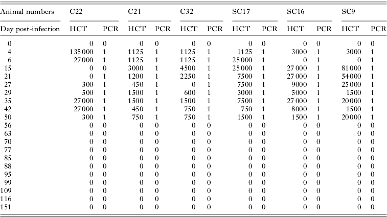
All the animals were inoculated via the jugular vein with 105 T. evansi on Day 0, and tested positive by microscopic observation of the buffy coat or direct blood observation and PCR from Day 4 post-infection and remained regularly positive until they were treated on D50 (Table 1). Parasitaemia was low, and did not exceed 135 000 parasites/ml, but it was almost always detectable by HCT, except for 6 out of 54 samples collected between Day 4 and Day 50. Parasitaemia fluctuated greatly from one animal to another and from day to day, and it was higher in the splenectomized animals (confidence interval, CI=14 532±7023 parasites/ml) than in the normal cattle (CI=8919±10 011 parasites/ml), but the difference was not statistically significant (t=0·90<1·96; degrees of freedom=52). Over that period, only 1 sample out of 54 (animal C22 on Day 15) was negative by PCR (Table 1).
Results of the HCT and PCR (TBR) in 3 normal and 3 splenectomized cattle experimentally infected by T. evansi (D0) and treated with melarsomine hydrochloride (D50)

Legend: for HCT: estimated numbers of trypanosomes/ml; for PCR: 0 negative; 1 positive.
After treatment, all the animals were confirmed negative by HCT and PCR up to the end of the experiment. BMIT and CSFMIT carried out on samples collected from all the cattle on D99 were all negative.
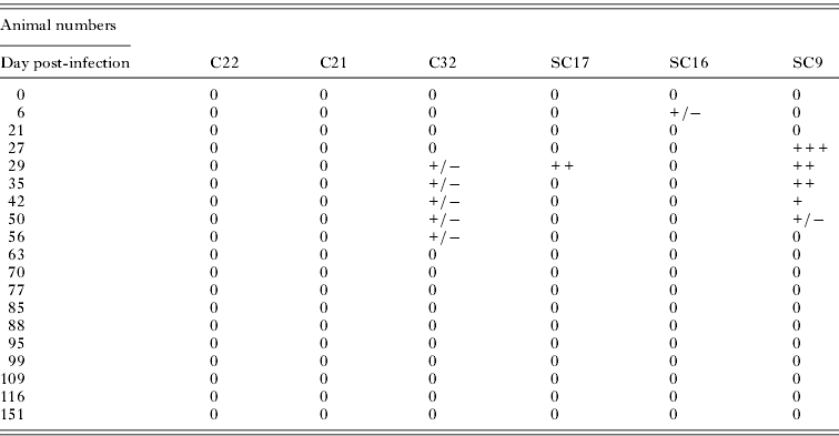
The CATT/T. evansi results were all negative prior to infection. After infection, CATT results remained negative in 2 animals (C21 and C22); they were doubtful in some instances in 2 other animals (C32 and SC16), and they were occasionally positive (5 occurrences) in the last 2 animals (SC17 and SC9), between Day 6 and Day 42. The results are presented in Table 2. After Day 56, all CATT values were negative in all the animals for 3 months, up to the end of the experiment. Due to the poor performance of the CATT, a new batch of CATT T. evansi was ordered (antigen lot 10A1B2; expiry in October 2011) and CATT tests were performed again with the same samples. The results obtained were very similar and showed a total of 6 positive results and 7 doubtful results.
Results of the CATT/T. evansi in 3 normal and 3 splenectomized cattle experimentally infected by Trypanosoma evansi (D0) and treated with melarsomine hydrochloride (D50)

Legend: 0, negative; +/−, doubtful; +, positive; ++, strong positive; +++, very strong positive.
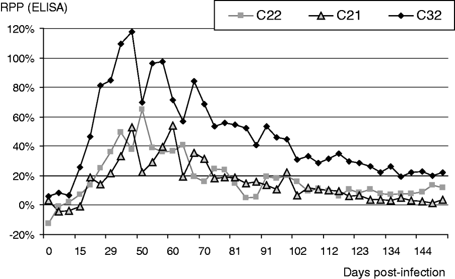
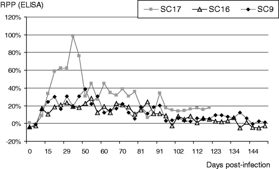
The results obtained by ELISA are indicated in Fig. 1 for the normal cattle and Fig. 2 for the splenectomized cattle. The animals became positive at 26 (±6) days post-infection in the normal cattle and 20 (±7) days post-infection in the splenectomized cattle, with an average of 21 (± 6) days post-infection (no significant differences). All samples remained positive throughout the infection, with the exception of 3 samples from 2 splenectomized cattle (see Fig. 2, before Day 50). Immune responses in the normal cattle were clearly higher (RPP>50%) than in the splenectomized animals, of which only 1 displayed RPP over 50%, between Day 22 and Day 42. After treatment (Day 50), RPP declined gradually. Samples were classified as negative by ELISA at 45 (±25) days post-treatment (p.t.) in the normal cattle, and at 25 (±15) days p.t. in the splenectomized cattle. From the 45th day p.t., all the animals were negative except one of the normal cattle (C32), which remained positive up to Day 131 (80 days p.t.) and was then irregularly negative but close to the positive threshold when the experiment ended. At the end of the experiment, the RPP of SC32 was regularly below 25% but not yet regularly negative.
Monitoring of ELISA results in 3 normal cattle experimentally infected with Trypanosoma evansi and treated with melarsomine hydrochloride. RPP, relative percentage of positivity (%) in ELISA; Day 0, date of infection; Day 50, date of treatment; Day 19, date of cerebrospinal fluid mouse inoculation test (CSFMIT) and blood mouse inoculation test (BMIT).

Monitoring of ELISA results in 3 splenectomized cattle experimentally infected with Trypanosoma evansi and treated with melarsomine hydrochloride. RPP, relative percentage of positivity (%) in ELISA; Day 0, date of infection; Day 50, date of treatment; Day 99 date of cerebrospinal fluid mouse inoculation test (CSFMIT) and blood mouse inoculation test (BMIT).

Only 1 of the splenectomized cattle (SC17) had a high RPP, and the others exhibited a low response in ELISA, being irregularly and weakly positive (Fig. 2). SC17 developed an anaplasmosis relapse on Day 77 and was treated with 10 mg/kg of oxytetracycline on Day 78. A. marginale disappeared from the blood observation after Day 81, but the animal never recovered from this infection; in the absence of fever or other obvious infectious signs, SC17 showed serious loss of appetite and weight, and finally died before the end of the experiment on Day 119. It was necropsied and additional examinations were carried out. BMIT, CSFMIT, and microscopic examination of the buffy coat, stained brain smears, as well as PCR and microscopic examination of pellets from CSF, joint fluid and aqueous humour of the eyes, were all negative for trypanosomes. The RPP in ELISA was below the positive threshold from Day 95 (45 days p.t.) to Day 119 when the animal died. Apart from a single positive result on Day 29, CATT/T. evansi remained negative in this animal.
By the end of the experiment, all animals had given negative results in all the parasitological, molecular and serological tests (apart from ELISA in heifer C32, which will be discussed in the next section) for a period of at least 3 weeks, including 1 negative BMIT and 1 negative CSFMIT.
DISCUSSION
In vitro protocols and in vivo rodent protocols have been developed to evaluate chemo‑resistance to trypanocides (Eisler et al. Reference Eisler, Brandt, Bauer, Clausen, Delespaux, Holmes, Ilemobade, Machila, Mbwambo, McDermott, Mehlitz, Murilla, Ndung'u, Peregrine, Sidibe, Sinyangwe and Geerts2001). However, the only way to assess the efficacy of a trypanocide in a given host is to carry out the experiment in that very host. For this reason, despite its high cost and substantial inconvenience, our experiment could only be carried out on dairy cattle. Based on previous observations (Dia and Desquesnes, Reference Dia and Desquesnes2007), it is already known that a dose of 0·25 mg/kg is not effective in cattle, so there was no need for an untreated control group in our study.
The evaluation of a trypanocide is intended to determine the precise effect of the treatment. Does it kill all parasites, leading to a fully curative treatment, or does it only kill most of the parasites, leaving some of them alive, which leads to healthy carriers and a possible infection relapse under particular conditions such as physical stress (restraining for vaccination), food reduction or drought? This was demonstrated under experimental conditions with Latin American T. evansi in sheep (Desquesnes, Reference Desquesnes2004). In the case of T. evansi, which tends to penetrate deep into the tissues and cross most biological barriers, it has even been reported that parasites can survive in extravascular foci such as cerebrospinal fluid or joint fluid (Schillinger and Röttcher, Reference Schillinger and Röttcher1986). In T. vivax, the aqueous humour of the eyes was also described as a potential extravascular focus (Whitelaw et al. Reference Whitelaw, Gardiner and Murray1988). Consequently, some of these foci were included in our study to ensure total elimination of the parasites.
Parasitaemia may sometimes remain undetectable in partially immune animals, especially in the case of T. evansi in cattle, since they naturally develop very low parasitaemia. It is therefore impossible to demonstrate the efficacy of a treatment based on parasitological examination alone. To determine if any animal is still harbouring parasites after treatment, protocols must include serological follow-up. If the infection persists, immune stimulation would also persist and the RPP values in ELISA should stay above the positive threshold.
In our study, in the group of 3 normal cattle, if the treatment had failed to eliminate the parasites, immune stimulation would have been detected through persisting high RPP values in ELISA. In our observations, the RPP regularly decreased, falling below the positive threshold 45 (±25) days after treatment. In 1 animal, which proved to give high serological responses (C32), the RPP was still fluctuating around the positive threshold when the experiment ended. However, these regularly decreasing ELISA results, combined with the negative results obtained in all the other tests, including observation under the microscope, PCR, CATT, BMIT and CSFMIT, strongly support the hypothesis of a fully curative treatment, without refuge. It might be advisable to extend the follow-up of such animals when BMIT and CSFMIT cannot be carried out, but in our study all the results gathered allowed us to conclude on the absence of remaining infection. However, in our case, CSF was not examined before treatment, so it was not possible to assess whether the treatment might cure CSF infection or not.
The CATT performed very poorly since only 2 animals gave scarce positive results; the mean of positive results obtained from the samples collected during the period of active infection (from Day 6 to Day 50) was as low as 12% (5/42). When doubtful results were included, it reached 26%. A new batch of CATT T. evansi gave similar results. The results in the normal cattle (4 out of 28 samples from infected animals were doubtful) were worse than in the splenectomized cattle (5 positive and 2 doubtful out of 28 samples from infected animals). With 86% false-negative results, the sensitivity of the CATT was too low to be useful in our study. Other authors had already recorded this lack of sensitivity in pigs (Holland et al. Reference Holland, Thanh, Do, Sangmaneedet, Goddeeris and Vercruysse2005) but not yet in bovines; the usefulness of the CATT in cattle is therefore still uncertain, especially its negative predictive value, which was very low in our experiment.
In the group of 3 splenectomized cattle, if the treatment had failed to completely eliminate the parasites, the serological response might not have been detected due to the immunosuppressive effects of the splenectomy, but the parasitaemia ought to have increased (due to the immunodeficiency) and have been easily detected. This was not the case, since all the parasitological (and serological) examinations remained negative. Similarly, no cerebrospinal fluid refuge could be detected. Moreover, in the animal that died of anaplasmosis, the extensive search for the parasite in cerebrospinal, joints and eye fluids was negative. The fact that no relapse could be observed in the splenectomized animals gives satisfactory cause for confidence that the treatment is suitable for application in the field, where animals are sometimes immunosuppressed by intercurrent diseases.
Based on all these results, we concluded that treatment of the 6 dairy cattle was fully curative and the parasites were completely eliminated. These observations are quite closely related to previously published results in cattle or buffaloes (Lun et al. Reference Lun, Min, Huang, Liang, Yang and Huang1991; Zweygarth et al. Reference Zweygarth, Ngeranwa and Kaminsky1992; Payne et al. Reference Payne, Sukanto, Partoutomo, Jones, Luckins and Boid1994b). In particular, a recent observation in African cattle experimentally infected with the African strain of T. evansi from the North of Burkina Faso demonstrated that the dose of 0·5 mg/kg was effective in curing the animals, while preliminary tests using 0·25 mg/kg showed relapses (Dia and Desquesnes, Reference Dia and Desquesnes2007).
The policy for trypanocidal drug use in Thailand calls for discussion. Although the manufacturer's leaflet recommends using diminazene aceturate (DA) at a dose of 7 mg/kg to control Trypanozoon infections, for unknown reasons and out of habit, veterinarians in Thailand are used to applying 3·5 mg/kg only. Moreover, under the pressure of farmers who believe that this treatment may induce shock and a febrile side effect, the booster dose is sometimes divided into 2×1·75 mg/kg subdoses injected 24 h apart. In such circumstances, DA has been extensively under-dosed in Thailand for years. The 3·5 mg/kg dose most probably does not kill all the parasites and then helps to boost the immune responses of the animals, leading to unhealthy and healthy enzootic parasite carriers. This situation was confirmed by the high prevalence observed on infected farms, in a recent dairy cattle survey (Desquesnes et al. Reference Desquesnes, Kamyingkird, Pruvot, Kengradomkij, Bossard, Sarataphan and Jittapalapong2009). This under-dosing has applied for years in horses, mules, buffaloes and cattle and is most probably responsible for drug resistance in the field. In equines, for example, up to 50% of relapses were observed early after 3·5 mg/kg DA treatments (Tuntasuvan et al. Reference Tuntasuvan, Jarabrum, Viseshakul, Mohkaew, Borisutsuvan, Theeraphan and Kongkanjana2003a). Similarly in buffaloes, treatment at 5 mg/kg DA was also unsuccessful (Pholpark et al. Reference Pholpark, Pholpark, Sarataphan, Khunpasi and Taboran1984). As the under-dosing of DA in Thailand has long been the rule, both in equines and bovines, veterinarians had to increase the dose to 7 mg/kg, after some strains were selected for their resistance to 3·5 mg/kg, in order to overcome the rapid relapse of clinical signs, which after a time was also no longer effective. Field observations have indicated that in some cases 10 mg/kg of DA is no longer effective, and relapses occur very rapidly after treatment (P. Rodtian, unpublished data). In such circumstances, it is strongly recommended that DA use be halted and that such strains be eliminated with a trypanocide of another chemical family. In this context, Thailand urgently needs to identify such effective trypanocides. From our results, melarsomine hydrochloride could be proposed as a drug of choice to replace DA, especially in cases of apparent resistance to DA observed in the field.
We concluded from our experiment that the treatment of normal and splenectomized cattle was fully curative and the parasites were completely eliminated. The evidence provided by the efficacy in splenectomized cattle demonstrated that the chemical itself was responsible for the curative action, even with a depressed immune system. This showed potential satisfying treatment security, even under field conditions where some animals might be immunosuppressed by concurrent diseases or by unfavourable zootechnical parameters (food or water supply, endo- or ectoparasitic infestation, etc.). Melarsomine hydrochloride at the dose of 0·5 mg/kg can be recommended for treatment of surra in dairy cattle in Thailand. Such a dosage has already been suggested in China to treat buffaloes (Lun et al. Reference Lun, Min, Huang, Liang, Yang and Huang1991). However, larger-scale studies would be welcome to provide further information on the efficacy of this treatment in bovines.
With the increasing impact of surra in Asia (Dargantes et al. Reference Dargantes, Mercado, Dobson and Reid2009; Dobson et al. Reference Dobson, Dargantes, Mercado and Reid2009), including India (Laha and Sasmal, Reference Laha and Sasmal2009), especially due to spontaneous abortion (Löhr et al. Reference Löhr, Pholpark, Siriwan, Leesirikul, Srikitjakarn and Staak1986; Kashiwazaki et al. Reference Kashiwazaki, Pholpark, Polsar and Pholpark1998), a reduction in milk production (Sarataphan et al. Reference Sarataphan, Nishikawa, Tuntasuvan, Sukhapesna, Markvishitr and Satjapitak1989; Pholpark et al. Reference Pholpark, Pholpark, Polsar, Charoenchai, Paengpassa and Kashiwazaki1999), and interference in the Foot and Mouth and Haemorrhagic Septicaemia vaccination programme (Onah et al. Reference Onah, Hopkins and Luckins1997; Holland et al. Reference Holland, My, Dung, Thanh, Tam, Vercruysse and Goddeeris2001; Singla et al. Reference Singla, Juyal and Sharma2010), this would be a good time to identify and make available effective tools for the chemical control of surra.
Although this work demonstrated the potential merits of melarsomine hydrochloride for treating cattle infected by T. evansi, it did not demonstrate the passage of melarsomine hydrochloride through the haematoencephalic barrier. Further work will be necessary to demonstrate the efficacy of the treatment in cattle proved to be CSF infected, before such treatment can be proposed as an alternative to culling cattle, should T. evansi be accidentally introduced into Europe, as recently seen in continental Spain and France (Gutierrez et al. Reference Gutierrez, Desquesnes, Touratier and Buscher2010).
ACKNOWLEDGEMENTS
We wish to express our sincere gratitude to the Thailand International Cooperation Agency (TICA) for the financial and diplomatic support contributed to the project. Thanks to Merial for providing Cymelarsan and partial support of the maintenance expenses for the cattle. This research was also funded by Kasetsart University Research and Development Institution (Kor-Sor-Dor. 65.51), Kasetsart University and CIRAD which kindly supported the maintenance expenses for the cattle.
CONFLICT OF INTEREST STATEMENT
All the authors declare that they have no financial and personal relationships with other people or organizations that could inappropriately influence (bias) their work, including employment, consultancies, stock ownership, honoraria, paid expert testimony, patent application/registration, and grant or other funding.