Introduction
Parathyroidectomy for primary hyperparathyroidism is a common, highly successful and relatively low risk procedure. Cure rates following both exploratory and minimally invasive surgery have been demonstrated at more than 95 per cent,Reference Udelsman, Lin and Donovan1,Reference Reeve, Babidge, Parkyn, Edis, Delbridge and Devitt2 with a complication rate of 4.4 per cent and a 30-day post-operative mortality rate of less than 1 per cent.Reference Ishii, Mihai, Watkinson and Kim3 Advances in imaging, surgical technique and intra-operative monitoring continue to improve these statistics.Reference Goldfarb and Singer4 Despite this, re-operative parathyroidectomy is required in a cohort of patients.
Patients requiring re-operative parathyroidectomy are divided into those with persistent or recurrent hyperparathyroidism. Persistent hyperparathyroidism is defined as hypercalcaemia identified within six months of surgery performed for primary hyperparathyroidism. Recurrent hyperparathyroidism is defined as hypercalcaemia presenting more than six months after surgery for primary hyperparathyroidism.Reference Guerin, Paladino, Lowery, Castinetti, Taieb and Sebag5 The incidence of recurrent and persistent hyperparathyroidism has been found to be 2.5 per cent in retrospective analysis.Reference Venkat, Kouniavsky, Tufano, Schneider, Dackiw and Zeiger6
Experienced parathyroid surgeons’ series suggest that the causes of persistent and recurrent hyperparathyroidism are attributed to a combination of factors. These include the failure to recognise an adenoma, which may be due to ectopic gland position and multiglandular disease.Reference Gough7 Other rarer causes include parathyroid carcinoma and parathyromatosis.Reference Kollmorgen, Aust, Ferreiro, McCarthy and van Heerden8 The experience of the operating surgeon has been demonstrated to be a major factor in persistent disease, with recurrence rates increasing to up to 30 per cent in surgeons who perform under 10 parathyroidectomies per year.Reference Mihai, Barczynski, Iacobone and Sitges-Serra9 The primary modality of treatment for persistent and recurrent hyperparathyroidism is re-operative parathyroidectomy.
Re-operative intervention is generally accepted to be a more difficult procedure, with higher complication rates resulting in part from the loss of distinct tissue planes and increased fibrotic change due to previous surgery.Reference Cham, Sepahdari, Hall, Yeh and Harari10 A recent consensus statement on the subject suggests that parathyroidectomy in the previously operated neck should be carried out by an experienced surgeon using precision pre-operative localisation, intra-operative parathyroid hormone (PTH) monitoring and a team approach.Reference Stack, Tolley, Bartel, Bilezikian, Bodenner and Camacho11 Unfortunately, not all technologies are available in all centres.
This study aimed to review the re-operative cases of an experienced parathyroid surgeon in a tertiary referral centre and compare these to a consecutive series of operations performed for primary hyperparathyroidism, in order to identify key differences between the two groups. Additionally, the utilisation and accuracy of localisation techniques, overall operative success rates, and complication rates were assessed.
Materials and methods
This was a retrospective cohort study undertaken in a single tertiary referral centre in the UK. A retrospective review of prospectively collected data was performed from the senior author's operative database between the years 2003 and 2020. All re-operative parathyroidectomy cases were identified and assessed for: demographics; indication for surgery; pre-operative imaging techniques and accuracy; histological findings, including specimen weight and diagnosis; biochemical findings, including pre- and post-operative calcium levels, and pre- and post-operative PTH levels; gland location at time of surgery; and any post-operative complications. These data were then compared to those for 100 consecutive primary parathyroidectomies performed by the same surgeon.
Exclusion criteria included any operative indication other than primary hyperparathyroidism. Additionally, patients who had undergone previous neck surgery for reasons other than parathyroidectomy were excluded.
Patient information was obtained via the consultant's own operative database and patients’ electronic healthcare records. Statistical analysis was performed using SPSS® software version 27.
Results
Demographics
Twenty-eight revision parathyroidectomies were identified, of which 18 were performed for persistent hyperparathyroidism and 7 for recurrent hyperparathyroidism. In three cases, these data were unavailable. In 46 per cent of cases, the initial parathyroidectomy was performed by a surgeon other than the senior author. Average patient age for revision parathyroidectomy was 58 years; 82 per cent of patients were female. There was no significant difference in demographics between the revision and primary surgery groups (Table 1).
Demographics

*n = 28; †n = 100. SD = standard deviation
Imaging
Pre-operative imaging was undertaken in the majority of both primary and revision surgery patients. Of the revision surgery patients, 39.3 per cent underwent a pre-operative thyroid ultrasound scan, compared to 54 per cent of primary surgery patients. There were no significant differences in: the presence of positive ultrasound findings (36.4 per cent positive in revision surgery, 42.6 per cent positive in primary surgery, p = 0.702 (chi-square test)), or correct lateralisation of the pathological gland (18.2 per cent of revision surgical procedures, 37 per cent of primary surgical procedures, p = 0.190 (chi-square test)).
Technetium-99m sestamibi scanning was performed in 85.7 per cent of revision surgery patients, compared to 67.3 per cent of primary surgery patients. There were no significant differences in the presence of positive technetium-99m sestamibi scanning findings (87.5 per cent positive in revision surgery, 73.5 per cent positive in primary surgery, p = 0.161 (chi-square test)), or correct lateralisation of the pathological gland (70.8 per cent of revision surgical procedures, 69.1 per cent of primary surgical procedures, p = 0.875 (chi-square test)).
Both pre-operative ultrasound scanning and technetium-99m sestamibi scanning were performed in 39.3 per cent of revision cases and in 46 per cent of primary cases. In the revision group, combination imaging was positive in 90.9 per cent of cases, and correctly lateralised the pathological gland in 81.8 per cent of cases. In the primary surgery group, combination imaging was positive in 69.6 per cent of cases, and correctly lateralised the pathological gland in 63 per cent of cases.
Biochemistry
Mean pre-operative adjusted calcium levels were significantly higher in the re-operative group (2.88 mmol/l vs 2.80 mmol/l, p = 0.021 (independent samples t-test)). There were no significant differences in post-operative calcium levels (2.43 mmol/l vs 2.41 mmol/l, p = 0.48), pre-operative PTH levels (14.05 pmol/l vs 13.85 pmol/l, p = 0.944) or post-operative PTH levels (4.77 pmol/l vs 5.01 pmol/l, p = 0.766).
Histology
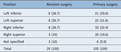
Gland position is shown in Table 2. There was no significant difference in gland position between the two groups (p = 0.545 (chi-square test)). The total gland number is higher than the total patient number as some patients had multiple pathological glands excised.
Gland position

Data represent numbers (and percentages) of cases
Mean gland weight was significantly higher in the revision surgery group (2758.3 mg vs 1153.7 mg, p = 0.006 (independent samples t-test)). There was a markedly larger variance in gland size in the revision surgery group (100–21 000 mg, standard deviation (SD) = 4847.0 mg vs 80–8100 mg, SD = 1461.4 mg). There was a 10 per cent increase in the incidence of gland ectopia in the revision group, but this increase did not achieve statistical significance (p = 0.261). Ectopic location is summarised in Table 3.
Gland ectopia and location

Data represent numbers (and percentages) of cases. *n = 28; †n = 100. ‡Chi-square test
Complications
The overall complication rate was significantly higher in the revision group (21.4 per cent vs 8 per cent, p = 0.044 (chi-square test)). Complications are summarised in Table 4. The post-operative mortality rate at three months was 0 per cent in both groups.
Post-operative complications

Data represent numbers (and percentages) of cases. *n = 28; †n = 100. RLN = recurrent laryngeal nerve
Discussion
The findings of this study correlate well with, and help build upon, existing data on parathyroid surgery, with primary surgery cure rates of 97 per cent and revision surgery cure rates of 89.3 per cent, in line with existing research.Reference Shin, Milas, Mitchell, Berber, Ross and Siperstein12 The current consensus on revision parathyroid surgery is that patients should undergo detailed pre-operative assessment, including localisation studies, and a review of the previous surgery and possible causes for its failure. Subsequent surgery should then be performed by an experienced surgeon and surgical team. Adjuncts to surgery including recurrent laryngeal nerve monitoring and intra-operative PTH monitoring are recommended when available.Reference Stack, Tolley, Bartel, Bilezikian, Bodenner and Camacho11 However, these adjuncts are not readily available in all centres and require cost–benefit analyses before implementation.
Pre-operative imaging of the patients in this series consisted predominantly of neck ultrasound and technetium-99m sestamibi scanning. Technetium-99m sestamibi scanning was the most frequently utilised, and gland identification and localisation rates were similar across both groups. The accuracy of technetium-99m sestamibi scanning has been demonstrated to be 42–88 per cent for primary surgeryReference Thomas, Bartel, Menda, Howe, Graham and Juweid13,Reference Chen, Mack and Starling14 and 67 per cent in revision surgery;Reference Thompson, Grant, Perrier, Harman, Hodgson and Ilstrup15 these findings are reinforced by the results of this study. Ultrasound provided less benefit, with only 18.2 per cent of glands correctly identified and lateralised in revision surgery, compared to 37 per cent in primary surgery; however, this difference was not significant. In existing literature, successful detection of parathyroid adenomas on ultrasound ranges from 33 per cent to 95 per cent.Reference Lal and Chen16,Reference Agha, Hornung, Schlitt, Stroszczynski and Jung17 Operator competency has been proposed as the primary reason for this wide range, and on-table ultrasound is now proposed by some. One such study found no difference in localisation accuracy between the surgeon and parathyroid radiologist in a series of 218 patients.Reference Soon, Delbridge, Sywak, Barraclough, Edhouse and Sidhu18
Combined ultrasound and technetium-99m sestamibi scanning has previously been demonstrated to be superior to a single imaging modality.Reference Scattergood, Marsden, Kyrimi, Ishii, Doddi and Sinha19 In this study, ultrasound used in combination with technetium-99m sestamibi scanning did not demonstrate any significant differences in localisation or lateralisation of pathological gland, in either the primary or revision surgery group, when compared to technetium-99m sestamibi scanning alone. These findings are limited by the small sample sizes of patients who underwent combination imaging.
The results of this study demonstrate substantial limitations in the pre-operative localisation of pathological glands in revision surgery using ultrasound and technetium-99m sestamibi scanning. Because of this, four-gland exploration is performed in most revision cases to reduce the risk of missed adenoma. Furthermore, some centres utilise additional imaging, with single-photon emission computed tomography (‘SPECT’), four-dimensional (4D) computed tomography (CT) and magnetic resonance imaging (MRI) all showing high diagnostic accuracy.Reference Thomas, Bartel, Menda, Howe, Graham and Juweid13,Reference Merchavy, Luckman, Guindy, Segev and Khafif20,Reference Hinson, Lee, Hobbs, Fitzgerald, Bodenner and Stack21 Research into 4D CT in particular has shown superiority of this imaging modality in the re-operative setting when compared to technetium-99m sestamibi scanning.Reference Mortenson, Evans, Lee, Hunter, Shellingerhout and Vu22 In the revision cohort in this study, two patients underwent MRI, with a 100 per cent pathological gland localisation rate. Three patients underwent CT, with a 66 per cent pathological gland localisation rate. Inaccuracy of imaging did not correlate with operative success, demonstrating the importance of surgeon competency in the performance of four-gland exploration.
Ectopic parathyroid glands occur because of the abnormal migration of parathyroid tissue during embryological development, or descent due to the deglutition effect. Their existence poses a surgical challenge, and knowledge of their common locations is vital to successful detection on pre-operative imaging. Consensus guidelines recommend that imaging cover from the neck down to below the aortic arch, to assess for the presence of ectopic glands in all pre-operative evaluations of both primary and revision parathyroidectomies.Reference Stack, Tolley, Bartel, Bilezikian, Bodenner and Camacho11 The incidence of the ectopic gland was 35.7 per cent in the revision group, compared to 25 per cent in the primary surgery group. Although there is a clear trend towards an increased incidence of ectopia in the re-operative group, this increase was not significant. The most common ectopic locations in revision surgery were thymic and retro-oesophageal ectopia, compared to para-oesophageal ectopia in the primary group. Glands being both deeper and lower in the neck in the revision surgery group may explain their missed identification on initial surgical exploration and lack of detection on imaging. Despite this, the majority of pathological glands excised in re-operative parathyroidectomy are found in normal anatomical locations.Reference McIntyre, Allen, Constantinides, Jackson, Tolley and Palazzo23
In this study, patients with persistent and recurrent disease had significantly higher pre-operative calcium levels and significantly larger glands excised. The clinical relevance of this is debatable; however, previous studies have found a correlation between calcium level and gland size,Reference Moretz, Watts, Virgin, Chin, Gourin and Terris24 a conclusion supported by the results of this study. Both parathyroid adenoma size and calcium level have been demonstrated to affect disease severity.Reference Rao, Honasoge, Divine, Phillips, Lee and Ansari25 Therefore, it can be argued that patients with recurrent and persistent disease present with increased severity of disease, which should be considered when deciding on the necessity and urgency of operative intervention.
• Revision parathyroid surgery is a complex procedure best carried out by an experienced surgeon with the assistance of a specialised multidisciplinary team
• In this study, sestamibi scanning was superior to ultrasound for gland localisation, in both primary and revision parathyroid surgery
• Revision parathyroid surgery patients present with higher pre-operative calcium and larger adenomas
• The presence of ectopic glands trends towards an increase in revision surgery, and ectopic locations differ between primary and revision surgery
• Patients undergoing revision parathyroid surgery are at higher risk of complications than those undergoing primary surgery, in particular persistent disease post-operatively
Expert opinion suggests that re-operative parathyroidectomy poses a higher risk of complications due to scarring of the neck and distortion of tissue planes,Reference Stack, Tolley, Bartel, Bilezikian, Bodenner and Camacho11 and existing audit data have demonstrated higher complication rates in revision surgery. However, few studies provide a direct comparison of complications between primary and revision surgery completed by the same surgeon or within the same centre. This study demonstrated a significantly higher complication rate in revision surgery, with the greatest increase being in the presence of persistent disease. All the patients with persistent hyperparathyroidism following revision surgery had persistent hyperparathyroidism prior to surgery, and all cases of recurrent hyperparathyroidism were cured following surgery. There were lower rates of temporary vocal fold palsy and recurrent laryngeal nerve injury in the re-operative group, in contrast to the anticipated risks of the re-operative neck.
Acknowledgement
The authors would like to thank Mrs Helen Tan for her contributions to this research.
Competing interests
None declared.