Introduction
Laryngeal carcinoma represents 2.2 per cent of all cancers.Reference Rosai1 The treatment modalities for laryngeal carcinoma, especially in its early stages, are mainly based on tumour eradication with conservation of function. In advanced-stage tumours, surgical treatment cannot achieve good functional results. Organ preservation protocols, including radiotherapy and chemotherapy, have been widely used as an alternative to surgical treatment in advanced-stage tumours.Reference Gillison and Forastiere2–Reference Moyer, Wolf and Bradford4 Surgical treatment as salvage surgery is preferred in the case of tumour recurrence.
Pre-operative radiotherapy gradually increases post-operative complications. The most common complication seen after salvage laryngectomy is pharyngocutaneous fistula formation.Reference Withrow, Rosenthal, Gourin, Peters, Magnuson and Terris5–Reference Sassler, Esclamado and Wolf7 The rate of pharyngocutaneous fistula formation increases if salvage surgery is performed within the first year after radiotherapy.Reference Gil, Gupta, Kummer, Cordeiro, Kraus and Shah8 Such a complication results in increased morbidity, and prolonged hospitalisation and greater hospital care expenditure.Reference Applebaum and Levine9, Reference Berry and Fischer10 The incidence of pharyngocutaneous fistula formation reported in the literature is extremely variable, ranging from 5 to 65 per cent.Reference Applebaum and Levine9–Reference Celikkanat, Koç, Akyol and Ozdem11 To deal with this significant post-operative morbidity, several authors have advocated the routine use of non-irradiated tissues to reinforce the neopharynx. Pectoralis major myofascial flaps, sternocleidomastoid muscle collar flaps, latissimus dorsi flaps, deltopectoral flaps and free flaps have been used for reinforcement.Reference Gil, Gupta, Kummer, Cordeiro, Kraus and Shah8, Reference Sousa, Castro, Porcaro-Salles, Soares, de Moraes and Carvalho12, Reference Tsou, Hua, Lin, Tseng, Tsai and Shaha13
Since it was first described in 1979, the pectoralis major myocutaneous flap has been used widely in head and neck reconstruction, and also in the reconstruction of pharyngocutaneous fistulas.Reference Ariyan14 In contrast, the pectoralis major myofascial flap has been used to prevent fistula formation in salvage laryngectomy. The main purpose of this study was to evaluate the effect of the pectoralis major myofascial flap on pharyngocutaneous fistula formation and time to oral feeding.
Materials and methods
This retrospective study reviewed 155 total laryngectomies performed between 2005 and 2012. All patients suffered from T3–T4-staged squamous cell carcinoma of the larynx. Subjects who underwent laryngopharyngectomy were not included in the study. Patients were divided into two groups according to their initial therapy. Group 1 included 110 patients who were treated primarily by total laryngectomy and unilateral or bilateral functional or modified radical neck dissection. Group 2 included 45 patients with recurrent disease after radical dose radiotherapy with cisplatin-based chemotherapy for T3–T4 laryngeal carcinoma and who were treated by salvage laryngectomy.
Since 2009, we began to use the pectoralis major myofascial flap as a second layer over the neopharynx after primary closure. Therefore, we divided group 2 into two subgroups: patients who underwent only primary closure (group 2a) and patients who underwent primary closure with a pectoralis major myofascial flap used to buttress the pharyngeal suture line (group 2b).
All data were gathered from patient charts and electronic medical records. Pharyngocutaneous fistula formation was categorised as wound dehiscence in the neopharynx region resulting in a fistula. Detailed demographic information was obtained including prior disease and surgeries, pre-operative treatment, duration and treatment of fistula, and time to oral feeding. The oral feeding day was considered to be the first day of oral nutrition after healing of the fistula.
Intra-operative and post-operative antibiotics – clindamycin phosphate 600 mg four times per day and amikacin 500 mg twice daily – were given to all patients for 7 days and additional doses or antibiotics were given when required during the treatment. Closure of the pharynx was performed in two layers, using 3-0 Vicryl® suture: an inner interrupted submucosal layer with inversion of the mucosa, and an outer interrupted layer with the inferior constrictor muscle. The suture line was T- or Y-shaped according to the less tension technique, and differed in relation to the extent of excised and remaining pharyngeal wall. A primary tracheoesophageal puncture was performed in all patients and a nasogastric feeding tube was inserted into the puncture site. Feeding started through the tube on the day after the operation. Oral feeding was attempted on the 10th day post-operatively in both groups. It was continued if there was no evidence of fistula and stopped if a fistula was suspected on clinical examination.
All of the cases with fistula were initially managed conservatively by stopping oral feeding, draining the wound by the removal of three or four stitches, debriding the fistula tract, administering broad-spectrum antibiotics recommended after wound culture was performed and applying a light compression dressing over the fistula site. If the defect was small (less than 2 cm), healing occurred or saliva leakage progressively decreased over the following week, conservative management continued and the patient began oral feeding 1 week after healing of the tract.
If the defect was large (greater than 2 cm) and the leakage persisted despite conservative management, the pharyngocutaneous fistula was reconstructed with a pectoralis major myocutaneous flap as a second procedure. Repair of the pharyngocutaneous fistula was performed after maturation of the fistula margins to try to ensure that further wound breakdown would not occur. A skin paddle with a length similar to that of the defect was cut into a trapezium shape. It was harvested with its intact vascular pedicle, then subcutaneous tunnelling was performed at the chest-neck junction and the flap was dragged carefully to the neck. The cutaneous part of the flap was inverted to close the defect and the margins of the lateral skin flap were sutured to the edges of the defect. The muscular parts of the flap were then sutured to the base of the tongue, the ipsilateral and contralateral sternocleidomastoid muscles and the anterolateral section of the cervical oesophagus.
After 2009, pectoralis major myofascial flap reinforcement was performed as a second layer over the pharyngeal closure sutures after primary closure of the neopharynx. At the same time as the total laryngectomy, the pectoralis major muscle was harvested from the chest and laid down over the suture line of the pharynx. The flap was sutured to the base of the tongue, the ipsilateral and contralateral sternocleidomastoid muscles and the anterolateral section of the cervical oesophagus.
All procedures contributing to this work complied with the ethical standards of the relevant national and institutional guidelines on human experimentation (Ankara Oncology Education and Research Hospital, Turkey) and with the 1975 version of the Declaration of Helsinki, as revised in Seoul in 2008.
Statistical analyses were performed using the two-tailed Fisher's exact test from a 2 × 2 contingency table or the chi-square test to determine whether the rate of pharyngocutaneous fistula formation was increased with previous chemoradiotherapy and reduced using the pectoralis major myofascial flap. Furthermore, we compared the pre-operative diseases, tracheotomy and post-operative oral feeding day in each group. Continuous variables were analysed with the t-test. For all analyses, a p value ≤0.05 was deemed statistically significant.
Results
Group 1 consisted of 110 patients while group 2 comprised 45 patients. The median ages of the patients were 58 years (range: 37–70 years) for group 1 and 52 years (range: 42–68 years) for group 2. The female/male ratio was 5/105 and 1/44 for group 1 and 2, respectively. All of the patients had T3–T4 squamous cell carcinoma of the larynx.
In group 1, 5 (4.5 per cent) patients were hypothyroid, 23 (20.9 per cent) had diabetes mellitus and 28 (25.4 per cent) had a pre-operative tracheotomy. Fourteen patients (12.7 per cent) had a previous failed attempt at partial laryngectomy. In group 2, 3 (6.6 per cent) patients were hypothyroid, 8 (17.7 per cent) had diabetes mellitus and 11 (24.4 per cent) had a pre-operative tracheotomy. None of the patients had a previous failed attempt at partial laryngectomy.
In group 1, 82 (74.5 per cent) patients underwent total laryngectomy with bilateral functional or modified radical neck dissection, while 28 (25.5 per cent) patients underwent total laryngectomy with unilateral radical neck dissection. In group 2, primary radiotherapy with curative intent in the range of 65–70 Gy was given, with primary cobalt radiotherapy laryngeal field only in 17 cases and laryngeal field with draining neck nodes in 28 cases, and with concomitant chemotherapy. Group 2a (primary closure only) consisted of 24 patients and group 2b (primary closure with pectoralis major myofascial flap) consisted of 21 patients.
Pharyngocutaneous fistula formation occurred between the 9th and 17th day post-operatively in 18 of the patients in group 1 (16.3 per cent). In 11 of these patients (61.1 per cent), complete healing of the defect occurred with conservative management. Seven patients (38.8 per cent) required a pectoralis major myocutaneous flap for reconstruction of the pharyngeal defect and this second operation was performed between the 17th and 24th day post-operatively. The flap was viable in all of the cases.
Pharyngocutaneous fistula formation occurred in 23 patients in group 2 (51.1 per cent). In group 2a, pharyngocutaneous fistula formation occurred in 14 (58.3 per cent) patients and 10 (71.4 per cent) of these required a pectoralis major myocutaneous flap for reconstruction of the pharyngeal defect. This second operation was performed between the 25th and 32nd day post-operatively. In group 2b, pharyngocutaneous fistula formation occurred in 9 (42.8 per cent) patients and all could be treated with conservative management. None of the patients in group 2b required a second flap for pharyngocutaneous fistula reconstruction. The rates of pharyngocutaneous fistula formation, the treatments used for each group and the post-operative oral feeding day are shown in Tables I and II.
Rates of pharyngocutaneous fistula formation in our patient cohort

*None of the patients in group 2b required a second pectoralis major myocutaneous flap for pharyngocutaneous fistula reconstruction. PMMC = pectoralis major myocutaneous
Post-operative oral feeding day of patients with pharyngocutaneous fistula
*None of the patients in group 2b required a second pectoralis major myocutaneous flap for pharyngocutaneous fistula reconstruction. PMMC = pectoralis major myocutaneous
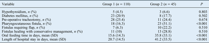
Hypothyroidism (p = 0.803), diabetes mellitus (p = 0.502) and pre-operative tracheotomy (p = 0.674) did not have a significant effect on pharyngocutaneous fistula formation. The data on pre-operative conditions, fistula formation and its post-operative course are shown in Table III. The data on the oral feeding day and length of hospital stay for each group and subgroup are shown in Table IV. Previous chemoradiotherapy was significantly associated with fistula formation (p < 0.001) (Table III).
Pre-operative conditions, fistula formation and post-operative course of fistulas in groups 1 and 2
SD = standard deviation
Post-operative course of fistulas in groups 2a and 2b
*None of the patients in group 2b required a second pectoralis major myocutaneous flap for pharyngocutaneous fistula reconstruction. N/A = not applicable; SD = standard deviation
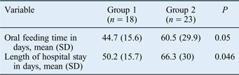
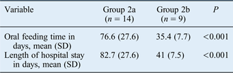
The use of a pectoralis major myofascial flap did not have a significant effect on pharyngocutaneous fistula formation (p = 0.376) in the salvage group (group 2). When comparing the oral feeding days of all patients included in the study, the differences between groups 1 and 2 (p < 0.001) and between groups 2a and 2b (p = 0.004) were statistically significant. The oral feeding day and length of hospital stay of patients with pharyngocutaneous fistula formation in each group are shown in Tables V and VI. When comparing the oral feeding day of patients with pharyngocutaneous fistula, the differences between groups 1 and 2 (p ≤ 0.05), groups 2a and 2b (p < 0.001) and groups 1 and 2a (p = 0.001) were statistically significant, whereas the differences between groups 1 and 2b were not statistically significant for the oral feeding day and length of hospital stay of patients with pharyngocutaneous fistula (p = 0.105 and p = 0.109, respectively).
Oral feeding time and length of hospital stay in patients with pharyngocutaneous fistulas in groups 1 and 2
SD = standard deviation
Oral feeding time and length of hospital stay in patients with pharyngocutaneous fistulas in groups 2a and 2b
SD = standard deviation
Discussion
Pre-operative radiotherapy followed by salvage laryngectomy has been shown to be the most significant risk factor contributing to the development of pharyngocutaneous fistula.Reference Paydarfar and Birkmeyer15 Pharyngocutaneous fistulas develop earlier, tend to be larger and persist longer in pre-operatively irradiated patients.Reference Paydarfar and Birkmeyer15 One of the major problems in salvage patients is further wound breakdown after pharyngocutaneous fistula formation. The maturation of the fistula margins occurs later than in primary laryngectomy patients, therefore the repair of pharyngocutaneous fistulas is performed later and the cost of the treatment gradually increases as a result of prolonged hospital stay and additional surgery. The fistulas of irradiated patients usually require surgical closure whereas most of the fistulas of non-irradiated patients heal with conservative management.Reference Paydarfar and Birkmeyer15 In patients irradiated pre-operatively and in fistulas larger than 1 cm in diameter, surgical closure with primary closure, local flaps or free tissue transfer is often necessary.Reference Gooi and Richmon16
Although radiotherapy with the concurrent administration of cisplatin-based chemotherapy is more effective than radiotherapy alone for larynx preservation and locoregional control, it is associated with a two-fold increase in the incidence of pharyngocutaneous fistula.Reference Gil, Gupta, Kummer, Cordeiro, Kraus and Shah8, Reference Ganly, Patel, Matsuo, Singh, Kraus and Boyle17, Reference Weber, Berkey, Forastiere, Cooper, Maor and Goepfert18 Tumour–node–metastasis (TNM) staging, associated comorbidities, neck clearance, low levels of haemoglobin pre- and post-operatively, low serum albumin, previous tracheotomy, type of closure, duration of surgery and hypothyroidism have also been determined to be risk factors for pharyngocutaneous fistula.Reference Sousa, Castro, Porcaro-Salles, Soares, de Moraes and Carvalho12, Reference White, Golden, Sweeny, Carroll, Magnuson and Rosenthal19 In our study, all of the tumours were at an advanced stage and closure of the pharynx was performed in two layers with a T- or Y-shaped suture line. We examined the effect of chemoradiotherapy, hypothyroidism, diabetes mellitus and previous tracheotomy on pharyngocutaneous fistula formation and only pre-operative chemoradiotherapy was significantly associated with pharyngocutaneous fistula formation. The rates of fistula formation that we observed in primary total laryngectomy and salvage laryngectomy were similar to those reported in other studies.Reference Patel and Keni20
In recent studies, the use of non-irradiated flaps during initial surgery has been recommended to reduce the incidence rates and morbidities associated with salivary fistulas in salvage laryngectomy, even when there is sufficient mucosa for primary closure.Reference Hanasono, Lin, Wax and Rosenthal21, Reference Furuta, Homma, Oridate, Suzuki, Hatakeyama and Suzuki22 Usually, the choice of using a flap or not is based on the intra-operative preference and judgement of the surgeon.Reference Sousa, Castro, Porcaro-Salles, Soares, de Moraes and Carvalho12 The main surgical principle is safe closure with well-vascularised tissue and without suture tension.
• Contrary to recent studies, our study showed that pectoralis major myofascial flap reinforcement did not decrease the rate of pharyngocutaneous fistula formation. Instead, it prevented the formation of large fistulas and provided similar time to oral feeding and length of hospital stay to primary laryngectomy patients
• In the salvage group with pectoralis major myofascial flap reinforcement, all of the fistulas healed with conservative management in a similar time to primary laryngectomy patients
• No significant difference was observed between the oral feeding day and length of hospital stay of patients who underwent primary laryngectomy and those who underwent salvage laryngectomy with pectoralis major myofascial flap
The pectoralis major myocutaneous flap has been demonstrated to be a reliable and versatile flap for reconstruction in the head and neck area, as it has been associated with a low incidence of complications.Reference Milenović, Virag, Uglesić and Aljinović-Ratković23 One of the consequences of using pectoralis major myofascial flap reinforcement is the change in timing, indications present and flap composition compared with the standard pectoralis major myocutaneous flap. In this procedure, the flap is no longer considered to be a secondary treatment used for closure of the defect; instead, it is used during the initial operation to prevent the formation of a fistula. In a pectoralis major myocutaneous flap, the vitality of the cutaneous part, which is supplied by the microvascular vessels of the underlying muscle, determines the success of the reconstruction. In a pectoralis major myofascial flap, the flap only contains muscle and fascia tissues that are directly supplied from its pedicle. When a pectoralis major myocutaneous flap is used as a second procedure for reconstruction, total flap necrosis is the only complication that requires another flap.Reference Milenović, Virag, Uglesić and Aljinović-Ratković23 However, when a pectoralis major myofascial flap is used during salvage laryngectomy, another cutaneous flap may be required in the case of formation of a large fistula. This is probably the most important disadvantage of the procedure. Although none of our patients required a second flap, the risk of a large fistula always exists in pre-operatively irradiated patients.
Righini et al. demonstrated a reduction in the rates of pharyngocutaneous fistula formation from 50 to 23 per cent by using pectoralis major myofascial flap reinforcement during salvage laryngectomy.Reference Righini, Lequeux, Cuisnier, Morel and Reyt24 Their findings failed to reach statistical significance due to their small sample size; however, they did suggest that the use of a pectoralis major myofascial flap would reduce the rate of pharyngocutaneous fistula formation in salvage surgery.Reference Righini, Lequeux, Cuisnier, Morel and Reyt24 Smith et al. compared their results for pectoralis major myofascial flaps with those of an historical cohort that was surgically treated without pectoralis major myofascial flaps and reported a significant reduction in the rate of pharyngocutaneous fistula formation (from 23 to 1 per cent). They concluded that the pectoralis major myofascial flap should become a standard technique in total laryngectomy for the prevention of pharyngocutaneous fistula.Reference Smith, Burrage, Ganguly, Kirby and Drover25 Similarly, Gil et al. reported that the pectoralis major myofascial flap should be used judiciously as a surgical adjunct for buttressing the pharyngeal suture line in patients who are at a high risk for pharyngocutaneous fistula formation.Reference Gil, Gupta, Kummer, Cordeiro, Kraus and Shah8
There are several limitations related to the retrospective design of our study. The small number of patients who were enrolled in our study limits our ability to draw strong conclusions from the data. Our study confirmed that a greater percentage of the pharyngocutaneous fistulas in primary laryngectomy patients healed with conservative management and in a shorter time than in the salvage group.Reference White, Golden, Sweeny, Carroll, Magnuson and Rosenthal19 In the salvage group with pectoralis major myofascial flap reinforcement, all of the fistulas healed with conservative management in a similar time to the primary laryngectomy patients. No significant difference was observed between the oral feeding day and length of hospital stay of patients who underwent primary laryngectomy and patients who underwent salvage laryngectomy with a pectoralis major myofascial flap. In the salvage group without a pectoralis major myofascial flap, 75 per cent of the fistulas required surgical closure with a pectoralis major myocutaneous flap after the initial surgery. This most probably can be attributed to the diminished healing capacity of the irradiated tissues secondary to decreased vascularity. Contrary to recent studies, our study demonstrated that pectoralis major myofascial flap reinforcement did not decrease the rate of pharyngocutaneous fistula formation. Instead, it prevented the formation of large fistulas, which would require surgical management, and it provided similar time to oral feeding and length of hospital stay to primary laryngectomy patients.