Introduction
In order to improve embryo production efficiency and embryo quality, co-culture systems using various homogeneous cell types have been used widely because of the embryotrophic factors secreted by various cells, such as human (Simón et al., Reference Simón, Mercader, Garcia-Velasco, Nikas, Moreno, Remohí and Pellicer1999; Spandorfer et al., Reference Spandorfer, Pascal, Parks, Clark, Veeck, Davis and Rosenwaks2004; Parikh et al., Reference Parikh, Nadkarni, Naik, Naik and Uttamchandani2006), mouse (Joo et al., Reference Joo, Kim, Na, Moon, Lee and Kim2001) and bovine (Goovaerts et al., Reference Goovaerts, Leroy, Rizos, Bermejo-Alvarez, Gutierrez-Adan, Jorssen and Bols2011) cells. Moreover, it has been reported that co-culture with human tube epithelium cells (Goldberg et al., Reference Goldberg, Khalifa, Friedman and Kim1991; Bongso et al., Reference Bongso, Ng, Fong, Anandakumar, Marshall, Edirisinghe and Ratnam1992; Frasor et al., Reference Frasor, Sherbahn, Soltes, Molo, Binor, Radwanska and Rawlins1996) or oviductal cells (Xu et al., Reference Xu, Chan, Lee, Lee and Yeung2004) could improve the development ability and quality of mouse embryo. These results indicate that the benefit of coculturing embryos with cells is not species specific.
Furthermore, many studies have reported that the proportion of mammalian denuded germinal vesicle oocytes (DOs) that developed to meiosis MII (MII) phase was increased when they were cocultured with homogeneous (Ge et al., Reference Ge, Han, Lan, Zhou, Liu, Zhang, Sui and Tan2008a, Reference Ge, Sui, Lan, Liu, Wang and Tanb) or heterogeneous (Anderiesz & Trounson, Reference Anderiesz and Trounson1995; Chin & Chye, Reference Chin and Chye2004; Lee et al., Reference Lee, Quaas, Wright, Toth and Teixeira2011) somatic cells. In mice, the cumulus cell monolayer has been shown to contribute greatly to maintaining normal cellular characteristics in DOs during in vitro maturation (IVM). Cumulus cells regulate the activity of maturation promoting factor (MPF), the transition from G2 to M phase, the distribution of cortical granules (CGs), spindle assembly, mitochondrial congregation and the exocytosis of CGs, which illustrate the mechanisms by which homogeneous cumulus cells improve the maturation and developmental potential of DOs (Ge et al., Reference Ge, Sui, Lan, Liu, Wang and Tan2008b). Until now, however, little information has been available about the mechanisms by which heterogeneous somatic cells influence the maturation and developmental potential of mammalian DOs.
Therefore, the present study was designed to investigate the effect of mouse cumulus cells (mCCs) on the nuclear and cytoplasmic maturation of bovine DOs, as well as on the expression of specific genes, zona pellucida (ZP) hardening and the developmental potential of the bovine MII oocytes obtained.
Materials and methods
Chemicals and media were purchased from Sigma Chemical Co. (St. Louis, MO, USA), unless otherwise indicated. All experimental protocols concerning the handling of animals were performed in accordance with the requirements of the Institutional Animal Care and Use Committee at the Chinese Academy of Agricultural Sciences.
Oocyte collection and IVM
Bovine ovaries were collected at a local abattoir and were transported to the laboratory in physiological saline solution (0.9% NaCl) with 50 μg/ml streptomycin and 75 μg/ml penicillin at 35°C within 2 h of collection. Cumulus–;oocyte complexes (COCs) were aspirated from follicles 2–;8 mm in diameter, and only COCs with at least three layers of compact cumulus cells were used for IVM. DOs were prepared by denuding COCs of cumulus cells by vortexing in 0.1% (w/v) hyaluronidase for 2–;3 min.
Before undergoing IVM, COCs and DOs were divided into four different experimental groups: COCs alone; DOs alone; DOs + bovine cumulus cells (bCCs) (DOs with 1 × 10⁶ cells/ml bCCs) and DOs + mCCs (DOs with 1 × 10⁶ cells/ml mCCs). Briefly, 50 COCs or DOs were cultured with either bCCs or mCCs (prepared as described below) in a separate well of four-well dishes in an incubator at 38.5°C with 5% CO2. In all experimental groups, IVM was performed for 22–;24 h in a total volume of 500 μl maturation media, comprising Medium 199 (Gibco BRL, Grand Island, NY, USA) supplemented with 10 μg/ml follicle-stimulating hormone, 10 μg/ml luteinizing hormone, 10 μg/ml estradiol, 10% (v/v) fetal bovine serum (FBS; HyClone; Gibco BRL, Paisley, Scotland, UK) and 10 μg/ml heparin.
Preparation of mCCs
Kunming white mice (Academy of Military Medical Sciences, Beijing, China) were housed at 20–;24°C under light-controlled conditions (lights on from 06:00–20:00). Female mice that were 6 to 8 weeks old were superovulated using 10 IU equine chorionic gonadotropin (Ningbo Hormone Product Co., Ltd, Ningbo, Zhejiang Province, China) administered via intraperitoneal injection followed 48 h later by 10 IU human chorionic gonadotropin (Ningbo Hormone Product Co., Ltd). The oviducts of superovulated female mice were excised 13 h after hCG administration of human chorionic gonadotropin, and the COCs were collected in M2 medium. mCCs were stripped from oocytes by pipetting COCs in M2 medium containing 0.1% (w/v) hyaluronidase.
The mCCs obtained were cultured in DMEM/F-12 supplemented with 10% (v/v) FBS in a 96-well plate in an incubator at 37.5°C with 5% CO2 until 80% confluent. Then the cells were trypsinized, centrifuged, diluted with bovine IVM media to a density of 1 × 10⁶ cumulus cells/ml, and pre-equilibrated in bovine IVM media for at least 2 h before IVM of bovine DOs.
Preparation of bCCs
bCCs were detached from bovine COCs by vortexing in 0.1% (w/v) hyaluronidase for 2–;3 min, and then cultured as described above.
Assessment of MII oocytes
After 22–24 h IVM, denuded oocytes were fixed in methanol for 10 min, mounted on a slide, and stained with 10 μg/ml Hoechst 33342. The presence or absence of the polar body was determined under the fluorescence microscopy (TE2000S; Nikon, Tokyo, Japan), and only those with the first polar body were considered as MII oocytes.
Assay of MII oocyte glutathione content
After IVM, COCs were denuded before the glutathione (GSH) assay. For collection of samples, 5 μl of 0.2 M sodium phosphate containing 10 mm ethylenediaminetetraacetic acid (EDTA) and 5 μl of 1.25 M phosphoric acid were added to a centrifuge tube which contained 20–;30 MII oocytes. Then, samples were frozen at −80°C. To ensure that oocytes were lysed, thawed oocytes were homogenized by repeated pipetting and freezing.
GSH content in samples was measured using the Total Glutathione Assay Kit (BiYunTian, Shanghai, China), according to the method described by Funahashi et al. (Reference Funahashi, Cantley, Stumpf, Terlouw and Day1994) with some modifications. Briefly, samples were placed into 96-well plates, 150 μl detection solution was added to each well, equilibrated at 25°C for 5 min, and then 50 μl 0.16 mg/ml nicotinamide adenine dinucleotide phosphate (NADPH) was added. GSH standards (25, 15, 10, 5 and 2 μm) and a sample blank without GSH were also assayed at the same time. The formation of 5-thio-2-nitrobenzonic acid was detected at 412 nm using a Beckman DU-40 spectrophotometer (Beckman Coulter, CA, USA) at 1 s intervals for 30 min, and the calculated GSH content was divided by the number of oocytes in each sample to obtain the mean GSH content per oocyte.
Quantitative real-time PCR
Samples containing 100 MII oocytes were selected randomly from each experimental group. Total RNA was isolated with TRIZOL reagent (Invitrogen, CA, USA) according to the manufacturer's instructions and dissolved in sterile water at –80°C until use. The total RNA was reversed to cDNA by the reverse transcription enzyme Superscript III (Invitrogen, CA, USA).
Polymerase chain reactions (PCR) were carried out in a 25-μl total volume, which contained 1.2 μl cDNA, 0.5 μl (10 μM) of each primer, and 0.5 μl 20× Master SYBR Green mix (Invitrogen, USA) at 95°C 2 min, followed by 45 cycles of 95°C for 10 s and 60°C for 30 s. The β-actin gene was used as a reference gene, and the primers for each gene were listed in Table 1. Real-time quantitative PCR was applied using the comparative Ct (2−ΔCt) method (Schmittgen & Livak, Reference Schmittgen and Livak2008) on the ABI 7500 SDS instrument (Applied Biosystems, CA, USA).
Primer sequences and expected fragment sizes of BMP-15, GDF-9 and β-actin genes

Assessment of ZP hardening
ZP hardening was assessed by the rate of dissolution of the ZP by α-chymotrypsin according to the method described by Matson et al. (Reference Matson, Graefling, Junk, Yovich and Edirisinghe1997). Oocytes were incubated in a 0.5% (w/v) solution of α-chymotrypsin dissolved in phosphate-buffered saline with bovine serum albumin (BSA) on a heated stage (37°C). The integrity of the ZP was monitored over time, with the end point being assessed when the limits of the ZP were no longer clearly visible.
Parthenogenetic activation of MII oocytes
After 22–;24 h of IVM, MII oocytes were treated with 5 μM Ca-ionophore A23187 in CR1aa for 5 min, followed by 2 μM 6-dimethylaminopurine in CR1aa with 3 mg/ml BSA for a further 4 h. After activation, oocytes were cultured in CR1aa for 48 h at 38.5°C in 5% CO2 in air. Then oocytes were subsequently cultured for an additional 5 days in CR1aa supplemented with 10% (v/v) FBS, and the medium was changed every 2 days throughout the culture period.
Statistical analysis
All experimental groups were repeated at least three times and all results were presented as mean ± standard error. The cumulus cell apoptotic rates were analyzed with chi-squared test. All other percentage data were subjected to arcsine transformation before statistical analysis, and analysed using the Statistical Analysis System software (SAS Institute Inc., Cary, NC, USA) to perform one-way analysis of variance (ANOVA) using Duncan's test. p-values <0.05 were considered statistically significant.
Results
Effect of mCCs on the MII rate of bovine DOs
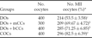
As shown in Table 2, the MII rate of the DOs group (53.5 ± 3.58%) was significantly lower than that of the COCs group (82.5 ± 6.39%; p <0.05). The MII rates of the DOs + mCCs group (69.67 ± 4.72%) and the DOs + bCCs group (71.25 ± 6.85%) were similar (p < 0.05), but significantly higher than that of the DOs group (p < 0.05).
Effect of mouse cumulus cells (mCCs) on the meiosis II (MII) rate of bovine denuded germinal vesicle oocytes (DOs)

a,b Values with different superscripts within the same column differ significantly (P < 0.05). *Values indicate mean ± standard error.
bCCs, bovine cumulus cells; COCs, cumulus–oocyte complexes.
Effect of mCCs on the GSH content of bovine MII oocytes
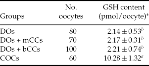
No significant difference was found between the GSH contents of the oocytes in either the DOs + mCCs group (2.17 ± 0.31 pmol/oocyte), the DOs + bCCs group (2.21 ± 0.74 pmol/oocyte) or the DOs group (2.14 ± 0.53 pmol/oocyte; p > 0.05).The GSH contents of the oocytes in these three groups were significantly lower than that in the COCs group (10.28 ± 1.32 pmol/oocyte; p < 0.05), as shown in Table 3.
The effect of mouse cumulus cells (mCCs) on the glutathione (GSH) content of bovine meiosis II (MII) oocytes

a,b Values with different superscripts within the same column differ significantly (P < 0.05). *Values indicate mean ± standard error.
bCCs, bovine cumulus cells; COCs, cumulus–oocyte complexes; DOs, denuded germinal vesicle oocytes.
Effect of mCCs on BMP-15 and GDF-9 mRNA expression in bovine MII oocytes
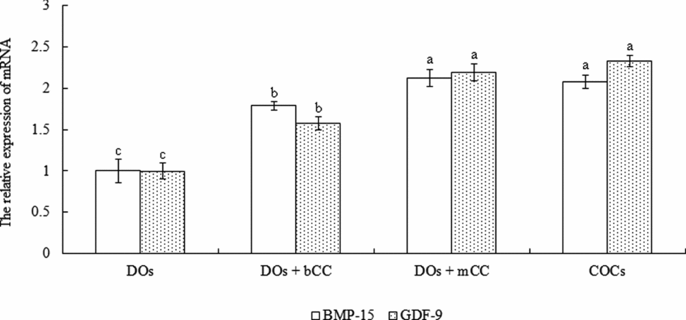
As shown in Fig. 1, the relative expression levels of BMP-15 and GDF-9 mRNA of the DOs + mCCs group and DOs + bCCs group were similar (p > 0.05), and significantly higher than those of DOs group (p < 0.05) but lower than those of the COCs group (p < 0.05).
Effect of mouse cumulus cells (mCCs) on the expression of BMP-15 and GDF-9 mRNA in bovine meiosis II (MII) oocytes. bCC, bovine cumulus cell; COCs, cumulus–oocyte complexes; DOs, denuded germinal vesicle oocytes. a,b,cFor the expression of the same gene, values with different superscripts between different groups differ significantly (p < 0.05).

Effect of mCCs on the ZP hardening of bovine MII oocytes
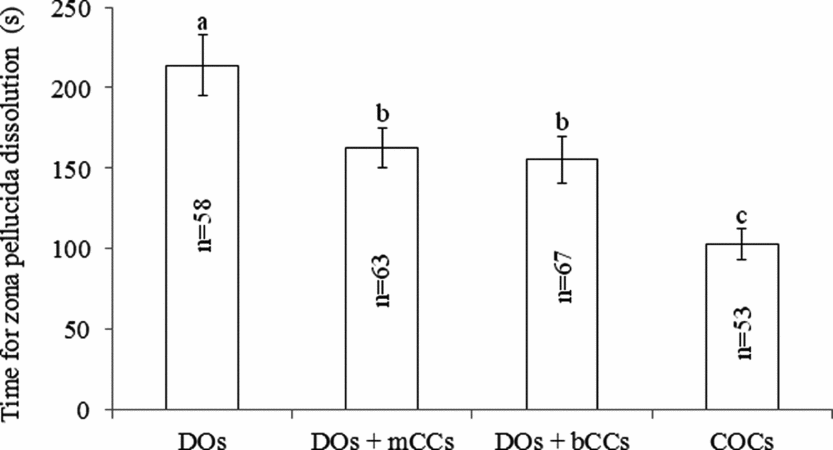
As shown in Fig. 2, the time for ZP dissolution of the oocytes in the DOs group (213.95 ± 18.87 s) was significantly higher than that of the oocytes in the DOs + mCCs group (162.49 ± 12.51 s), DOs + bCCs group (155.17 ± 14.29 s) or COCs group (102.68 ± 9.43 s; p < 0.05). Meanwhile, the time for ZP dissolution of the DOs + mCCs group (162.49 ± 12.51 s) and DOs + bCCs group (155.17 ± 14.29 s) were similar and significantly higher than that of COCs group (102.68 ± 9.43 s; p < 0.05).
Effect of mouse cumulus cells (mCCs) on the ZP hardening of bovine MII oocytes. bCCs, bovine cumulus cells; COCs, cumulus–oocyte complexes; DOs, denuded germinal vesicle oocytes. a,b,cValues with different superscripts between different groups differ significantly (p < 0.05).

Effect of mCCs on the parthenogenetic development of bovine MII oocytes
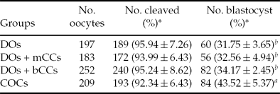
No significant difference was found between the blastocyst rates of the DOs + mCCs group (32.56 ± 4.94%), DOs + bCCs group (34.17 ± 2.45%) and DOs group (31.75 ± 3.65%), which were all lower than that of the COCs group (43.52 ± 5.37%; p < 0.05), as shown in Table 4.
The effect of mouse cumulus cells (mCCs) on the parthenogenetic development of bovine meiosis II (MII) oocytes

a,b Values with different superscripts within the same column differ significantly (P < 0.05). *Values indicate mean ± standard error.
bCCs, bovine cumulus cells; COCs, cumulus–oocyte complexes; DOs, denuded germinal vesicle oocytes.
Discussion
Effect of mCCs on the MII rate of bovine DOs
In agreement with previous results in bovine (Zhang et al., Reference Zhang, Jiang, Wozniak, Yang and Godke1995; Geshi et al., Reference Geshi, Takenouchi, Yamauchi and Nagai2000), porcine (Maedomari et al., Reference Maedomari, Kikuchi, Ozawa, Noguchi, Kaneko, Ohnuma, Nakai, Shino, Nagai and Kashiwazaki2007) and mouse oocytes (Ge et al., Reference Ge, Han, Lan, Zhou, Liu, Zhang, Sui and Tan2008a, Reference Ge, Sui, Lan, Liu, Wang and Tan2008b), our experiment showed that the removal of cumulus cells from bovine oocytes prior to IVM negatively affected the MII rates of bovine oocytes, which may be attributed to the destruction of the gap junction communication (GJC) in DOs as the GJC was required for the coordination of nuclear and cytoplasmic meiotic competence (Carabatsos et al., Reference Carabatsos, Sellitto, Goodenough and Albertini2000). However, the rate of bovine oocytes reaching MII phase significantly increased when bCCs were added to the medium, in accordance with observations in mouse oocytes (Ge et al., Reference Ge, Han, Lan, Zhou, Liu, Zhang, Sui and Tan2008a, Reference Ge, Sui, Lan, Liu, Wang and Tan2008b).
The accurate mechanism by which bCCs promoted the maturation of DOs is not understood well. bCCs may produce a diffusible factor(s) to support DOs maturation in a cumulus cell–oocyte junctional communication-dependent manner (Ge et al., Reference Ge, Han, Lan, Zhou, Liu, Zhang, Sui and Tan2008a) and possibly exert on DOs via the regulation of MPF activity and meiotic progression (Ge et al., Reference Ge, Sui, Lan, Liu, Wang and Tan2008b). Additionally, as a promotion factor of oocyte maturation, melatonin may act directly on bovine oocyte maturation via a receptor-dependent mechanism as melatonin synthesis occurs in bovine cumulus cells and melatonin receptors are expressed in both oocytes and cumulus cells (El-Raey et al., Reference El-Raey, Geshi, Somfai, Kaneda, Hirako, Abdel-Ghaffar, Sosa, El-Roos and Nagai2011).
Interestingly, despite the species difference, mCCs were able to support the nuclear maturation of bovine oocytes, as the rate of oocytes reaching MII in the DOs + mCCs group was similar to the DOs + bCCs group and significantly increased compared with the DOs group. It has been reported that porcine (Chin & Chye, Reference Chin and Chye2004) and human mural granulosa cell monolayers (Anderiesz & Trounson, Reference Anderiesz and Trounson1995) could enhance IVM of mouse DOs. Recently, it has also been proven that the maturation rates of murine germinal vesicle-stage oocytes to MII at 12 and 24 h significantly increased by co-culture with human tubal epithelial cells and immortalized hTECS, compared with human tubal fluid media alone (Lee et al., Reference Lee, Quaas, Wright, Toth and Teixeira2011). These studies and our experiments indicated that the effects of somatic cells on oocyte nuclear maturation were not tissue or species dependent.
Effect of mCCs on the GSH content of bovine MII oocytes
Cumulus cells are structurally and metabolically linked to oocytes by GJC via which they are reported to regulate the synthesis and accumulation of GSH in the ooplasm (de Matos et al., Reference de Matos, Furnus and Moses1997). As an important indicator of cytoplasmic maturation in oocytes (Funahashi et al., Reference Funahashi, Cantley, Stumpf, Terlouw and Day1994; Abeydeera et al., Reference Abeydeera, Wang, Cantley, Rieke and Day1998; Zuelke et al., Reference Zuelke, Jeffay, Zucker and Perreault2003), the GSH content in COCs group (10.28 ± 1.32 pmol/oocyte) and DOs group (2.14 ± 0.53 pmol/oocyte) in our experiment was similar to that (9.46 ± 0.52 pmol/oocyte and 1.85 ± 0.0.16 pmol/oocyte, respectively) reported by Luciano et al. (Reference Luciano, Lodde, Beretta, Colleoni, Lauria and Modina2005).
Similar to previous research in bovine (de Matos et al., Reference de Matos, Furnus and Moses1997) and porcine (Maedomari et al., Reference Maedomari, Kikuchi, Ozawa, Noguchi, Kaneko, Ohnuma, Nakai, Shino, Nagai and Kashiwazaki2007) oocytes, our study showed that the GSH content in the DOs, DOs + mCCs or DOs + bCCs group was significantly lower than that of the COCs group. As GJC played an important role in GSH synthesis(de Matos et al., Reference de Matos, Furnus and Moses1997), our finding may be explained by the fact that GJC was completely destroyed in the DOs, DOs + mCCs and DOs + bCCs group while remaining intact in the COCs group. Although co-culture of DOs with COCs increased the GSH content in DOs (Luciano et al., Reference Luciano, Lodde, Beretta, Colleoni, Lauria and Modina2005), co-culture of DOs with bCCs or mCCs did not significantly increase the GSH content in our experiment. Further experiments need be conducted to investigate the reasons underlying this difference.
Effect of mCCs on BMP-15 and GDF-9 mRNA expression in bovine MII oocytes
Compared with the DOs and DOs + bCCs groups, the relative expression levels of BMP-15 and GDF-9 in oocytes increased significantly in the DOs + mCCs groups (Fig. 1), which indicated that mCCs did exert an influence on DOs at the gene expression level.
Both BMP-15 and GDF-9 positively regulated the function of cumulus cell, which in turn improved the quality of oocytes IVM via GJC (Gilchrist et al., Reference Gilchrist, Lane and Thompson2008). The developmental competence of oocytes in mice had been enhanced by the addition of exogenous BMP-15 and/or GDF-9 to the media in which COCs were matured (Hussein et al., Reference Hussein, Thompson and Gilchrist2006; Yeo et al., Reference Yeo, Gilchrist, Thompson and Lane2008). However, in our experiment, we showed that the GJC, via which cumulus cells promoted the IVM of oocytes, had been destroyed in the DOs, DOs + mCCs and DOs + bCCs groups. Taken together, the evidence indicated that although the expression of BMP-15 and GDF-9 increased significantly in the DOs + mCCs group, this increase probably could not lead to the improved IVM of DOs due to the absence of GJC.
Effect of mCCs on the ZP hardening of bovine MII oocytes
Similar to the results reported by Ge et al. (Reference Ge, Sui, Lan, Liu, Wang and Tan2008b), we also showed that the ZP hardening of MII oocytes from the DOs group was significantly higher than that of oocytes in the COCs group, It was possible that the removal of cumulus cells before IVM caused a precocious exocytosis of CGs, leading to the ZP hardening of oocytes (Ge et al., Reference Ge, Sui, Lan, Liu, Wang and Tan2008b).
However, co-culture with bCCs significantly decreased the ZP hardening of MII oocytes from the DOs group, a finding that was similar to the results of previous experiments in mice (Ge et al., Reference Ge, Sui, Lan, Liu, Wang and Tan2008b). It was reported that the presence of cumulus cells and serum could significantly increase the penetrability of rat oocytes by spermatozoa (Vanderhyden & Armstrong, Reference Vanderhyden and Armstrong1989), and cumulus cells could inhibit the premature exocytosis of CGs during the aging of mouse oocytes (Miao et al., Reference Miao, Liu, Qiao, Miao, Luo and Tan2005). Moreover, cumulus cells have been shown to inhibit both premature CG exocytosis and the conversion of the ZP2 glycoprotein ZP2 to ZP2f in mice (Ge et al., Reference Ge, Sui, Lan, Liu, Wang and Tan2008b). All evidence could help to explain the decreased ZP hardening in the DOs + mCCs group.
Interestingly, the ZP hardening of the MII oocytes obtained was also decreased when the DOs were cocultured with mCCs, indicating that heterogeneous cumulus cells could also protect the ZP from hardening during the IVM of DOs. The mechanisms by which heterogeneous and homogenous cumulus cells decreased the ZP hardening may be similar.
Effect of mCCs on the parthenogenetic development of bovine MII oocytes
Our study showed that the removal of cumulus cells before IVM significantly decreased the developmental potential of bovine oocytes, in accordance with the results of previous studies in bovine (Zhang et al., Reference Zhang, Jiang, Wozniak, Yang and Godke1995; Luciano et al., Reference Luciano, Lodde, Beretta, Colleoni, Lauria and Modina2005) and mouse (Ge et al., Reference Ge, Han, Lan, Zhou, Liu, Zhang, Sui and Tan2008a) oocytes. Similar to the results reported by Luciano et al. (Reference Luciano, Lodde, Beretta, Colleoni, Lauria and Modina2005), we also observed that a correlation between the GSH content of oocytes in different groups and their ability to reach the blastocyst stage after parthenogenetic activation. Cytoplasmic GSH has been shown to improve the fertilization of oocytes and in vitro embryonic development (Maedomari et al., Reference Maedomari, Kikuchi, Ozawa, Noguchi, Kaneko, Ohnuma, Nakai, Shino, Nagai and Kashiwazaki2007) by protecting oocytes from oxygen stress (Luberda, Reference Luberda2005), a finding that could explain the results of our experiments.
Meanwhile, the response of mature oocytes to inositol 1,4,5-trisphosphate (IP3) is thought to be a cytoplasmic indicator of their developmental ability. Mature oocytes, in which a greater increase in intracellular Ca2+ is induced by IP3, have increased Ca2+ oscillation at fertilization and improved developmental ability (Machaty et al., Reference Machaty, Funahashi, Day and Prather1997). The sensitivity to IP3 of mature porcine oocytes derived from DOs was lower than that of oocytes obtained from COCs, and the former had a decreased activation rate and development potential (Amano et al., Reference Amano, Mori, Matsumoto, Iritani and Watanabe2005). This difference in IP3 sensitivity partially explained the lower developmental potential of DOs compared with that of COCs in our experiment.
Co-culture with bCCs or mCCs increased the MII rates of bovine DOs compared with DOs cultured alone. However, the ability of bCCs or mCCs to enhance bovine oocyte nuclear maturation did not translate into an improved developmental competence, similar to the effects observed when mouse oocytes were cocultured with porcine granulosa cells (Chin & Chye, Reference Chin and Chye2004). Although bCCs or mCCs exerted many beneficial functions on bovine DOs as mentioned above, they failed to improve significantly the developmental potential of the MII oocytes obtained, indicating that GJC was essential for the maturation and development potential of oocytes.
In conclusion, this study demonstrated that despite the species difference, mCCs could increase the MII rate of DOs and the expression of specific genes in the MII oocytes obtained, decrease the ZP hardening of MII oocytes, but could not improve their GSH content and developmental potential.
Acknowledgements
The work was supported by the Basic Research Fund of IAS, CAAS (2010jc-3-1), the Genetically Modified Organisms Breeding Major Projects of China (2009ZX08011-031B) and the Key Projects in the National Science & Technology Pillar Program (2011BAD19B04–6 and 2011BAD19B02).