




No CrossRef data available.
Article contents
Mapping fucosylated epitopes on glycoproteins and glycolipids of Schistosoma mansoni cercariae, adult worms and eggs
Published online by Cambridge University Press: 13 December 2004
Abstract
The developmental expression of the antigenic fucosylated glycan motifs Fucα1-3GalNAcβ1-4GlcNAc (F-LDN), Fucα1-3GalNAcβ1-4(Fucα1-3)GlcNAc (F-LDN-F), GalNAcβ1-4(Fucα1-3)GlcNAc (LDN-F), Galβ1-4(Fucα1-3)GlcNAc (Lewis X), and GalNAcβ1-4(Fucα1-2Fucα1-3)GlcNAc (LDN-DF) in Schistosoma mansoni cercariae, adult worms and eggs, was surveyed using previously defined anti-carbohydrate monoclonal antibodies (mAbs). Lewis X was found both on glycolipids and glycoproteins, yet with completely different expression patterns during the life-cycle: on glycolipids, Lewis X was mainly found in the cercarial stage, while protein-conjugated Lewis X was mainly present in the egg stage. Also protein-conjugated LDN-F and LDN-DF were most highly expressed in the egg-stage. On glycolipids LDN-DF was found in all three examined stages, whereas LDN-F containing glycolipids were restricted to adult worms and eggs. The motifs F-LDN and F-LDN-F were found both on glycoproteins and glycolipids of the cercarial and egg stage, while in the adult stage, they appeared to occur predominantly on glycolipids. Immunofluorescence assays (IFA) showed that these F-LDN and F-LDN-F containing glycolipids were localized in a yet undefined duct or excretory system of adult worms. Murine infection serum showed major reactivity with this adult worm duct-system, which could be fully inhibited by pre-incubation with keyhole limpet haemocyanin (KLH). Clearly, the use of defined mAbs provides a quick and convenient way to map expression profiles of carbohydrate epitopes.
Information
- Type
- Research Article
- Information
- Copyright
- © 2004 Cambridge University Press
INTRODUCTION
Schistosomiasis is the second most important human parasitic disease after malaria. In tropical and subtropical countries, over 200 million people are infected with schistosomes. One of the major human schistosome species is Schistosoma mansoni, of which the adult worms live as pairs in the mesenteric vessels where they produce hundreds of eggs per day. It has been estimated that about half of the eggs of S. mansoni leave the body with the faeces, whilst the rest of the eggs are deposited in the liver and other organs of the host where they cause granulomatous inflammation. Miracidia that hatch from the eggs in fresh water are able to infect the intermediate host: snails of the genus Biomphalaria. Cercariae are formed by asexual replication and are released in the water to infect the definitive human host (Jordan, Webbe & Sturrock, 1993; Ross et al. 2002).
Throughout their intriguing and complex life-cycle schistosomes express a complex set of glycoproteins and glycolipids that play a major role in the immunology of schistosomiasis (Hokke & Deelder, 2001, and references cited therein). These glycoconjugates display exceptional glycan motifs, including carbohydrate chains of the gut-associated antigens CAA and CCA (Bergwerff et al. 1994; van Dam et al. 1994), (Fucα1-2)1-2Fucα1-3-elements (Khoo et al. 1995; Khoo et al. 1997a; Wuhrer et al. 2002) and the Fuc(α1-3)GalNAc-motif (Kantelhardt et al. 2002), that induce high antibody responses in humans and primates (van Remoortere et al. 2001, 2003; Kantelhardt et al. 2002; Naus et al. 2003). The more widely expressed glycans GalNAcβ1-4GlcNAc (LDN), Galβ1-4(Fucα1-3) GlcNAc (Lewis X) and GalNAcβ1-4(Fucα1-3)GlcNAc (LDN-F), which are shared between schistosomes and their mammalian hosts (Fox et al. 1983; Bergwerff et al. 1992; Yan, Chao & van Halbeek, 1993; De Graaf et al. 1993) generally induce weaker antibody responses (van Remoortere et al. 2000, 2003), but may have immunomodulatory effects. In addition, schistosome infections induce antibodies to keyhole limpet haemocyanin (KLH) (Eberl et al. 2001; Kantelhardt et al. 2002). It has been shown that Fucα1-3GalNAc-moieties in the N-linked glycans of the highly immunogenic KLH glycoprotein are the main schistosome cross-reactive epitopes (Kantelhardt et al. 2002; Geyer et al. 2004) which enable the use of KLH for serodiagnostic purposes (Grzych et al. 1987; Hamilton et al. 1999).
Fucosylated glycans have been shown to be able to induce the Th2 type immune responses that are characteristic for infections with schistosomes and other helminthes (Okano et al. 1999; Faveeuw et al. 2002; Gause, Urban Jr. & Stadecker, 2003; Thomas & Harn, Jr. 2004). The fucosylated glycan lacto-N-fucopentaose (LNFPIII) that contains Galβ1-4(Fucα1-3) GlcNAc- (LeX) has often been used as a model-glycan to study the immunomodulatory effects of schistosome glycans. When conjugated to a protein carrier for multivalent presentation it induces Th2-responses in naive mice (Okano et al. 2001; Thomas et al. 2003). Peripheral blood mononuclear cells (PBMC) from humans infected with S. mansoni, but not of uninfected individuals, respond to the LNFP-III-HSA conjugate by increased proliferation and IL-10 production (Velupillai et al. 2000). A synthetic LDN-DF conjugate has been shown to be a potent cytokine inducer on PBMCs of naive human donors (Van der Kleij et al. 2002). Moreover, in the induction of liver granulomas in in vivo mouse models, the glycosylation of egg antigens appears to play a crucial role (Weiss, Aronstein & Strand, 1987; El Ridi, Velupillai & Harn, 1996; Jacobs et al. 1998; Jacobs, Deelder & Van Marck, 1999).
The expression of (fucosylated) glycans on glycoproteins or glycolipids is developmentally regulated (Nyame, Yoshino & Cummings, 2002) but intense cross-reactivity occurs between the various schistosome stages based on the shared glycan elements (Strand, McMillan & Pan, 1982; Weiss, Magnani & Strand, 1986; Dunne & Bickle, 1987; van Remoortere et al. 2000). These glycan motifs may be present on many different lipid and protein carriers (Weiss & Strand, 1985; Weiss et al. 1986; van Dam et al. 1993; Deelder et al. 1996; van Remoortere et al. 2000). In this study, the expression profiles of 5 major fucosylated epitopes: Fucα1-3GalNAcβ1-4GlcNAc (F-LDN), Fucα1-3GalNAcβ1-4(Fucα1-3) GlcNAc (F-LDN-F), GalNAcβ1-4(Fucα1-3)GlcNAc (LDN-F), Galβ1-4(Fucα1-3)GlcNAc (Lewis X), and GalNAcβ1-4(Fucα1-2Fucα1-3)GlcNAc (LDN-DF) (Table 1) were mapped utilizing defined mAbs that specifically recognized these epitopes. S. mansoni cercarial, adult worm and egg preparations were analysed combining several techniques; glycoproteins were analysed on Western blot, simultaneously glycolipids were analysed using the HPTLC-overlay technique as well as ELISA and additionally localization of the epitopes was studied using fluorescence microscopy.
Table 1. An overview of the carbohydrate structures included in this study giving the abbreviations and the defined mAbs that recognize these glycan epitopes (van Remoortere et al. 2000; C. H. Hokke, A. van Remoortere, H. J. Vermeer and A. M. Deelder, unpublished observations.)

MATERIALS AND METHODS
Antigens and antibodies
The life-cycle of S. mansoni parasites was maintained using Golden hamsters (HsdHan-Aura). Worms were collected by perfusion 7 weeks after infection with 1200 cercariae. Eggs were isolated from livers as previously described (Dalton et al. 1997). Proteins were extracted from cercariae, adult worms and eggs by heating for 10 min at 95 °C in 0·1 M Tris-HCl, pH 6·8, containing 20% glycerol, 2% SDS and 2% 2-mercaptoethanol. S. mansoni glycolipids were prepared as described previously (Wuhrer et al. 1999). The pre-stained, broad-range precision protein standard from Bio-Rad (Veenendaal, The Netherlands) was used and the globoside standard was purchased from Matreya (State College, PA).
Monoclonal antibodies (mAbs) were produced as described previously (van Dam et al. 1993; Nourel Din et al. 1994; Nibbeling et al. 1998). Monoclonal antibody 291-4D10-A is specific for Galβ1-4(Fucα1-3) GlcNAc (Lewis X), mAb 290-2E6-A recognizes GalNAcβ1-4(Fucα1-3) GlcNAc (LDN-F), mAbs 290-4A8-A and 114-5B1-A bind to GalNAcα1-4(Fucα1-2Fucα1-3) GlcNAc (LDN-DF) (van Remoortere et al. 2000), mAb 291-5D5-A recognizes Fucα1-3GalNAcβ1-4GlcNAc (F-LDN) and mAb 128-1E7-C is specific for Fucα1-3GalNAcβ1-4(Fucα1-3)GlcNAc (F-LDN-F) (C. H. Hokke, A. van Remoortere, H. J.Vermeer and A. M. Deelder, unpublished observations). For glycoprotein analysis, the murine infection serum (MIS) was taken from a 15-week-infected outbred Swiss mouse that had been exposed to 70 cercariae and the human infection sera (HIS) originated from heavily infected schistosomiasis patients from Senegal (Stelma et al. 1993). For glycolipid analysis, different MIS and HIS were used (Kantelhardt et al. 2002).
Western blotting
Approximately 1 μg protein extracts from S. mansoni cercariae, adult worms and eggs were subjected to SDS-PAGE on 10% gels using the Mini-Protean 3 Cell system (Bio-Rad, Veenendaal, The Netherlands). The proteins were transferred onto a nitrocellulose membrane (Schleicher & Schuell BioScience GmbH, Dassel, Germany) in a Bio-Rad Criterion Blotter system according to the manufacturer's instructions. The protein blots were blocked in phosphate-buffered saline/5% BSA (PBS-B) at 4 °C overnight. The blots were washed with PBS/0·5%BSA/0·05%Tween (PBS-BT) and subsequently incubated with the primary anti-carbohydrate mAb, or with infection serum, for 1 h at room temperature (RT). Hybridoma culture supernatant was used 1[ratio ]2 diluted in PBS-BT, infection serum was used in a 1[ratio ]1000 dilution in PBS-BT. After extensive washing, the blots were incubated with AP-labelled goat anti-mouse Ig (1[ratio ]2000) (Zymed Laboratories, San Francisco, CA) in the case of primary incubation with mAbs or murine infection serum. For detection of Ig in human infection serum we combined biotinylated goat anti-human IgG and -IgM (1[ratio ]1000) (Nordic Immunological Laboratories, Tilburg, The Netherlands) with Streptavidin-AP (1[ratio ]2000) (Zymed). Finally, the blots were washed with PBS and stained using X-phosphate/5-Bromo-4-chloro-3-indolyl-phosphate (BCIP) (Roche Diagnostics, Mannheim, Germany) and 4-nitro blue tetrazolium chloride (NBT) (Roche).
High performance TLC (HPTLC) and ELISA
S. mansoni glycolipids were separated by HPTLC using chloroform/methanol/0·25% aqueous KCl (50[ratio ]40[ratio ]10, by volume) as running solvent, followed by either orcinol/H2SO4 staining or HPTLC immuno-staining (Wuhrer et al. 1999). The primary incubation step was carried out using mAbs (hybridoma culture supernatant 1[ratio ]25 in PBS-BT) or human and murine infection sera (1[ratio ]1000 in PBS-BT). ELISA with S. mansoni glycolipids (10 ng carbohydrate/well) as adsorbed antigens and KLH (10 μg/100 μl per well) as an inhibitor was performed as outlined previously (Wuhrer et al. 2000). As secondary antibodies for both HPTLC and ELISA, alkaline-phosphatase-conjugated goat anti-mouse Ig (Dako Diagnostics, Hamburg, Germany) and goat anti-human Ig (Dianova, Hamburg, Germany) were used, diluted 1[ratio ]1000.
Immunofluorescence assay (IFA)
Immunofluorescence assays were performed on frozen gut sections (6 μm thick) of S. mansoni infected hamsters as reported previously (van Dam et al. 1993). Briefly, slides were fixed in ice-cold acetone and dried. To remove glycolipids, slides were incubated for 30 min in chloroform/methanol 1[ratio ]1 and dried again. Subsequently, slides were incubated with a mAb (undiluted culture supernatant) or with 1[ratio ]10 diluted murine or human infection serum for 1 h at 37 °C. In KLH-inhibition studies, mAbs or infection sera were incubated with KLH (Sigma-Aldrich, The Netherlands) 1 mg/ml, for 2 h at RT, prior to incubation with the slides. Subsequently, the slides were washed and incubated with FITC-labelled goat anti-mouse Ig (1[ratio ]40), or swine anti-human Ig (Nordic, The Netherlands) in a conjugate solution containing Evans blue. Para-phenylenediamine was used as anti-fading agent and slides were analysed using a Leica DMRA microscope equipped with an Hamamatsu Orca-ER digital camera and Improvision software.
RESULTS
Fucose-containing glycans are differentially expressed on schistosomal glycoproteins or glycolipids
To survey the developmental expression of antigenic fucosylated glycan motifs in schistosomes (Table 1), we have analysed glycoproteins and glycolipids of S. mansoni cercariae, adult worms and eggs by Western blotting, HPTLC overlays and IFAs using previously characterized anti-carbohydrate mAbs (van Remoortere et al. 2000); C. H. Hokke, A. van Remoortere, H. J. Vermeer and A. M. Deelder, unpublished observations) or infection sera. Staining with mAb 291-4D10-A showed that the Lewis X epitope was expressed on glycoproteins of all 3 examined life-cycle stages, but most extensively on egg-glycoproteins (Fig. 1A). Lewis X was also expressed on cercarial and adult worm glycolipids, but could hardly be detected on egg glycolipids (Fig. 2A), as shown previously (Kantelhardt et al. 2002; Wuhrer et al. 2002). Like Lewis X, LDN-F was expressed on glycoproteins of all examined stages and highest in the egg-stage (Fig. 1B). In contrast to Lewis X, however, LDN-F expression on glycolipids was restricted to the adult worm- and egg-stage (Fig. 2B) and could not be detected on cercarial glycolipids.

Fig. 1. Western blots and silver staining of Schistosoma mansoni glycoproteins. Cercarial (Ce), adult worm (Ad) and egg (Eg) protein extracts were applied to a 10% gel and visualized by silver staining or immunostaining using various mAbs directed against carbohydrate epitopes (A–F) and human- (HIS1,2) or murine (MIS) infection sera (G–I). An isotype control is shown (J) along the pre-stained broad range protein marker (H).

Fig. 2. HPTLC-immunostaining of Schistosoma mansoni glycolipids. HPTLC-resolved cercarial (Ce), adult (Ad) and egg (Eg) complex glycolipids (50 ng of carbohydrate per lane; 300 ng carbohydrate per lane in E, L and M) were visualized by immunostaining using the same panel of anti-carbohydrate mAbs as was used for the Western blots (Fig. 1) and human- (HIS) or murine (MIS) infection sera (L,M). The migration positions of orcinol/H2SO4-stained globoside standards (CDH, CTH and CtetH) are indicated. Pictures marked by * are reproduced with permission from Kantelhardt et al. 2002, © The Biochemical Society.
The LDN-DF motif was present in all 3 examined stages on glycoproteins as well as on glycolipids (Figs 1C,D and 2C,D) and, again, the most intense staining was observed for glycoproteins of the egg-stage. The two anti-LDN-DF mAbs 290-4A8-A and 114-5B1-A revealed distinct band patterns on Western blots and on HPTLC, indicating that these mAbs recognize different natural epitopes. The mAbs 291-5D5-A (anti-F-LDN) and 128-1E7-C (anti-F-LDN-F) yielded rather different staining patterns on HPTLC, but in contrast displayed very similar staining patterns on Western blot. The F-LDN and F-LDN-F epitopes were highly expressed on glycoproteins of eggs and cercariae, but almost entirely absent in the glycoprotein preparation of the adult worm-stage (Fig. 1E,F). On glycolipids, however, these epitopes were expressed in all 3 stages (Fig. 2E,F). The band pattern on HPTLC obtained with anti-F-LDN-F mAb 128-1E7-C (Fig. 2F) showed striking similarity with the band pattern that was observed for anti-LDN-DF mAb 114-5B1-A (Fig. 2D).
Staining of Western blots and HPTLC with human- or murine infection sera resulted in different complex band patterns for each serum and each examined life-cycle stage. In these patterns some bands recognized by the serum occurred in every stage of the parasite, whereas other (glyco-) proteins were only stage-specifically expressed (Fig. 1,G–I). Strikingly, on HPTLC, staining with HIS or MIS resulted in highly comparable patterns (Fig. 2L,M), as depicted previously (Kantelhardt et al. 2002).
Cross-reactivity between schistosomes and KLH
In the paper of Kantelhardt (Kantelhardt et al. 2002) the terminal Fuc(α1-3)GalNAc-motif was found to be the dominant epitope on S. mansoni glycolipids that causes cross-reactivity between schistosome infection sera and KLH. In order to see whether other fucosylated epitopes than the Fuc(α1-3)GalNAc-motif are involved in the cross-reactivity between S. mansoni and KLH, we have examined the potential of KLH to inhibit the binding of the different mAbs to S. mansoni glycolipids in an ELISA experiment. Reactivity of anti-F-LDN mAb 291-5D5-A as well as anti-F-LDN-F mAb 128-1E7-C with S. mansoni glycolipids was strongly inhibited by KLH (Fig. 3E,F), confirming that F-LDN(-F) epitopes are present on KLH. Reactivity of the anti-Lewis X, anti-LDN-F and anti-LDN-DF mAbs with glycolipids of all 3 examined life-cycle stages was unaffected by the presence of KLH (Fig. 3A–D), which showed that these carbohydrate motifs were not present on KLH. For Lewis X and LDN-F, the ELISA results confirmed the HPTLC-results regarding stage-specific expression of glycolipids exhibiting Lewis X and LDN-F epitopes.

Fig. 3. ELISA-detection of Schistosoma mansoni glycolipids by various monoclonal antibodies and infection sera in the presence or absence of KLH as an inhibitor. S. mansoni glycolipids from cercariae (Ce), adults (Ad) and eggs (Eg) were probed with anti-carbohydrate mAbs (A-F) and human- (HIS) or murine (MIS) infection sera (L,M) with (hatched columns) or without (black columns) KLH as an inhibitor. Pictures marked by * are reproduced with permission from Kantelhardt et al. 2002, © The Biochemical Society.
F-LDN(-F) in S. mansoni adult worms is mainly expressed on glycolipids
To further examine the expression of the major antigenic F-LDN and F-LDN-F motifs IFA was performed on frozen sections of S. mansoni adult worms and eggs. Results are shown for anti-F-LDN-F mAb 128-1E7-C (Fig. 4a–d), but staining with anti-F-LDN mAb 291-5D5-A gave the same fluorescence patterns (data not shown). Incubation of slides with mAb 128-1E7-C resulted in fluorescence of spots in the parenchyma of the adult worm and in the secreted egg antigens and eggshell (Fig. 4a,c).

Fig. 4. Immunofluorescence patterns of Schistosoma mansoni sections with or without chloroform/methanol treatment. Frozen sections of adult worms and eggs were stained using anti-F-LDN-F mAb 128-1E7-C (a–d) or murine infection serum (MIS) (e–h) followed by an FITC-labelled anti-mouse Ig conjugate. Use of anti-F-LDN mAb 291-5D5-A gave the same results as mAb 128-1E7-C and an isotype control showed no fluorescence (data not shown). After incubation with mAb 128-1E7-C fluorescence was observed in parenchymal ducts of adult worms (a) and in egg-secreted antigens and egg-shell (c). After chloroform/methanol treatment F-LDN-F related fluorescence patterns were lost in adult worms (b), but not in eggs (d). MIS recognized adult worm tegument (T), gut (G) and parenchymal spots (PS) in adult worms (e, g). Only the gut fluorescence remained after chloroform/methanol treatment (f). When MIS was pre-incubated with KLH (1 mg/ml) fluorescence in the parenchymal structures disappeared (h).
Chloroform/methanol treatment of the slides to remove lipids and glycolipids prior to incubation with mAb 128-1E7-C resulted in a complete loss of fluorescence in adult worms (Fig. 4a,b). Together with the limited staining of adult worm glycoproteins with the F-LDN(-F) mAbs on Western blots (Fig. 1E,F) this indicated that F-LDN(-F) epitopes in adult worms are predominantly glycolipid conjugated. In addition, pre-incubation of mAb 128-1E7-C with KLH before IFA resulted in the same loss of fluorescence in the parenchymal spots of adult worms as caused by chloroform/methanol treatment (data not shown), which is consistent with the finding in ELISA that KLH fully inhibits the binding of the anti-F-LDN(-F) mAbs to schistosome glycolipids. In contrast to our observations in adult worms, in the egg-stage reactivity of mAb 128-1E7-C remained unchanged after chloroform/methanol treatment (Fig. 4c,d), which, together with intensive staining of egg-glycoproteins on Western blots, showed that the F-LDN(-F) motif in this life-cycle stage is expressed at least for a significant part on glycoproteins.
Immunofluorescence staining with MIS showed fluorescence of the adult worm parenchymal spots, tegument and gut (Fig. 4e,g) and of egg-secreted antigens, the miracidium, and the egg-shell (data not shown). After chloroform/methanol treatment, reactivity of MIS with the spots in the parenchyma of adult worms and the tegument completely disappeared and only the gut remained brightly positive (Fig. 4e,f). This suggests that antigens in the tegument and the parenchymal spots recognized by the serum antibodies are of (glyco-) lipid origin, whereas antigens in the gut are for a significant part of (glyco-) protein origin. Reactivity with the parenchymal spots was also lost when MIS was pre-incubated with KLH (Fig. 4g,h). In combination with the observation that the reactivity of MIS to these spots disappears after (glyco-) lipid extraction, this shows that MIS contains antibodies directed against F-LDN(-F) containing worm-glycolipids. In contrast to the findings with adult worms, for eggs and egg secretions no differences could be observed between the MIS fluorescence patterns with or without chloroform/methanol treatment, showing that a substantial part of the antibodies in MIS recognize egg-glycoproteins. Also, no effect of pre-incubation of MIS with KLH was observed for egg fluorescence. Although Western blots, HPTLC overlay data and ELISA show that significant amounts of F-LDN(-F) are produced in schistosome eggs, this suggests that F-LDN(-F) is a less prominent epitope for MIS in eggs than in adult worms.
DISCUSSION
This study on the expression of the fucosylated antigenic glycan motifs F-LDN, F-LDN-F, LDN-F, Lewis X and LDN-DF revealed their prevalence and distribution on proteins and lipids of S. mansoni cercariae, adult worms and eggs. The global expression patterns of the glycan motifs observed here were mostly in agreement with earlier mass spectrometry based structural studies performed on isolated glycans of diverse parasite preparations [as reviewed by (Hokke & Deelder, 2001)], showing that the use of defined anti-carbohydrate mAbs provides a convenient alternative to study the expression of specific glycan epitopes in schistosomes.
A major focus of this study was the F-LDN and F-LDN-F glycan motifs, which occur in schistosomes and keyhole limpet haemocyanin, but have never been described in mammals (Khoo et al. 1995, 1997a; Kantelhardt et al. 2002; Wuhrer et al. 2002; Geyer et al. 2004). In IFA, incubation with 291-5D5-A (F-LDN) and 128-1E7-C (F-LDN-F) resulted in similar staining of spots in the parenchyma of adult worms. These IFA pictures show fluorescence patterns rather similar to those described by Thors and Linder who found that KLH cross-reactive components are present in a schistosome adult worm duct-system (Thors & Linder, 1998, 2003), of which the function remains unknown so far. The disappearance of fluorescence after chloroform/methanol treatment of adult worm sections, together with the faint staining of adult worm glycoproteins on Western blot indicates that the F-LDN(-F) element in these parenchymal spots, or ducts, is mainly glycolipid conjugated. In contrast to adult worms, cercariae and eggs express F-LDN and F-LDN-F not only on glycolipids, but also extensively on glycoproteins. The Western blotting data suggest that F-LDN and F-LDN-F are present on the same sets of glycoproteins. SDS-PAGE does not easily give rise to the separation of the different glycoforms of the protein, in particular when neutral carbohydrate chains are involved. Notably, the band patterns on 291-5D5-A or 128-1E7-C stained HPTLC overlays differed from each other, showing that these mAbs have different specificities and that the different glycolipids bound by the mAbs are separated by HPTLC. Because F-LDN is a partial structure of F-LDN-F the sets of glycolipids recognized by mAbs 291-5D5-A and 128-1E7-C are partly overlapping. These glycolipid subsets seem to be situated in the same parenchymal spots in the adult worm as no differences in the localization of the subsets could be observed in our IFA experiments (data not shown).
KLH inhibition experiments in ELISA and IFA showed that the schistosomal F-LDN(-F) motif is the motif cross-reactive with KLH, which corroborated the earlier findings (Kantelhardt et al. 2002) that Fuc(α1-3)GalNAc- is the major antigenic motif of S. mansoni glycolipids implicated in KLH cross-reactivity. This current observation is also in line with the recent report (Geyer et al. 2004) which describes that the anti-schistosomal mAb M2D3H recognizes the tetrasaccharide deoxyhexose-HexNAc-[Deoxyhexose]-HexNAc on KLH. It is conceivable that the complete structure of this tetrasaccharide is Fucα1-3GalNAcβ1-4(Fucα1-3) GlcNAc (F-LDN-F).
Another highly antigenic glycan that plays a major role in the immunobiology of schistosomiasis is LDN-DF, against which similar high antibody titres are observed as to F-LDN(-F) (van Remoortere et al. 2003). There are no indications that LDN-DF, or other known strongly immunogenic glycans like polyLeX (CCA) and CAA occur on KLH: Mabs reactive with these epitopes do not bind to KLH (A. van Remoortere, unpublished observations). A previous study has shown that 114-5B1-A and 290-4A8-A bind to synthetic LDN-DF, and both mAbs stain an adult worm excretory system in IFA (van Remoortere et al. 2000). The current study shows that the naturally occurring epitopes of these mAbs differ and that they are present on different sets of glycoproteins and glycolipids. To be able to map in more detail the expression of schistosome glycoconjugates it will be of significant interest to deduce what these natural epitopes are.
The antigens recognized by mAb 114-5B1-A (LDN-DF) are localized in a duct- or excretory system in adult worms that has been well-defined in comprehensive microscopic studies (Bogers et al. 1994, 1995; van Remoortere et al. 2000). Thors & Linder (2003) suggested that this system was the same as they observed after staining schistosome adult worms with rabbit anti-KLH polyclonal antibodies. This may be the case, since anti-LDN-DF mAb 114-5B1-A and the KLH reactive anti-F-LDN-F mAb 128-1E7-C (Van de Vijver et al. 2004) yielded similar reaction patterns in HPTLC immunostaining for the adult worm stage, indicating that the F-LDN-F and LDN-DF motifs occur as combined structural elements on the same glycolipids. In line with this observation, the (Fucα1-3GalNAcβ1-4[Fucα1-2Fucα1-3]GlcNAcβ1-) (F-LDN-DF) sequence that combines the two motifs has previously been identified in S. mansoni cercarial glycocalyx (Khoo et al. 1995) and on O-glycans and glycolipids of eggs (Khoo et al. 1997a,b; Wuhrer et al. 2002). The current results imply that glycolipids with this structure are also present in adult worms. However, in contrast to the F-LDN motif, in adult worms LDN-DF is not exclusively present on glycolipids, since numerous LDN-DF containing glycoproteins were observed on Western blots of all 3 examined stages. Moreover, fluorescence patterns of mAb 114-5B1-A in adult worms and eggs were not lost after chloroform/methanol treatment (data not shown), indicating that mainly glycoproteins were recognized.
The expression of Lewis X and LDN-F on schistosome glycoproteins in the cercarial, adult worm, and most abundantly in the egg-stage is in line with the observations of Nyame et al. (2003), who also found that Lewis X, LDN-F and in addition non-fucosylated LDN are more extensively expressed on egg glycoproteins compared to other developmental stages. However, no detailed comparisons can be made between the band patterns on the Western blots in the two studies since different antibodies and methods for sample preparation were used. Remarkably, the current study shows that on the glycolipid level of Lewis X was hardly detectable in eggs (Kantelhardt et al. 2002; Wuhrer et al. 2002). The single Lewis X containing glycolipid observed as a weak band on HPTLC is in line with the finding of a Lewis X containing glycolipid in eggs in a previous study (Wuhrer et al. 2002). This would imply that fluorescence observed with anti-Lewis X mAb 291-4D10-A (van Remoortere et al. 2000) of the egg-shell and secreted egg antigens in S. mansoni is mainly based on glycoprotein conjugated Lewis X.
In the same study of van Remoortere et al. (2000), it has been shown that Lewis X is only present in the oral sucker of cercariae, but not on the surface. In total cercarial extracts, we found numerous cercarial glycoproteins and glycolipids that contain Lewis X. Lewis X containing structures appear on the surface after shedding of the cercarial glycocalyx during the process of transformation to schistosomula (Koster & Strand, 1994; Nyame et al. 2003). Probably, the numerous Lewis X containing glycoproteins and glycolipids we observed are already assembled but not yet expressed on the cercarial surface prior to transformation.
The biosynthesis of the many different fucosylated glycoconjugates in Schistosoma spp. is catalysed by various fucosyltransferases, of which the expression is developmentally regulated (Marques Jr et al. 2001). Marques et al. (2001) found a 50-fold higher total fucosyltransferase-specific activity in egg-extracts than in cercariae and adult worms. In agreement with their study, we observed that the fucosylated epitopes Lewis X, LDN-F, LDN-DF, F-LDN and F-LDN-F were significantly more abundantly expressed on egg glycoproteins than on glycoproteins from cercariae and adult worms. This increase in fucosylation in eggs is not clearly visible in the case of the glycolipids. Notably, in contrast to egg glycoproteins, the Lewis X epitope is almost entirely absent in glycolipids of eggs. This difference in the expression of the different fuco-oligosaccharides may reflect the specific activities of the different fucosyltransferases involved.
Although several studies on the structural characterization of schistosome glycans have been reported, only very few studies have addressed identification of the protein backbones that carry the different glycan epitopes so far described. The present study showed that panels of monoclonal anti-glycan antibodies are useful tools for identification of glycosylation patterns. In this respect, the current quick expansion of the schistosome genomic and proteomic databases open up new possibilities for the large-scale identification of antigenic schistosome glycoproteins by targeted approaches using the specific, anti-carbohydrate mAbs described in this and previous studies.
References
REFERENCES
BERGWERFF, A. A.,
THOMAS-OATES, J. E.,
VAN OOSTRUM, J.,
KAMERLING, J. P. &
VLIEGENTHART, J. F. (
1992).
Human urokinase contains GalNAc beta (1-4)[Fuc alpha ((1-3)]GlcNAc beta (1–2) as a novel terminal element in N-linked carbohydrate chains.
FEBS Letters
314,
389–394.CrossRefGoogle Scholar
BERGWERFF, A. A.,
VAN DAM, G. J.,
ROTMANS, J. P.,
DEELDER, A. M.,
KAMERLING, J. P. &
VLIEGENTHART, J. F. (
1994).
The immunologically reactive part of immunopurified circulating anodic antigen from Schistosoma mansoni is a threonine-linked polysaccharide consisting of - ->6)-(beta-D-GlcpA-(1- ->3))-beta-D-GalpNAc-(1- ->repeating units.
Journal of Biological Chemistry
269,
31510–31517.Google Scholar
BOGERS, J. J.,
NIBBELING, H. A.,
SINGH, S. K.,
DEELDER, A. M. &
VAN MARCK, E. A. (
1995).
Ultrastructural immunolocalization of two circulating egg antigens in miracidia of Schistosoma mansoni.
Parasitology
110,
365–370.CrossRefGoogle Scholar
BOGERS, J. J.,
NIBBELING, H. A.,
VAN MARCK, E. A. &
DEELDER, A. M. (
1994).
Immunofluorescent visualization of the excretory and gut system of Schistosoma mansoni by confocal laser scanning microscopy.
American Journal of Tropical Medicine and Hygiene
50,
612–619.CrossRefGoogle Scholar
DALTON, J. P.,
DAY, S. R.,
DREW, A. C. &
BRINDLEY, P. J. (
1997).
A method for the isolation of schistosome eggs and miracidia free of contaminating host tissues.
Parasitology
115,
29–32.CrossRefGoogle Scholar
DE GRAAF, T. W.,
VAN DER STELT, M. E.,
ANBERGEN, M. G. &
VAN DIJK, W. (
1993).
Inflammation-induced expression of sialyl Lewis X-containing glycan structures on alpha 1-acid glycoprotein (orosomucoid) in human sera.
Journal of Experimental Medicine
177,
657–666.CrossRefGoogle Scholar
DEELDER, A. M.,
VAN DAM, G. J.,
KORNELIS, D.,
FILLIE, Y. E. &
VAN ZEYL, R. J. (
1996).
Schistosoma: analysis of monoclonal antibodies reactive with the circulating antigens CAA and CCA.
Parasitology
112,
21–35.CrossRefGoogle Scholar
DUNNE, D. W. &
BICKLE, Q. D. (
1987).
Identification and characterization of a polysaccharide-containing antigen from Schistosoma mansoni eggs which cross-reacts with the surface of schistosomula.
Parasitology
94,
255–268.CrossRefGoogle Scholar
EBERL, M.,
LANGERMANS, J. A.,
VERVENNE, R. A.,
NYAME, A. K.,
CUMMINGS, R. D.,
THOMAS, A. W.,
COULSON, P. S. &
WILSON, R. A. (
2001).
Antibodies to glycans dominate the host response to schistosome larvae and eggs: is their role protective or subversive?
Journal of Infectious Diseases
183,
1238–1247.Google Scholar
EL RIDI, R.,
VELUPILLAI, P. &
HARN, D. A. (
1996).
Regulation of schistosome egg granuloma formation: host-soluble L-selectin enters tissue-trapped eggs and binds to carbohydrate antigens on surface membranes of miracidia.
Infection and Immunity
64,
4700–4705.Google Scholar
FAVEEUW, C.,
ANGELI, V.,
FONTAINE, J.,
MALISZEWSKI, C.,
CAPRON, A.,
VAN KAER, L.,
MOSER, M.,
CAPRON, M. &
TROTTEIN, F. (
2002).
Antigen presentation by CD1d contributes to the amplification of Th2 responses to Schistosoma mansoni glycoconjugates in mice.
Journal of Immunology
169,
906–912.CrossRefGoogle Scholar
FOX, N.,
DAMJANOV, I.,
KNOWLES, B. B. &
SOLTER, R. D. (
1983).
Immunohistochemical localization of the mouse stage-specific embryonic antigen-1 in human tissues and tumors.
Cancer Research
43,
669–678.Google Scholar
GAUSE, W. C.,
URBAN, J. F. Jr. &
STADECKER, M. J. (
2003).
The immune response to parasitic helminths: insights from murine models.
Trends in Immunology
24,
269–277.CrossRefGoogle Scholar
GEYER, H.,
WUHRER, M.,
KUROKAWA, T. &
GEYER, R. (
2004).
Characterization of keyhole limpet hemocyanin (KLH) glycans sharing a carbohydrate epitope with Schistosoma mansoni glycoconjugates.
Micron
35,
105–106.CrossRefGoogle Scholar
GRZYCH, J. M.,
DISSOUS, C.,
CAPRON, M.,
TORRES, S.,
LAMBERT, P. H. &
CAPRON, A. (
1987).
Schistosoma mansoni shares a protective carbohydrate epitope with keyhole limpet hemocyanin.
Journal of Experimental Medicine
165,
865–878.CrossRefGoogle Scholar
HAMILTON, J. V.,
CHIODINI, P. L.,
FALLON, P. G. &
DOENHOFF, M. J. (
1999).
Periodate-sensitive immunological cross-reactivity between keyhole limpet haemocyanin (KLH) and serodiagnostic Schistosoma mansoni egg antigens.
Parasitology
118,
83–89.CrossRefGoogle Scholar
HOKKE, C. H. &
DEELDER, A. M. (
2001).
Schistosome glycoconjugates in host-parasite interplay.
Glycoconjugate Journal
18,
573–587.CrossRefGoogle Scholar
JACOBS, W.,
BOGERS, J. J.,
TIMMERMANS, J. P.,
DEELDER, A. M. &
VAN MARCK, E. A. (
1998).
Adhesion molecules in intestinal Schistosoma mansoni infection.
Parasitology Research
84,
276–280.CrossRefGoogle Scholar
JACOBS, W.,
DEELDER, A. &
VAN MARCK, E. (
1999).
Schistosomal granuloma modulation. II. Specific immunogenic carbohydrates can modulate schistosome-egg-antigen-induced hepatic granuloma formation.
Parasitology Research
85,
14–18.CrossRefGoogle Scholar
JORDAN, P.,
WEBBE, G. &
STURROCK, R. F. (
1993).
Human Schistosomiasis.
Cambridge University Press,
Cambridge.
KANTELHARDT, S. R.,
WUHRER, M.,
DENNIS, R. D.,
DOENHOFF, M. J.,
BICKLE, Q. &
GEYER, R. (
2002).
Fuc(alpha1->3)GalNAc-: major antigenic motif of Schistosoma mansoni glycolipids implicated in infection sera and keyhole limpet hemocyanin cross-reactivity.
The Biochemical Journal
366,
217–223.CrossRefGoogle Scholar
KHOO, K. H.,
CHATTERJEE, D.,
CAULFIELD, J. P.,
MORRIS, H. R. &
DELL, A. (
1997 a).
Structural characterization of glycosphingolipids from the eggs of Schistosoma mansoni and Schistosoma japonicum.
Glycobiology
7,
653–661.Google Scholar
KHOO, K. H.,
CHATTERJEE, D.,
CAULFIELD, J. P.,
MORRIS, H. R. &
DELL, A. (
1997 b).
Structural mapping of the glycans from the egg glycoproteins of Schistosoma mansoni and Schistosoma japonicum: identification of novel core structures and terminal sequences.
Glycobiology
7,
663–677.Google Scholar
KHOO, K. H.,
SARDA, S.,
XU, X.,
CAULFIELD, J. P.,
McNEIL, M. R.,
HOMANS, S. W.,
MORRIS, H. R. &
DELL, A. (
1995).
A unique multifucosylated -3GalNAc beta 1- ->4GlcNAc beta 1- ->3Gal alpha 1-motif constitutes the repeating unit of the complex O-glycans derived from the cercarial glycocalyx of Schistosoma mansoni.
Journal of Biological Chemistry
270,
17114–17123.CrossRefGoogle Scholar
KOSTER, B. &
STRAND, M. (
1994).
Schistosoma mansoni: immunolocalization of two different fucose-containing carbohydrate epitopes.
Parasitology
108,
433–446.CrossRefGoogle Scholar
MARQUES, E. T. Jr
., ICHIKAWA, Y.,
STRAND, M.,
AUGUST, J. T.,
HART, G. W. &
SCHNAAR, R. L. (
2001).
Fucosyltransferases in Schistosoma mansoni development.
Glycobiology
11,
249–259.CrossRefGoogle Scholar
NAUS, C. W.,
VAN REMOORTERE, A.,
OUMA, J. H.,
KIMANI, G.,
DUNNE, D. W.,
KAMERLING, J. P.,
DEELDER, A. M. &
HOKKE, C. H. (
2003).
Specific antibody responses to three schistosome-related carbohydrate structures in recently exposed immigrants and established residents in an area of Schistosoma mansoni endemicity.
Infection and Immunity
71,
5676–5681.CrossRefGoogle Scholar
NIBBELING, H. A.,
KAHAMA, A. I.,
VAN ZEYL, R. J. &
DEELDER, A. M. (
1998).
Use of monoclonal antibodies prepared against Schistosoma mansoni hatching fluid antigens for demonstration of Schistosoma haematobium circulating egg antigens in urine.
American Journal of Tropical Medicine and Hygiene
58,
543–550.CrossRefGoogle Scholar
NOUREL DIN, M. A.,
KORNELIS, D.,
VAN ZEYL, R. J. &
DEELDER, A. M. (
1994).
Immunologic characterization of two monoclonal antibodies reactive with repetitive carbohydrate epitopes of circulating Schistosoma mansoni egg antigen.
American Journal of Tropical Medicine and Hygiene
50,
487–498.CrossRefGoogle Scholar
NYAME, A. K.,
LEWIS, F. A.,
DOUGHTY, B. L.,
CORREA-OLIVEIRA, R. &
CUMMINGS, R. D. (
2003).
Immunity to schistosomiasis: glycans are potential antigenic targets for immune intervention.
Experimental Parasitology
104,
1–13.CrossRefGoogle Scholar
NYAME, A. K.,
YOSHINO, T. P. &
CUMMINGS, R. D. (
2002).
Differential expression of LacdiNAc, fucosylated LacdiNAc, and Lewis x glycan antigens in intramolluscan stages of Schistosoma mansoni.
Journal of Parasitology
88,
890–897.CrossRefGoogle Scholar
OKANO, M.,
SATOSKAR, A. R.,
NISHIZAKI, K.,
ABE, M. &
HARN, D. A. (
1999).
Induction of Th2 responses and IgE is largely due to carbohydrates functioning as adjuvants on Schistosoma mansoni egg antigens.
Journal of Immunology
163,
6712–6717.Google Scholar
OKANO, M.,
SATOSKAR, A. R.,
NISHIZAKI, K. &
HARN, D. A. Jr. (
2001).
Lacto-N-fucopentaose III found on Schistosoma mansoni egg antigens functions as adjuvant for proteins by inducing Th2-type response.
Journal of Immunology
167,
442–450.CrossRefGoogle Scholar
ROSS, A. G.,
BARTLEY, P. B.,
SLEIGH, A. C.,
OLDS, G. R.,
LI, Y.,
WILLIAMS, G. M. &
McMANUS, D. P. (
2002).
Schistosomiasis.
New England Journal of Medicine
346,
1212–1220.CrossRefGoogle Scholar
STELMA, F. F.,
TALLA, I.,
POLMAN, K.,
NIANG, M.,
STURROCK, R. F.,
DEELDER, A. M. &
GRYSEELS, B. (
1993).
Epidemiology of Schistosoma mansoni infection in a recently exposed community in northern Senegal.
American Journal of Tropical Medicine and Hygiene
49,
701–706.CrossRefGoogle Scholar
STRAND, M.,
McMILLAN, A. &
PAN, X. (
1982).
Schistosoma mansoni: reactivity with infected human sera and monoclonal antibody characterization of a glycoprotein in different developmental stages.
Experimental Parasitology
54,
145–156.CrossRefGoogle Scholar
THOMAS, P. G.,
CARTER, M. R.,
ATOCHINA, O.,
DA'DARA, A. A.,
PISKORSKA, D.,
McGUIRE, E. &
HARN, D. A. (
2003).
Maturation of dendritic cell 2 phenotype by a helminth glycan uses a toll-like receptor 4-dependent mechanism.
Journal of Immunology
171,
5837–5841.CrossRefGoogle Scholar
THOMAS, P. G. &
HARN, D. A. Jr. (
2004).
Immune biasing by helminth glycans.
Cellular Microbiology
6,
13–22.CrossRefGoogle Scholar
THORS, C. &
LINDER, E. (
1998).
Cross reacting antibodies against keyhole limpet haemocyanin may interfere with the diagnostics of acute schistosomiasis.
Parasite Immunology
20,
489–496.CrossRefGoogle Scholar
THORS, C. &
LINDER, E. (
2003).
Localization and identification of Schistosoma mansoni/KLH-crossreactive components in infected mice.
Journal of Histochemistry and Cytochemistry
51,
1367–1373.CrossRefGoogle Scholar
VAN DAM, G. J.,
BERGWERFF, A. A.,
THOMAS-OATES, J. E.,
ROTMANS, J. P.,
KAMERLING, J. P.,
VLIEGENTHART, J. F. &
DEELDER, A. M. (
1994).
The immunologically reactive O-linked polysaccharide chains derived from circulating cathodic antigen isolated from the human blood fluke Schistosoma mansoni have Lewis x as repeating unit.
European Journal of Biochemistry
225,
467–482.Google Scholar
VAN DAM, G. J.,
KORNELIS, D.,
VAN ZEYL, R. J.,
ROTMANS, J. P. &
DEELDER, A. M. (
1993).
Schistosoma mansoni: analysis of monoclonal antibodies reactive with gut-associated antigens.
Parasitology Research
79,
55–62.Google Scholar
VAN DER KLEIJ, D.,
VAN REMOORTERE, A.,
SCHUITEMAKER, J. H.,
KAPSENBERG, M. L.,
DEELDER, A. M.,
TIELENS, A. G.,
HOKKE, C. H. &
YAZDANBAKHSH, M. (
2002).
Triggering of innate immune responses by schistosome egg glycolipids and their carbohydrate epitope GalNAc beta 1-4(Fuc alpha 1-2Fuc alpha 1-3)GlcNAc.
Journal of Infectious Diseases
185,
531–539.Google Scholar
VAN REMOORTERE, A.,
HOKKE, C. H.,
VAN DAM, G. J.,
VAN DIE, I.,
DEELDER, A. M. &
VAN DEN EIJNDEN, D. H. (
2000).
Various stages of schistosoma express Lewis(x), LacdiNAc, GalNAcbeta1-4 (Fucalpha1-3)GlcNAc and GalNAcbeta1-4(Fucalpha1-2Fucalpha1-3)GlcNAc carbohydrate epitopes: detection with monoclonal antibodies that are characterized by enzymatically synthesized neoglycoproteins.
Glycobiology
10,
601–609.CrossRefGoogle Scholar
VAN REMOORTERE, A.,
VAN DAM, G. J.,
HOKKE, C. H.,
VAN DEN EIJNDEN, D. H.,
VAN DIE, I. &
DEELDER, A. M. (
2001).
Profiles of immunoglobulin M (IgM) and IgG antibodies against defined carbohydrate epitopes in sera of Schistosoma-infected individuals determined by surface plasmon resonance.
Infection and Immunity
69,
2396–2401.CrossRefGoogle Scholar
VAN REMOORTERE, A.,
VERMEER, H. J.,
VAN ROON, A. M.,
LANGERMANS, J. A.,
THOMAS, A. W.,
WILSON, R. A.,
VAN DIE, I.,
VAN DEN EIJNDEN, D. H.,
AGOSTON, K.,
KEREKGYARTO, J.,
VLIEGENTHART, J. F.,
KAMERLING, J. P.,
VAN DAM, G. J.,
HOKKE, C. H. &
DEELDER, A. M. (
2003).
Dominant antibody responses to Fucalpha1-3GalNAc and Fucalpha1-2Fucalpha1-3GlcNAc containing carbohydrate epitopes in Pan troglodytes vaccinated and infected with Schistosoma mansoni.
Experimental Parasitology
105,
219–225.CrossRefGoogle Scholar
VAN DE VIJVER, K. K.,
HOKKE, C. H.,
VAN REMOORTERE, A.,
JACOBS, W.,
DEELDER, A. M. &
VAN MARCK, E. A. (
2004).
Glycans of Schistosoma mansoni and keyhole limpet haemocyanin induce hapatic granulomas in vivo.
International Journal for Parasitology
34,
951–961.CrossRefGoogle Scholar
VELUPILLAI, P.,
DOS REIS, E. A.,
DOS REIS, M. G. &
HARN, D. A. (
2000).
Lewis(x)-containing oligosaccharide attenuates schistosome egg antigen-induced immune depression in human schistosomiasis.
Human Immunology
61,
225–232.CrossRefGoogle Scholar
WEISS, J. B.,
ARONSTEIN, W. S. &
STRAND, M. (
1987).
Schistosoma mansoni: stimulation of artificial granuloma formation in vivo by carbohydrate determinants.
Experimental Parasitology
64,
228–236.CrossRefGoogle Scholar
WEISS, J. B.,
MAGNANI, J. L. &
STRAND, M. (
1986).
Identification of Schistosoma mansoni glycolipids that share immunogenic carbohydrate epitopes with glycoproteins.
Journal of Immunology
136,
4275–4282.Google Scholar
WEISS, J. B. &
STRAND, M. (
1985).
Characterization of developmentally regulated epitopes of Schistosoma mansoni egg glycoprotein antigens.
Journal of Immunology
135,
1421–1429.Google Scholar
WUHRER, M.,
DENNIS, R. D.,
DOENHOFF, M. J.,
BICKLE, Q.,
LOCHNIT, G. &
GEYER, R. (
1999).
Immunochemical characterisation of Schistosoma mansoni glycolipid antigens.
Molecular and Biochemical Parasitology
103,
155–169.CrossRefGoogle Scholar
WUHRER, M.,
DENNIS, R. D.,
DOENHOFF, M. J. &
GEYER, R. (
2000).
A fucose-containing epitope is shared by keyhole limpet haemocyanin and Schistosoma mansoni glycosphingolipids.
Molecular and Biochemical Parasitology
110,
237–246.CrossRefGoogle Scholar
WUHRER, M.,
KANTELHARDT, S. R.,
DENNIS, R. D.,
DOENHOFF, M. J.,
LOCHNIT, G. &
GEYER, R. (
2002).
Characterization of glycosphingolipids from Schistosoma mansoni eggs carrying Fuc(alpha1-3)GalNAc-, GalNAc(beta1-4)[Fuc(alpha1-3)]GlcNAc- and Gal(beta1-4)[Fuc(alpha1-3)]GlcNAc- (Lewis X) terminal structures.
European Journal of Biochemistry
269,
481–493.CrossRefGoogle Scholar
YAN, S. B.,
CHAO, Y. B. &
VAN HALBEEK, H. (
1993).
Novel Asn-linked oligosaccharides terminating in GalNAc beta (1- ->4)[Fuc alpha (1- ->3)]GlcNAc beta (1- ->.) are present in recombinant human protein C expressed in human kidney 293 cells.
Glycobiology
3,
597–608.Google Scholar

Table 1. An overview of the carbohydrate structures included in this study giving the abbreviations and the defined mAbs that recognize these glycan epitopes

Fig. 1. Western blots and silver staining of Schistosoma mansoni glycoproteins. Cercarial (Ce), adult worm (Ad) and egg (Eg) protein extracts were applied to a 10% gel and visualized by silver staining or immunostaining using various mAbs directed against carbohydrate epitopes (A–F) and human- (HIS1,2) or murine (MIS) infection sera (G–I). An isotype control is shown (J) along the pre-stained broad range protein marker (H).
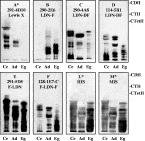
Fig. 2. HPTLC-immunostaining of Schistosoma mansoni glycolipids. HPTLC-resolved cercarial (Ce), adult (Ad) and egg (Eg) complex glycolipids (50 ng of carbohydrate per lane; 300 ng carbohydrate per lane in E, L and M) were visualized by immunostaining using the same panel of anti-carbohydrate mAbs as was used for the Western blots (Fig. 1) and human- (HIS) or murine (MIS) infection sera (L,M). The migration positions of orcinol/H2SO4-stained globoside standards (CDH, CTH and CtetH) are indicated. Pictures marked by * are reproduced with permission from Kantelhardt et al. 2002, © The Biochemical Society.

Fig. 3. ELISA-detection of Schistosoma mansoni glycolipids by various monoclonal antibodies and infection sera in the presence or absence of KLH as an inhibitor. S. mansoni glycolipids from cercariae (Ce), adults (Ad) and eggs (Eg) were probed with anti-carbohydrate mAbs (A-F) and human- (HIS) or murine (MIS) infection sera (L,M) with (hatched columns) or without (black columns) KLH as an inhibitor. Pictures marked by * are reproduced with permission from Kantelhardt et al. 2002, © The Biochemical Society.
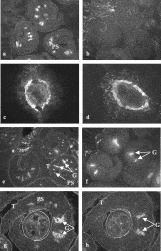
Fig. 4. Immunofluorescence patterns of Schistosoma mansoni sections with or without chloroform/methanol treatment. Frozen sections of adult worms and eggs were stained using anti-F-LDN-F mAb 128-1E7-C (a–d) or murine infection serum (MIS) (e–h) followed by an FITC-labelled anti-mouse Ig conjugate. Use of anti-F-LDN mAb 291-5D5-A gave the same results as mAb 128-1E7-C and an isotype control showed no fluorescence (data not shown). After incubation with mAb 128-1E7-C fluorescence was observed in parenchymal ducts of adult worms (a) and in egg-secreted antigens and egg-shell (c). After chloroform/methanol treatment F-LDN-F related fluorescence patterns were lost in adult worms (b), but not in eggs (d). MIS recognized adult worm tegument (T), gut (G) and parenchymal spots (PS) in adult worms (e, g). Only the gut fluorescence remained after chloroform/methanol treatment (f). When MIS was pre-incubated with KLH (1 mg/ml) fluorescence in the parenchymal structures disappeared (h).
 You have
Access
- 55
- Cited by