Introduction
The prevalence of non-communicable diseases (NCD) such as type-2 diabetes and cardiovascular disease (CVD) is increasing globally at an alarming rate; the World Health Organization predicts that they will be responsible for 73% of all deaths by 2020. Much of this increase will occur in developing nations as they undergo socio-economic improvement.Reference Ramachandran and Snehalatha 1 Fixed genomic variations such as single nucleotide polymorphisms and copy number variations only account for a small proportion of the variation in NCD risk.Reference Morris, Voight and Teslovich 2 Environmental factors such as diet and level of physical activity are likely to play a major role in the development of NCDs and in particular there is growing evidence that the early life environment can play an important role in influencing the risk of developing a wide range of NCDs in later life.Reference Godfrey, Inskip and Hanson 3 The developmental environment can alter later phenotype through the altered epigenetic regulation of genes and this review focuses on the evidence that perinatal influences on epigenetic processes, particularly maternal diet, can lead to persistent phenotypic changes and an altered risk of NCDs in later life.
Early life environment and future disease risk
Following on from the proposal by ForsdahlReference Forsdahl 4 that undernutrition during childhood and adolescence might increase the risk of later CVD, subsequent studies found an inverse relationship between birth weight and increased CVD mortalityReference Barker, Winter, Osmond, Margetts and Simmonds 5 and that babies born at the highest birth weights are also at an increased risk of later NCDs.Reference Pettitt and Jovanovic 6 , Reference Ong 7 In such studies birth weight is thought to be a proxy measurement of the intrauterine environment, which may have been compromized through a variety of maternal, environmental or placental factors.Reference Hanson and Gluckman 8
Studies of the Dutch Hunger Winter provide evidence that maternal nutrition influences offspring health in later life and suggest that the timing of the nutritional constraint is important; first trimester famine exposure increased the risk of obesity and CVD, whereas exposure in the later stages of gestation increased the risk of later insulin resistance and hypertension.Reference Roseboom, de Rooij and Painter 9 , Reference Painter, Roseboom and Bleker 10 Comparable findings are now well established in a variety of animal models where nutrition can be precisely controlled. Early animal studies focussed on the effects of global maternal undernutrition or an isocaloric low protein diet. With the growing epidemic of maternal obesity in both industrialized and developing countries, animal models have been established to investigate the effect of energy-rich maternal diets on the health of the offspring.Reference Hoile, Irvine and Kelsall 11 – Reference Howie, Sloboda, Reynolds and Vickers 15 Interestingly, offspring born to mothers fed these different diets exhibit similar features to human cardiometabolic diseases including hypertension, dyslipidemia, obesity and insulin resistance in later life. The experimental studies in animals implicate altered epigenetic regulation of genes as a major mechanism through which the developmental environment induces altered phenotypes.
Epigenetics
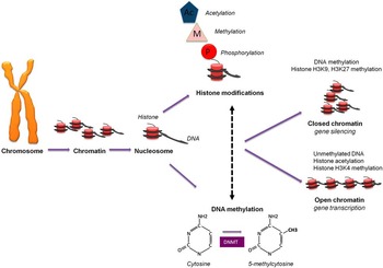
Epigenetic processes, such as DNA methylation and histone modifications, induce heritable changes in gene expression without a change in gene sequenceReference Bird and Macleod 16 (Fig. 1). The epigenome can therefore be regarded as a molecular record of life events, which accumulates over a lifetime. For example, monozygotic twins have been shown to be epigenetically most similar at birth but their epigenomes diverge with age at a rate that is lessened if the twins share a common environment.Reference Fraga, Ballestar and Paz 17 Fine-tuning of phenotype by the developmental environment has adaptive value since it allows the fetus to predict and prepare for the environment to be experienced later.Reference Gluckman, Hanson and Spencer 18 Elucidation of these epigenetic processes has the potential to enable early intervention strategies to improve early development and later health and the study of epigenetic biomarkers is a rapidly advancing field.
DNA methylation
DNA methylation is a common modification in eukaryotic organisms. Typically it involves the transfer of a methyl group to the 5' carbon position of cytosine, creating 5-methylcytosine (5-mC).Reference Kumar, Cheng and Klimasauskas 19 In mammals, methylation of cytosine mainly occurs within the dinucleotide sequence CpG, where a cytosine is immediately 5' to a guanine (the p denotes the intervening phosphate group), although non-CpG methylation is also prevalent in embryonic stem (ES) cells.Reference Ramsahoye, Biniszkiewicz and Lyko 20 DNA methylation is a stable epigenetic mark that is transmitted through mitotic DNA replication and cell division.Reference Bird 21 CpG dinucleotides are not randomly distributed throughout the genome but are clustered at the 5' end of genes in regions known as CpG islands, with hypermethylation and hypomethylation of these islands often associated with gene silencing and activation, respectively.Reference Irizarry, Ladd-Acosta and Wen 22 , Reference Song, Smith and Kimura 23 DNA methylation can act directly to block binding of transcription factors to the DNA or by recruiting a myriad of other repressive factors, such as methyl CpG binding protein 2 (MeCP2), which in turn mediate local chromatin changes.Reference Fuks, Hurd and Wolf 24
Methylation of CpGs is largely established during embryogenesis and the perinatal period. Following fertilization, DNA methylation marks on the maternal and paternal genomes are largely erased (with the exception of the imprinted genes and other specific genomic regions), followed by a wave of de novo methylation within the inner cell mass just before blastocyst implantation.Reference Santos, Hendrich, Reik and Dean 25 , Reference Okano, Bell, Haber and Li 26 The de novo methylation of DNA is catalysed by DNA methyltransferases (DNMT) 3a and 3bReference Okano, Bell, Haber and Li 26 and is maintained through mitosis by methylation of hemi-methylated DNA by DNMTI.Reference Bacolla, Pradhan, Roberts and Wells 27 This gives rise to lineage-specific methylation patterns that are maintained in differentiated tissues. The failure to identify a DNA demethylase or mechanism for DNA demethylation led to the thought that DNA methylation patterns were relatively stable and generally maintained throughout life. However, this concept has now been challenged as in 2009 the existence of another epigenetic modification, 5-hydroxymethylcytosine (5hmC), was described as present in high levels in neurons and ES cells.Reference Tahiliani, Koh and Shen 28 5hmC arises from the oxidation of 5-mC by the enzymes of the TET family and has been proposed to act as a specific epigenetic mark opposing DNA methylation, rather than a passive intermediate in the demethylation pathway.Reference Guibert and Weber 29 The high levels found in the brain and neurons indicate a role in the control of neuronal differentiation and neuronal plasticity.Reference Santiago, Antunes, Guedes, Sousa and Marques 30
Histone modifications
In eukaryotic cells, DNA is wrapped around a core of eight histone proteins; two molecules of each of the four histone proteins H2A, H2B, H3 and H4 combine together to form a nucleosome, the most basic unit of chromatin. Each nucleosome is composed of 146 base pairs of DNA wrapped 1.65 times around the histone core. One molecule of the fifth histone protein, H1, is bound to the DNA as it enters each nucleosome core particle and is known as the linker histone. Each nucleosome is then folded upon itself to form a 30-nm chromatin fibre which is then compacted progressively into larger fibres.Reference Venkatesh and Workman 31 The folding of the chromatin is necessary to reduce the effective size of DNA but it has now become clear that the histones also play a critical role in regulating gene expression. Histone proteins contain two domains, a globular domain and an N terminal tail domain. The unstructured histone tails are subject to modifications including acetylation, methylation, ubiquitination, phosphorylation and attachment of Small Ubiquitin-like Modifier proteins (SUMOylation).Reference Turner 32 Histone modification can directly affect chromatin structure and also provide binding sites for proteins involved in gene regulation. Together, histone modifications and DNA methylation control chromatin structure and therefore the accessibility and functional role of the underlying DNA sequence.Reference Cedar and Bergman 33
Non-coding RNAs (ncRNAs)
The ENCODE project has shown that, although only 1–2% of the genome encodes for proteins, over 74% of the eukaryotic genome has functional elements.Reference Djebali, Davis and Merkel 34 RNAs arising from these functional elements that are not transcribed have been termed ncRNAs. They can be grouped into two classes; long ncRNAs (those longer than 200 nucleotides) and short ncRNAs (those <200 nucleotides, including the microRNAs (miRNAs), small interfering RNAs and PIWI-interacting RNAs).Reference Morris and Mattick 35 The ncRNAs are central components of the transcriptional regulation machinery of the cell. Short ncRNAs can induce mRNA degradation or translational repression and when targeted to the promoter region of a gene can induce both DNA methylation and repressive histone modifications.Reference Hawkins, Santoso, Adams, Anest and Morris 36 Large ncRNAs such as Xist, which plays a pivotal role in X chromosome inactivation, act by coating large regions of a chromosome creating repressive domains.Reference Reik and Lewis 37 , Reference Mercer, Dinger and Mattick 38
Early life nutrition and the epigenome
Originally it was thought that, once established in the blastocyst, DNA methylation is largely maintained throughout the life course. However, there is now growing evidence that the epigenome is particularly susceptible to a number of environmental factors during the prenatal and early postnatal periods and that changes during this time can lead to long-term phenotypic alterations in the offspring.
A clear example of how nutrition can alter phenotype through the altered epigenetic regulation of genes is seen in studies of the honeybee. Female larvae fed on Royal Jelly for 6 days develop into fertile queen bees, while those fed the jelly for 3 days become sterile worker bees, even though they are genetically identical.Reference Kucharski, Maleszka, Foret and Maleszka 39 Knockdown of DNMT3, the major DNMT in bees, increased the proportion of larvae developing into queen bees,Reference Kucharski, Maleszka, Foret and Maleszka 39 which suggests that the effect of nutrition on developmental fate is mediated through the altered methylation of DNA.
A classic example of maternal nutrition influencing DNA methylation in mammals is in the agouti mouse model, where coat colour is influenced by the methylation status of the 5' end of the Agouti gene. Differences in the mother’s intake of dietary methyl donors and co-factors (including folic acid, vitamin B12, betaine and choline) were shown to alter DNA methylation of the Agouti gene and induce differences in the coat colour of the offspring.Reference Morgan, Sutherland, Martin and Whitelaw 40 , Reference Waterland and Jirtle 41
Studies in other animal models have also shown that perturbations in maternal diet are associated with persistent metabolic changes in the offspring, accompanied by epigenetic changes in key metabolic genes or genes involved in appetite control. For example, feeding rats a protein restricted diet during pregnancy induced hypomethylation of the glucocorticoid receptor (GR) and peroxisome proliferator-activated receptor alpha (PPARα) receptor in the livers of juvenile and adult offspring, accompanied by an increased gene expression and a persistent change in the metabolic processes that these nuclear receptors control.Reference Lillycrop, Phillips, Jackson, Hanson and Burdge 42 , Reference Lillycrop, Phillips and Torrens 43 Increased expression of PPARα was also associated with an increase in histone marks that facilitate transcription and a decrease in those that suppress transcription.Reference Lillycrop, Slater-Jefferies and Hanson 12 In contrast, global dietary restriction (giving dams a 70% reduction in total nutrient intake during pregnancy) decreased methylation and increased expression of the GR and PPARα promoters in the offspring liver.Reference Gluckman, Lillycrop and Vickers 44 Thus, the effects of maternal nutrition on the epigenome of the offspring depend upon the nature of the maternal nutrient challenge.
Given the growing concern over the energy-rich Western diet a number of studies have also explored the effects of maternal high-fat feeding on DNA methylation in the offspring. Maternal high-fat feeding during pregnancy in rats leads to the reduced expression of FADS2, the rate-limiting enzyme in polyunsaturated fatty acid synthesis, along with altered methylation of key CpG nucleotides within its promoter in the liver of the offspring.Reference Hoile, Irvine and Kelsall 11 Maternal obesity and diabetes in mice have also been reported to induce widespread changes in DNA methylation in the offspring liver.Reference Li, Young and Maloney 45
It has also become apparent that the period of epigenetic plasticity may extend further into postnatal life. Overfeeding in rat pups induces hypermethylation of two CpG nucleotides within the proopiomelanocortin (POMC) promoter, which plays a key role in appetite control, and hypermethylation of the gene prevented upregulation of POMC expression despite high plasma levels of both leptin and insulin.Reference Plagemann, Harder and Brunn 46 Folic acid supplementation in the juvenile-pubertal period has been shown to induce hypermethylation of the PPARα gene, with decreased PPARα expression and levels of fatty acid β-oxidation,Reference Burdge, Lillycrop and Phillips 47 while Ly et al. Reference Ly, Lee and Chen 48 showed that folic acid supplementation during the peri-pubertal period led to an increased risk of mammary adenocarcinomas along with a decrease in DNMT activity. Plasticity may also extend into adult life; feeding adult rats a diet deficit in choline, folate, methionine and vitamin B12 for 4 weeks induced hypomethylation of the proto-oncogenes c-Myc, c-Fos and c-Ras, with this effect persisting 3 weeks after re-feeding.Reference Christman, Sheikhnejad, Dizik, Abileah and Wainfan 49 Feeding adult rats a fish oil enriched diet for 9 weeks led to a transient decrease in the expression of FADS2 coupled with an increase in FADS2 promoter methylation, with the effects reversed following feeding of a standard diet for 4 weeks.Reference Hoile, Irvine and Kelsall 11 Hence, while the epigenome may be most susceptible to environmental factors in early life, there is some plasticity maintained in later life and this offers the potential opportunity for intervention to reverse marks associated with disease risk.
Most studies have focused on identifying the effects of early life environment on changes in DNA methylation but there is now growing evidence that persistent changes can also be induced in both histone modifications and ncRNAs. Aaggard-Tillery et al. Reference Aagaard-Tillery, Grove and Bishop 50 established an obese pregnant monkey model where the offspring were both obese and found to have site-specific alterations in foetal hepatic H3 acetylation. A 50% protein reduction throughout gestation increased acetylation of histones H3 and H4 in the promoter region of C/EBPβ, a central regulator of energy metabolism, in the skeletal muscle of female rat offspring, along with increased gene and protein expression.Reference Zheng, Rollet and Pan 51 Maternal protein restriction during pregnancy in sheep has also been shown to program marked changes in miRNA expression in both the liverReference Lie, Morrison and Williams-Wyss 52 and skeletal muscleReference Lie, Morrison and Williams-Wyss 53 of the offspring.
The father’s diet can also have an effect on the epigenome and phenotype of the offspring. Offspring of fathers who were fed a low protein diet before mating showed widespread modest changes in DNA methylation (10–20%) compared with control offspring, including a substantial increase in methylation at an intergenic CpG island 50 kb upstream of the PPARα gene.Reference Carone, Fauquier and Habib 54 Ng et al. Reference Ng, Lin and Laybutt 55 also showed that a chronic paternal high-fat diet led to β-cell dysfunction in the female offspring. Experimental studies of paternal-diet-induced intergenerational metabolic reprogramming are an area of increasing research interest, with a Drosophila model showing effects mediated through histone changes and alterations in chromatin state.Reference Öst, Lempradl and Casas 56
DNA methylation is not completely erased during early embryogenesis and as such some methylated sites survive along with associated histones, providing one mechanism for epigenetic transgenerational inheritance.Reference Lange and Schneider 57 For a true transgenerational effect it must extend to the F3 generation since environmental exposure of an F0 female exposes the F1 embryo and the F2 germline present within that embryo; any traits present in the F2 generation cannot be considered a true transgenerational trait and are better described as a multigenerational effect.Reference Desai, Jellyman and Ross 58 In males multigenerational exposure is limited to the F0 and F1 generations since F2 offspring from the male line are not exposed to the F0 uterine environment. Maternal undernutrition in the F0 generation can alter glucose metabolism in the third generation, despite normal nutrition during pregnancy in the F1 and F2 generations.Reference Benyshek, Johnston and Martin 59 There are also gender-specific transgenerational effects; maternal high-fat feeding during pregnancy and lactation increased body size in F1 and F2 generations, with transmission via both the maternal and paternal lineages; however, transmission of the increased body size to the F3 generation was restricted to females and transmitted through the paternal lineage only.Reference Gregory and Tracy 60
Early life nutrition and the human epigenome
Alterations have been reported in the methylation of a number of genes in DNA isolated from whole blood from individuals whose mothers were exposed to famine during the Dutch Hunger Winter. Periconceptual exposure to famine was associated with a small decrease in CpG methylation of the imprinted IGF2 gene and an increase in methylation of leptin, IL-10, MEG3 and ABCA4,Reference Tobi, Lumey and Talens 61 while late gestation famine exposure had no effect on methylation, again showing that the timing of nutritional constraint is important. These measurements were made 60 years after the famine exposure, suggesting that maternal nutritional constraint induced long-term epigenetic changes in key metabolic regulatory genes and pointing to a mechanism through which fetal famine exposure has an effect on adult metabolism. Steegers-Theunissen et al. Reference Steegers-Theunissen, Obermann-Borst and Kremer 62 have also shown altered methylation of specific CpG sites in the IGF2 gene in the peripheral blood cells of children whose mothers did or did not take 400 μg of folic acid per day in the periconceptional period. Plasticity in the human epigenome may persist into adulthood; short-term high-fat overfeeding in healthy young men was shown to induce methylation changes in over 6000 skeletal muscle genes, with those changes only partially reversed after 6–8 weeks of a normocaloric diet.Reference Jacobsen, Brøns and Bork-Jensen 63
Epigenetic biomarkers to predict later disease risk
If the early life environment induces altered epigenetic regulation of genes then it should be possible to detect these altered epigenetic marks and use them as predictors of future metabolic capacity and disease risk. The development of new technologies to measure DNA methylation is enabling discovery of epigenetic biomarkers on a truly genome-wide scale. This increased coverage is likely to uncover many new genomic regions that contain specific epigenetic alterations outside those already studied.
In two independent cohorts Godfrey et al. Reference Godfrey, Sheppard and Gluckman 64 reported that the methylation status of a single CpG site in the promoter region of the nuclear receptor RXRA from umbilical cord tissue was strongly related to childhood adiposity in both boys and girls, with the methylated RXRA CpG explaining over 25% of the variance in age and sex adjusted fat mass in children at 6 and 9 years of age. Epigenetic alterations may therefore contribute a far greater proportion to an individual’s risk for NCD than previously thought.
Epigenetic biomarkers, unlike genetic biomarkers, are thought to have some tissue specificity. However, it is often impractical to study diseased and/or the appropriate tissues for epigenetic studies, so accessible surrogate tissues such as umbilical cord and blood are often used instead, with a further advantage being that these biomarkers can then be measured repeatedly. Although DNA methylation patterns can be tissue specific a number of studies have also now shown inter-tissue methylation correlations. For some genomic regions methylation appears to be largely independent of the tissue of origin, while for other regions there is a tissue-specific dependence.Reference Ollikainen, Smith and EJ-H 65 Methylation across differentially methylated regions for the imprinted genes H19, MEST and PEG10 did not significantly differ across a range of tissues (buccal, brain, eye intestine, liver, lung, muscle and umbilical cord blood).Reference Murphy, Huang and Hoyo 66 The methylation levels of a number of non-imprinted genes measured in blood were also equivalent in buccal cells despite these cell types originating from different germ layers (mesoderm and ectoderm, respectively).Reference Talens, Boomsma and Tobi 67 Methylation changes induced by maternal diet can be similar in the umbilical cord and liver.Reference Burdge, Hanson, Slater-Jefferies and Lillycrop 68 These studies suggest that methylation levels in more readily available tissues, such as blood, buccal or umbilical cord, may provide useful proxy markers of methylation in metabolically relevant tissues.
However, data showing that some DNA methylation marks can be dynamically regulated in response to postnatal environmental stimuli mean that much work will need to be done to demonstrate the utility of perinatal epigenetic predictive markers. For example, an acute burst of physical activity induced hypomethylation of the peroxisome proliferator-activated receptor γ, coactivator 1 alpha (PGC1α) and mitochondrial transcription factor A (TFAM) promoters in muscle tissue.Reference Barres, Yan and Egan 69 One study that examined DNA methylation stability over time in children found that the methylation levels of the genes MAOA, DRD4 and SLC6A4 was highly dynamic between the ages of 5 and 10 years.Reference Wong, Caspi and Williams 70 In contrast, Clarke-Harris et al. Reference Clarke-Harris, Wilkin and Hosking 71 reported year on year stability of seven CpG sites within the PGC1α promoter in peripheral blood cells in children from 5 to 14 years of age, suggesting that for these CpG sites methylation levels are set up in early life and stability maintained. Moreover, seven of the CpG sites analysed at age 5–7 years were predictive of adiposity in the children at ages 9–14, which is further evidence that developmentally induced methylation marks may be significant contributors to later phenotype. The differences in the stability of the PGC1α methylation between the two studies may reflect differences in the location of the CpGs or tissue-specific differences between blood and muscle (PGC1α has a muscle-specific transcript, although it is unclear which promoter region was analysed by Barres et al.). Whether the changes in methylation in response to exercise occur on top of a developmentally induced methylation change is also not known.
Interventions and developmental epigenetic changes
A number of intervention studies suggest that the effects of the early life environment on the epigenome and phenotype can be prevented and/or reversed. In their rat model of maternal protein restriction Lillycrop et al. Reference Lillycrop, Phillips, Jackson, Hanson and Burdge 42 showed that supplementation of the restricted diet with glycine or folic acid prevented hepatic GR and PPARα hypomethylation and with altered metabolic phenotype, suggesting that impaired 1-carbon metabolism plays a central role in this model. In neonatal rats whose mothers were exposed to a 70% in utero global nutrient restriction postnatal leptin treatment reversed promoter hypermethylation and increased expression of PPARα and GR in the liver.Reference Gluckman, Lillycrop and Vickers 44 Thus, it may be possible to not only identify those at risk of developing later disease, through the use of epigenetic biomarkers early in life, but to develop intervention strategies which target and reverse these epigenetic changes.
Conclusions
There is now considerable evidence that our genotype and later environment are not the only determinant of NCD risk, and that epigenetic marks induced by the early life environment are associated with altered gene expression patterns in important metabolic tissues, leaving to altered susceptibility to disease in later life.
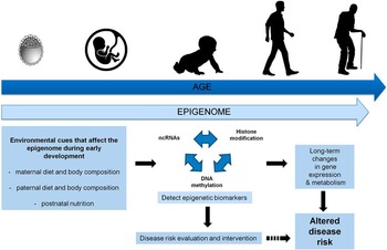
Demonstration of a role for altered epigenetic regulation of genes in the development of NCDs together with the identification of potential epigenetic biomarkers of future disease risk raises the possibility of preventive medicine. Individuals identified as at risk at an early stage in the life course could receive nutritional or lifestyle interventions, allowing a more effective strategy of preventive treatment. This would both improve quality of life and reduce the economic burden associated with current treatment strategies. Further understanding of the mechanisms by which nutrition can modify the epigenome and the periods of epigenetic susceptibility will aid development of novel intervention strategies to reverse this current global epidemic of NCDs (Fig. 2).
DNA methylation and histone modifications are two epigenetic modifications that determine the chromatin structure and the ability for genes to be transcribed. The processes interact and can lead to open chromatin and gene transcription or to closed chromatin and gene silencing. DNMT, DNA methyltransferase.

Epigenetic biomarkers; detecting early life epigenetic biomarkers that are associated with disease risk could lead to stratified interventions and reduced susceptibly to chronic disease. ncRNA, non-coding RNA.

Acknowledgement
None.
Financial Support
This work was supported by grants from the Medical Research Council (MC_U147585827, MC_ST_U12055) and the NIHR Southampton Biomedical Research Centre, University of Southampton and University Hospital Southampton NHS Foundation Trust. K.M.G. is supported by the European Union’s Seventh Framework Programme (FP7/2007-2013), project EarlyNutrition under grant agreement no. 289346.
Conflicts of Interest
K.M.G. has received reimbursement for speaking at conferences sponsored by companies selling nutritional products and K.M.G., P.M.C. and K.A.L. are part of an academic consortium that has received research funding from Abbott Nutrition, Nestec and Danone.