Introduction
The in vitro culture system of mammalian embryos is widely used in assisted reproductive technology (ART) and the physiological study of preimplantation embryo development. Successful in vitro culture of mammalian embryos is dependent on the culture microenvironment. Although the formulations of culture media have been optimized in the past several decades, in vitro culture environment cannot be compared equally with the female reproductive tract (Bavister, Reference Bavister1995, Reference Bavister2000; Knijn et al., Reference Knijn, Gjorret, Vos, Hendriksen, Van Der Weijden, Maddox-Hyttel and Dieleman2003; Summers & Biggers, Reference Summers and Biggers2003). Mammalian embryos will develop to morphologically normal blastocysts under a wide range of culture conditions, yet their intrinsic programmes can be disturbed by the adverse medium constituents (Khosla et al., Reference Khosla, Dean, Brown, Reik and Feil2001a,Reference Khosla, Dean, Reik and Feilb; Lane & Gardner, Reference Lane and Gardner2003; Fernandez-Gonzalez et al., Reference Fernandez-Gonzalez, Moreira, Bilbao, Jimenez, Perez-Crespo, Ramirez, De Fonseca, Pintado and Gutierrez-Adan2004; Mann et al., Reference Mann, Lee, Doherty, Verona, Nolen, Schultz and Bartolomei2004). Embryo culture is often associated with a stress response in metabolism, gene expression (e.g. overexpression of heat shock genes) and apoptosis (Christians et al., Reference Christians, Campion, Thompson and Renard1995, Reference Christians, Michelob and Renard1997; Niemann & Wrenzycki, Reference Niemann and Wrenzycki2000; Schultz & Williams, Reference Schultz and Williams2002; Rizos et al., Reference Rizos, Gutierrez-Adan, Perez-Garnelo, De La Fuente, Boland and Lonergan2003). Consequently, the embryonic and fetal development may be impaired; even the postnatal behaviour and physiological health of offspring are also influenced (Khosla et al., Reference Khosla, Dean, Brown, Reik and Feil2001a; Schultz & Williams, Reference Schultz and Williams2002; Lane & Gardner, Reference Lane and Gardner2003; Fernandez-Gonzalez et al., Reference Fernandez-Gonzalez, Moreira, Bilbao, Jimenez, Perez-Crespo, Ramirez, De Fonseca, Pintado and Gutierrez-Adan2004). These considerations deliver the necessity to explore further the effects of specific components on embryos in culture system.
Antibiotics, which are not nutritionally necessary, are routinely included in embryo culture systems in order to avoid contamination from microorganisms. Supplementation with 100 IU/ml penicillin and 50 μg/ml streptomycin is adopted as standard, which is putatively considered nontoxic to embryos. However, this assumption is based on cell culture testing, which is quite different from embryo culture systems. Even in cell culture systems, the combination of penicillin and streptomycin or only streptomycin can inhibit DNA replication and protein synthesis, and results in destruction of chromatin integrity along with a decline of cell proliferation (Amonn et al., Reference Amonn, Baumann, Wiesmann, Hofmann and Herschkowitz1978; Stemp et al., Reference Stemp, Pascoe and Gatehouse1989; Manuvakhova et al., Reference Manuvakhova, Keeling and Bedwell2000). After all, antibiotics are biologically active molecules, and embryos are never exposed to antibiotics under natural conditions in vivo. In human assisted reproductive procedures, antibiotic supplementation, even in reduced concentrations, has a deleterious effect on the growth rate of preimplantation embryos (Magli et al., Reference Magli, Gianaroli, Fiorentino, Ferraretti, Fortini and Panzella1996). When antibiotics were present, in vitro development of hamster embryos was significantly impaired (Zhou et al., Reference Zhou, McKiernan, Ji and Bavister2000). Therefore, the effects of antibiotics on embryos in culture might be more severe than we previously assumed.
Although the roles of medium components on gene expression and viability of embryos have been studied extensively (Ho et al., Reference Ho, Wigglesworth, Eppig and Schultz1995; Khosla et al., Reference Khosla, Dean, Reik and Feil2001b; Lane & Gardner, Reference Lane and Gardner2003; Rizos et al., Reference Rizos, Gutierrez-Adan, Perez-Garnelo, De La Fuente, Boland and Lonergan2003; Fernandez-Gonzalez et al., Reference Fernandez-Gonzalez, Moreira, Bilbao, Jimenez, Perez-Crespo, Ramirez, De Fonseca, Pintado and Gutierrez-Adan2004; Sturmey et al., Reference Sturmey, Brison and Leese2008), the potential effects of routine use of antibiotics on the intrinsic health of mammalian embryos remain to be elucidated. In the present study, we first examined the development and differentiation of mouse zygotes cultured with or without antibiotics. Because morphological assessment cannot entirely reflect the embryo quality (Rizos et al., Reference Rizos, Gutierrez-Adan, Perez-Garnelo, De La Fuente, Boland and Lonergan2003; Vergouw et al., Reference Vergouw, Botros, Roos, Lens, Schats, Hompes, Burns and Lambalk2008; Sturmey et al., Reference Sturmey, Hawkhead, Barker and Leese2009), we further investigate the effects of antibiotics on expression of zygotically activated genes, DNA damage and apoptosis of cultured embryos.
Materials and Methods
All chemicals and reagents were purchased from Sigma Chemical Co. unless otherwise stated.
Animals
We declare our respect to the right and dignity of animals in the experiments. ICR mice were maintained with food and water ad libitum on a 12 h light/dark cycle under controlled temperature (23–25 °C).
Collection of zygotes
Female mice (6–8 weeks old) were superovulated with consecutive injection of 5 IU pregnant mare serum gonadotropin and 5 IU human chorionic gonadotropin (HCG) 48 h apart. Then they were caged with male mice (12 weeks old). The zygotes were recovered from oviducts at 16 h post-HCG. The medium for collection of zygotes was the modified potassium simplex optimized medium (KSOM) containing 20 mM HEPES, 4 mM NaHCO3 and 1 mg/ml BSA.
In vitro culture of mouse zygotes
A batch of KSOM with essential and non-essential amino acids (KSOMaa) containing 8 mg/ml BSA was prepared with the only exception of adding antibiotics. The same batch was divided into two aliquots: the first aliquot was free of antibiotics (antibiotic free); the second was supplemented with 100 IU/ml penicillin and 50 μg/ml streptomycin (antibiotic plus). Every 12–20 zygotes were cultured in a 40 μl droplet (Paria & Dey, Reference Paria and Dey1990; Gandolfi, Reference Gandolfi1994) of antibiotic-free or antibiotic-plus medium at 37.5 °C under a humidified atmosphere of 5% CO2 in air (this time point was defined as ‘0 h’ in the present study). The development to 2-cell, morula and blastocyst stage was examined at 24 h, 72 h and 96 h, respectively.
Differential staining of blastocysts
Differential staining of inner cell mass (ICM) and trophectoderm (TE) cells was performed as previously described (Tang et al., Reference Tang, Wang, Zhang, Gao, Ma, Yin, Sun, Liu and Zhang2009) with some modifications. Briefly, blastocysts were first incubated in 1% Triton X-100 and 100 μg/ml propidium iodide (PI) in phosphate-buffered saline (PBS) for 30 s. Then blastocysts were fixed for 3 h in absolute ethanol with 50 μg/ml of Hoechst 33342 stain at 4 °C. The blastocysts were mounted on slides and observed using epifluorescence microscopy. TE cells labelled with PI and Hoechst 33342 were identified by their pink fluorescence and ICM cells labelled with Hoechst 33342 appeared blue.
Real-time RT-PCR
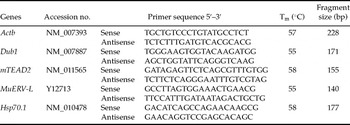
Effects of antibiotics on the expression of zygotically activated genes in 2-cell embryos cultured with or without antibiotics were determined by real-time RT-PCR. Relative quantification was performed using the SuperScript III Platinum CellsDirect Two-Step qRT-PCR Kit with SYBR Green (Invitrogen). Total RNA extraction and first-strand cDNA synthesis were performed according to the manufacturer's manual. Quantitative PCR was carried out on the SmartCycler system (Cepheid) with primers listed in Table 1. The programme for quantitative PCR was as follows: 95 °C × 2 min, followed by 40 cycles of 95 °C × 15 s, 55–58 °C × 30 s, 72 °C × 30 s. Specificity of the reaction was verified by both melting curve analysis and appropriate restriction digests of amplification products (data not shown). Real-time RT-PCR experiment was performed in triplicate and normalized to beta-actin (Actb) by the ΔΔCt method (Livak et al., Reference Livak, Flood, Marmaro, Giusti and Deetz1995). The transcriptional levels of in vivo 2-cell embryos (40 h post-HCG) were used as control and have an arbitrary value of 1. The expression profiles of zygotically activated genes in 2-cell embryos from antibiotic-free and antibiotic-plus groups were presented as multiples of the control.
Primers for real-time RT-PCR

Detection of the nuclear status
Embryos were fixed for 1 h in 4% paraformaldehyde diluted in PBS. Embryos were repeatedly washed at least three times in PBS plus 1 mg/ml polyvinylpyrrolidone-40 (PBS/PVP) for 5 min. Then embryos were incubated in 25 μg/ml RNase A for 30 min. The nuclei were counterstained with 25 μg/ml PI for 30 min in the dark. Embryos were mounted on slides for confocal microscopy.
Apoptosis detection
The number of apoptotic cells in blastocysts was determined using a DeadEnd Fluorometric TUNEL System (Promega). All manipulations were done at room temperature unless otherwise stated. Between two steps, embryos were washed at least three times in PBS/PVP for 5 min, unless otherwise stated. Blastocysts were fixed in 4% paraformaldehyde in PBS/PVP and permeabilized in 0.2% Triton X-100 in PBS for 5 min. Blastocysts were equilibrated in equilibration buffer (Promega) for 8 min, and then incubated with FITC-conjugated dUTP and terminal deoxynucleotidyl transferase in equilibration buffer at 37 °C for 60 min in the dark (hereafter, all manipulations were done in the dark). The tailing reaction was terminated in 2× SSC for 15 min. Then embryos were incubated in 25 μg/ml RNase A for 30 min; and without wash, the nuclei were counterstained with 25 μg/ml PI for 30 min. Blastocysts were mounted on slides for confocal microscopy.
Confocal microscopy
Fluorescence was detected under a Carl Zeiss LSM 510 laser confocal scanning microscope. The nuclei were identified by their red fluorescence. The merged images double-labelled with FITC and PI appeared yellow.
Statistical analysis
Each experiment was independently performed at least three times. The data were analysed by using SigmaStat 3.5 software (Systat Software). In vitro development of zygotes in antibiotic-free and antibiotic-plus groups was compared by chi-squared analysis. The other comparisons were performed with Student's t-test. Apoptosis index indicated the incidence of apoptotic cells in blastocysts and was calculated via the formula: (apoptotic cell number/total cell number) × 100. Values of p < 0.05 were considered significantly different.
Results
Effect of antibiotics on in vitro development of mouse embryos
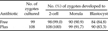
Mouse zygotes cultured to 2-cell, morula and blastocyst stage in antibiotic-free and antibiotic-plus media were used to investigate the effects of antibiotics on in vitro development of mouse embryos. Cleavage rates of the embryos were not significantly either in the absence or presence of antibiotics. Most of the zygotes developed to blastocysts in antibiotic-free (84.8%) or antibiotic-plus (83.3%) groups at 96 h. No significant differences were observed in developmental rates between the two experimental groups (Table 2).
In vitro development of mouse zygotes cultured with or without antibiotics

Zygotes were cultured in antibiotic-free or antibiotic-plus KSOMaa medium containing 8 mg/ml BSA. The development to 2-cell, morula and blastocyst stages was examined at 24 h, 72 h and 96 h, respectively. No significant differences in the developmental rates were observed between two experimental groups. Data were analysed by chi-squared analysis.
Differential cell counting in blastocysts
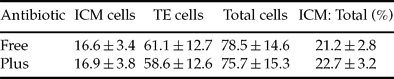
To investigate whether antibiotic supplementation had an impact on ICM, TE differentiation and total cell number of blastocysts, the ICM and TE cells were counted by differential staining. No significant differences were observed in ICM, TE, total cell number and the ratio of ICM to total cells between two experimental groups (Table 3).
ICM, TE, total cell number, and the ratio of ICM to total cells (mean ± SD) in blastocysts developed from mouse zygotes cultured with or without antibiotics
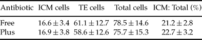
Zygotes were cultured in antibiotic-free or antibiotic-plus KSOMaa medium containing 8 mg/ml BSA for 96 h. Inner cell mass (ICM) and trophectoderm (TE) cells were counted by differential staining with propidium iodide and Hoechst 33342 stain. No significant differences were observed between two experimental groups. Data were analysed by Student's t-test. Thirty embryos were analysed for each group.
Disturbed expression of zygotically activated genes in 2-cell embryos cultured with antibiotics
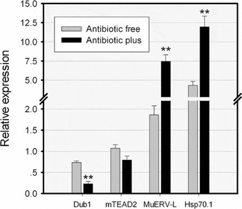
The mRNA levels of four zygotically activated genes, namely, deubiquitinating enzyme 1 (Dub1), murine TEA domain family member 2 (mTEAD2), murine endogenous retrovirus-like (MuERV-L) and 70 kDa heat shock protein family member 1 (Hsp70.1), were quantified to investigate the effect of antibiotics on gene expression (Fig. 1). The relative abundance of Dub1 transcript was significantly lower in 2-cell embryos cultured in antibiotic-plus medium than that in antibiotic-free medium (p < 0.01). There was no significant difference in mTEAD2 expression between the two groups. The relative expression of MuERV-L and Hsp70.1 was significantly higher in 2-cell embryos in the antibiotic-plus group than that in antibiotic-free group (p < 0.01).
Disturbance of zygotic gene activation (ZGA) in 2-cell embryos cultured with antibiotics. The expression of zygotically activated genes was determined by real-time RT-PCR. The mRNA transcriptional levels of in vivo 2-cell embryos were used as control and have an arbitrary value of 1. The expression profiles of zygotically activated genes in 2-cell embryos cultured in antibiotic-free and antibiotic-plus media were presented as multiples of the control. Each column denotes mean ± SEM. **p < 0.01; Difference between antibiotic-free and antibiotic-plus groups were analysed by Student's t-test.

Effects of antibiotics on chromatin damage and blastomere fragmentation in 2-cell embryos
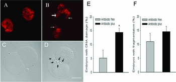
After 24 h of in vitro culture, 2-cell embryos were labelled by PI to display chromatin integrity. The formation of micronuclei and lobulated nuclei was considered as chromatin damages (Fig. 2A, B). The percentage of embryos with chromatin damages rose significantly in the antibiotic-plus group versus antibiotic-free group (Fig. 2E; p < 0.05). The percentage of embryos with blastomere fragmentation increased in antibiotic plus group (Fig. 2C, D), but there was no significant difference between two groups (Fig. 2F).
Effects of antibiotics on chromatin damages and blastomere fragmentation in mouse 2-cell embryos. The nuclei of embryos were stained with propidium iodide (PI). (A) Two-cell nuclei in an embryo cultured without antibiotics. (B) Two-cell nuclei contained chromatin abnormalities: micronuclei (white thin arrow) and lobulated nuclei (white thick arrow) in an embryo cultured with antibiotics. (C, D) Optical scanning images to (A) and (B), respectively. (C) Two-cell embryo with no fragmentation. (D) Two-cell embryo with fragmentation (black arrow). Scale bar represented 20 μm. (E) The proportion of embryos with chromatin damages was significantly increased in the antibiotic-plus group versus antibiotic-free group. (F) The proportion of embryos with fragmentation was increased in the antibiotic-plus group versus the antibiotic-free group. However, there was no significant difference between two groups. Each column denoted mean ± SEM. *p < 0.05; data were analysed by Student's t-test. From three replications, 54 embryos for antibiotic-free group and 55 embryos for antibiotic-plus group were analysed. See online for a colour version of this figure.

Effect of antibiotics on apoptosis in mouse blastocysts
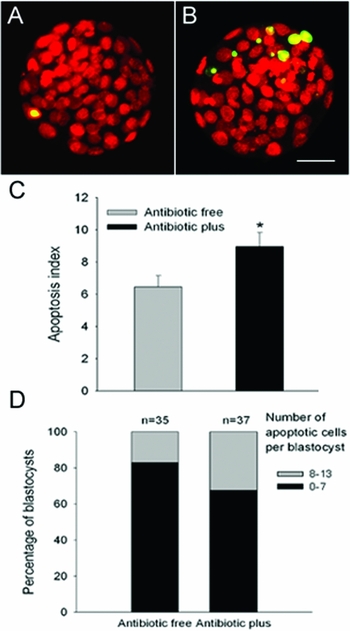
The nuclear DNA fragmentation in blastocysts from antibiotic-free and antibiotic-plus groups was detected by TUNEL assay. Increased apoptotic cells were observed in blastocysts from the antibiotic-plus group (Fig. 3A, B). The incidence of apoptosis was shown in Fig. 3C, blastocysts cultured in antibiotic-plus medium had more apoptotic cells than blastocysts cultured in antibiotic-free medium (p < 0.05). The proportion of blastocysts with different apoptotic cell numbers was shown in Fig. 3D. In the antibiotic-free group, 82.9% blastocysts had less than seven apoptotic cells; yet in the antibiotic-plus group, the percentage is only 67.6%.
Effect of antibiotics on apoptosis in mouse blastocysts. (A) Blastocyst developed from a zygote cultured in antibiotic-free medium. (B) Blastocyst developed from a zygote cultured in antibiotic-plus medium. Note the increased nuclear DNA fragmentation detected by TUNEL assay (yellow). Scale bar represented 20 μm. (C) The apoptosis index of blastocysts was dramatically increased in the antibiotic-plus group versus antibiotic-free group. The formula for apoptosis index was (apoptotic cell number/total cell number) × 100. Each column denoted mean ± SEM. *p < 0.05; data were analysed by Student's t-test. (D) The proportional distribution of blastocysts with different apoptotic cell number in the above two groups. From three replicates, 35 blastocysts were analysed for the antibiotic-free group and 37 blastocysts for the antibiotic-plus group. See online for a colour version of this figure.

Discussion
For a long time, penicillin and streptomycin were regarded as safe drugs in embryo culture systems. The results in this study question this viewpoint and provide evidence that, when cultured with antibiotics, embryos with normal appearance may possess intrinsic physiological and genetic abnormalities.
In contrast to previous studies (Magli et al., Reference Magli, Gianaroli, Fiorentino, Ferraretti, Fortini and Panzella1996; Zhou et al., Reference Zhou, McKiernan, Ji and Bavister2000), we did not observe impairment of antibiotic supplementation on in vitro development of cultured embryos, one explanation being the different employment of animal models between this study and the previous. With the rapid progress of embryo culture technology, several culture media have been designed and optimized. In vitro culture of mouse zygotes to blastocysts can reach 95–100% of the developmental rate (Fernandez-Gonzalez et al., Reference Fernandez-Gonzalez, Moreira, Bilbao, Jimenez, Perez-Crespo, Ramirez, De Fonseca, Pintado and Gutierrez-Adan2004; Takenaka et al., Reference Takenaka, Horiuchi and Yanagimachi2007). Therefore, the adverse effects of antibiotics on cultured embryos have long been ignored. Although in vitro development of human and hamster embryos was impaired in antibiotic-plus medium (Magli et al., Reference Magli, Gianaroli, Fiorentino, Ferraretti, Fortini and Panzella1996; Zhou et al., Reference Zhou, McKiernan, Ji and Bavister2000), the experimental evidence for antibiotic toxicity to cultured embryos is not enough. With updated culture system, embryos are easily able to develop to morphologically normal blastocysts (Table 1) with proper cell number (Table 2). However, for their intrinsic normality, this situation is not necessarily the case. In contrast to the normal developmental rate and cell number, the presence of antibiotics significantly disturbs expression of zygotically activated genes, damages chromatin integrity and increases apoptosis in blastocysts of cultured embryos.
Zygotic gene activation (ZGA) is a critical event that mediates the transition from maternal to zygotic control of preimplantation development following fertilization (Schultz, Reference Schultz1993; Schultz et al., Reference Schultz, Davis, Stein and Svoboda1999). This process comprises a dramatic reprogramming of global gene expression, which is a necessary prerequisite for successful embryonic development. Characterization of zygotic gene expression patterns and their relationship to other parameters (e.g. embryo viability, DNA damage and apoptosis etc.) provides a useful tool for defining optimized embryo culture conditions (Niemann & Wrenzycki, Reference Niemann and Wrenzycki2000; Kanka, Reference Kanka2003). mTEAD2 is the only member of the TEAD transcription factor family expressed in early mouse embryos, where its presence most likely accounts for the TEAD-dependent enhancer activity (Kaneko et al., Reference Kaneko, Cullinan, Latham and DePamphilis1997). In the four selected genes, only mTEAD2 transcription was not significantly different in both groups. Dub1 has deubiquitinating activity to remove ubiquitin from protein and continuous expression of Dub1 induces developmental arrest in the G1 phase of the cell cycle (Zhu et al., Reference Zhu, Carroll, Papa, Hochstrasser and D'Andrea1996). As global protein synthesis is largely changed during the preimplantation stages, Dub1 may play some roles in mouse embryonic development by controlling protein stability (Suzuki et al., Reference Suzuki, Minami, Kono and Imai2006). MuERV-L is one of the earliest transcribed genes in mouse ZGA and its expression affects the expression of other genes involved in the major phase of ZGA. The higher expression of MuERV-L has been implicated to play an important role in early embryonic development from the 2-cell to 4-cell stage (Kigami et al., Reference Kigami, Minami, Takayama and Imai2003). Hsp70.1, the most extensively investigated member of the inducible heat shock family, is highly transcribed at the onset of ZGA (Christians et al., Reference Christians, Campion, Thompson and Renard1995, Reference Christians, Michelob and Renard1997). Hsp70.1 upregulation exhibits a stress response pattern modulated by suboptimal in vitro culture conditions (Christians et al., Reference Christians, Campion, Thompson and Renard1995, Reference Christians, Michelob and Renard1997; Fiorenza et al., Reference Fiorenza, Bevilacqua, Canterini, Torcia, Pontecorvi and Mangia2004). The above observations indicate that the alteration of zygotic gene expression has a tendency to relieve the growth suppression on embryos and promote developmental transitions. The disturbed expression of zygotically activated genes reflects that the presence of antibiotics augments environmental stress in embryo culture system.
DNA damage caused by genotoxic agents can impact virtually any cellular process due to its ability to affect gene expression and subsequent gene products (Vinson & Hales, Reference Vinson and Hales2002). The inhibitory effect of antibiotics upon DNA replication results in destruction of chromatin integrity (Amonn et al., Reference Amonn, Baumann, Wiesmann, Hofmann and Herschkowitz1978; Stemp et al., Reference Stemp, Pascoe and Gatehouse1989; Manuvakhova et al., Reference Manuvakhova, Keeling and Bedwell2000). The presence of antibiotics in culture medium damages chromatin integrity as early as 2-cell stage, when embryos just complete the first replication cycle. The formation of micronuclei and lobulate nuclei in 2-cell embryos provides direct evidence for DNA damage (Fig. 2B). This destruction of chromatin structure may partially account for the disturbance of zygotic gene expression (Kanka, Reference Kanka2003; Felsenfeld et al., Reference Felsenfeld, Boyes, Chung, Clark and Studitsky1996; Vinson & Hales, Reference Vinson and Hales2002; Schultz & Worrad, Reference Schultz and Worrad1995). Under our experimental conditions, the proportion of embryos with blastomere fragmentation has a limited rise when cultured with antibiotics. It has been reported that blastomere fragmentation in embryos has a positive correlation with the incidence of programmed cell death (Jurisicova et al., Reference Jurisicova, Varmuza and Casper1996). Although difference is not significant, high levels of DNA damage and apoptosis are still observed in this study.
Apoptosis in blastocysts is a natural event that plays an important role in elimination of abnormal cells with aberrant developmental potential during normal development (Hardy, Reference Hardy1997). In addition, when embryos grow under environmental pressure, the cumulative damages will produce more cells with abnormality so that the proportion of apoptotic cells in blastocysts increases under suboptimal culture conditions (Hardy, Reference Hardy1997; Gjorret et al., Reference Gjorret, Knijn, Dieleman, Avery, Larsson and Maddox-Hyttel2003; Knijn et al., Reference Knijn, Gjorret, Vos, Hendriksen, Van Der Weijden, Maddox-Hyttel and Dieleman2003; Lane & Gardner, Reference Lane and Gardner2003; Xie et al., Reference Xie, Wang, Zhong, Puscheck, Shen and Rappolee2006). The present study demonstrates that antibiotics in culture medium are harmful to the chromatin integrity of cultured embryos and disturb the expression of zygotically activated genes. Further, these adverse effects produce more abnormal cells with aberrant developmental potential; and then result in rising levels of cell death in blastocysts (Hardy, Reference Hardy1997; Lane & Gardner, Reference Lane and Gardner2003). These impacts indicate that the presence of antibiotics, not as previously assumed, is adverse to embryos in culture.
A primary concern about antibiotic supplementation is to prevent bacterial and fungal contaminations. However, now integral asepsis can be implemented in any embryology laboratory. Antibiotics are no longer indispensable to maintain sterility in embryo culture system with modern aseptic techniques (McKieman & Bavister, Reference McKieman and Bavister1990, Reference McKieman and Bavister2000; Magli et al., Reference Magli, Gianaroli, Fiorentino, Ferraretti, Fortini and Panzella1996; Zhou et al., Reference Zhou, McKiernan, Ji and Bavister2000). In the present study, after the embryos developed to blastocysts, we aspirated the culture droplets and spread the medium on microbial culture plates. No microbial colonies formed in both groups after overnight incubation (data not shown).
In the mouse, the culture process interferes with normal development of preimplantation embryos and produces abnormalities during fetal and postnatal development (Khosla et al., Reference Khosla, Dean, Brown, Reik and Feil2001a; Lane & Gardner, Reference Lane and Gardner2003; Fernandez-Gonzalez et al., Reference Fernandez-Gonzalez, Moreira, Bilbao, Jimenez, Perez-Crespo, Ramirez, De Fonseca, Pintado and Gutierrez-Adan2004; Watkins et al., Reference Watkins, Platt, Papenbrock, Wilkins, Eckert, Kwong, Osmond, Hanson and Fleming2007). In ART, embryos are also incubated in culture medium until transfer into surrogate mothers. It has been documented that ART is associated with a significant increase in birth defects compared with natural conception (Ericson & Kallen, Reference Ericson and Kallen2001; Hansen et al., Reference Hansen, Kurinczuk, Bower and Webb2002, Reference Hansen, Bower, Milne, de Klerk and Kurinczuk2005; Summers & Biggers, Reference Summers and Biggers2003; Olson et al., Reference Olson, Keppler-Noreuil, Romitti, Budelier, Ryan, Sparks and Van Voorhis2005). In these cases, they share a common feature that all embryo culture media contain antibiotics. The antibiotic damage to embryos during in vitro culture step, however small, should not be ignored. Until now, there is no direct evidence that postnatal defects are results of routine use of antibiotics as their effects are not as serious as the other components. Yet some intrinsic alteration and genetic aberration might be recessive (Aitken, Reference Aitken2008). At least, antibiotics add another undesirable variable to the experiments.
This investigation suggests that, unless necessary (e.g. treatment of infected semen samples), antibiotic supplementation is not essential for sterility of embryo culture system and can produce intrinsic abnormality in chromatin integrity, apoptosis and zygotic gene expression in cultured embryos. Therefore, antibiotic supplementation can be abolished during embryo culture. Especially, the use of antibiotics for ART should be considered prudently.
Acknowledgements
We thank Ms Lili Ren for the help in statistical analysis. We would like to thank Guoliang Pei for the assistance with the confocal microscopy. We are also grateful to Dr Mingtao Zhao for his critical reading of the paper.