Introduction
Idiopathic Parkinson's disease (PD) is a neurodegenerative disease characterized by the progressive loss of dopaminergic neurons of the mesencephalic substantia nigra and its projections to the striatum (nigrostriatal pathway). This loss is responsible for motor dysfunctions, initially asymmetrical, including slowness of movements, rigidity, tremor, and posture and gait alterations. This degenerative process is now known to be more extensive and to progressively involve multiple neuronal systems related to a wide variety of non-motor manifestations, including cognitive, mood, sleep, smell, and autonomic disorders (Braak et al., Reference Braak, Del Tredici, Rub, De Vos, Jansen Steur and Braak2003).
The degree to which cognitive deficits in early-stage PD reflect dopaminergic depletion or the pathological involvement of other neuronal systems has long been a subject of major interest. A few studies of PD patients “off” and “on” levodopa have addressed this question (Cools, Barker, Sahakian, & Robbins, Reference Cools, Barker, Sahakian and Robbins2001, Reference Cools, Barker, Sahakian and Robbins2003; Lange et al., Reference Lange, Robbins, Marsden, James, Owen and Paul1992; Lewis, Slabosz, Robbins, Barker, & Owen, Reference Lewis, Slabosz, Robbins, Barker and Owen2005; Malapani, Pillon, Dubois, & Agid, Reference Malapani, Pillon, Dubois and Agid1994; Nadeau & Crosson, Reference Nadeau and Crosson1997; Skeel, Crosson, Nadeau, Algina, Bauer, & Fennell, Reference Skeel, Crosson, Nadeau, Algina, Bauer and Fennell2001). However, given the complexity of the problem and the difficulties of evaluating cognition in PD subjects, the issue has never been definitively resolved. In this regard, the neuropsychological study of patients who develop hemiparkinsonism after acute vascular damage of the contralateral nigrostriatal pathway may yield useful data. Several such patients have been reported, but neuropsychological descriptions have not been available (Defer et al., Reference Defer, Remy, Malapert, Ricolfi, Samson and Degos1994; Geny et al., Reference Geny, N'Guyen, Cesaro, Goujon, Brugieres and Degos1995; Ghaemi, Krauss, & Nakamura, Reference Ghaemi, Krauss and Nakamura2009; Inci, Celik, Soylemezoglu, & Ozgen, Reference Inci, Celik, Soylemezoglu and Ozgen2007; Kudo et al., Reference Kudo, Goto, Nishikawa, Hamasaki, Soyama, Ushio and Hirata2001; Leung, Fan, & Ho, Reference Leung, Fan and Ho1999; Li & Zhong, Reference Li and Zhong2007; Padilla Parrado, Campos Arillo, Martinez del Valle Torres, & Ortega Lozano, Reference Padilla Parrado, Campos Arillo, Martinez del Valle Torres and Ortega Lozano2007; Pahwa, Lyons, Kempf, Wilkinson, & Koller, Reference Pahwa, Lyons, Kempf, Wilkinson and Koller2002; Peralta et al., Reference Peralta, Werner, Holl, Kiechl, Willeit, Seppi and Poewe2004; Samadani et al., Reference Samadani, Umemura, Jaggi, Colcher, Zager and Baltuch2003).
With this background, we present the case of a relatively young man who developed hemiparkinsonism secondary to a mesencephalic hemorrhage that destroyed the right nigrostriatal pathway, who underwent a detailed neuropsychological study at 7 years after the acute lesion.
Case Study
Case Record
A 35-year-old right-handed male with left hemiparkinsonism and motor complications refractory to medical treatment was referred to our center for pre-surgical assessment. He had no family or personal history of interest and had a medium-high educational level (diploma).
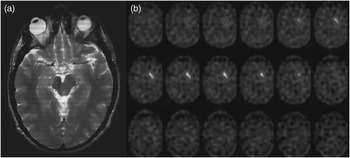
An initial report on this case was previously published (Padilla Parrado et al., Reference Padilla Parrado, Campos Arillo, Martinez del Valle Torres and Ortega Lozano2007). Briefly, at the age of 27 years, he suffered from a right mesencephalic hemorrhage attributed to the rupture of a cavernoma. The clinical picture was consistent with an incomplete Weber's syndrome, with a mild left facio-brachio-crural motor deficit (pyramidal pathway) and impaired supraversion of the right eye (oculomotor nuclei). A favorable outcome was obtained in a few weeks, with only a slight clumsiness in the left hand remaining and no documentation of cognitive impairment. Two months later, however, he began with shaking, rigidity, and increased motor difficulty in left hemibody. These symptoms were progressive, and he was diagnosed with secondary hemiparkinsonism 2 years later. Magnetic resonance imaging (MRI) scan revealed a hypointense signal in the right mesencephalon, while 123I-ioflupane single photon emission computed tomography (SPECT) scan (DaTscan) showed absence of nigrostriatal dopaminergic terminals in the right striatum (Figure 1). Treatment was initiated with levodopa at 300 mg/day, and a major clinical improvement was observed. After 1 year of levodopa treatment, however, he developed “end-of-dose deterioration” and severe “peak-dose” dyskinesias in left hemibody. Motor fluctuations became complex over the next few years, and no satisfactory benefit was obtained from different dosage regimens of levodopa and other dopaminergic agents. The patient voluntarily suspended all treatments and developed a severe depression that was only resolved after the reintroduction of medication. His optimal drug regimen was levodopa/carbidopa/entacapone (100/25/200 mg) 5 times a day during waking hours. At 7 years after onset (5 years with treatment), he was evaluated for deep brain stimulation surgery.
Neuroimaging studies 2 years after a right mesencephalic hemorrhage. a: T2-weighted magnetic resonance image scan showing atrophy of the right cerebral peduncle and reduced signal in the vicinity of the right substantia nigra and midbrain tegmentum consistent with hemosiderin deposition from old hemorrhage. b: 123I-ioflupane single photon emission computerized tomography (SPECT) scan or DaTscan showing normal distribution of dopamine transporter in the left striatum but absence of nigrostriatal dopaminergic terminals in the right striatum.

Neurological Examination
In basal “off” state (in the morning after 12 hr without medication), the patient had severe bradykinesia, rigidity, and tremor in the left hemibody, especially the arm, with a Unified PD Rating Scale Part III (UPDRS-III) score of 30. A levodopa challenge with his usual morning dose (100 mg) showed an improvement after 25 min, with a UPDRS-III score of 8 (73% improvement). The effect lasted for approximately 2 hr; in the middle of this 2-hr period, “peak-dose” dyskinesias were observed in the left hemibody, particularly the arm.
Neuropsychological Examination
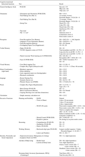
A neuropsychological examination was conducted to rule out the presence of cognitive impairment or behavioral manifestations that relatively or absolutely contraindicate surgery. The test batteries (Table 1) evaluated a wide range of cognitive functions following the recommendations of the Movement Disorder Society (Dubois et al., Reference Dubois, Burn, Goetz, Aarsland, Brown, Broe and Emre2007), and psychopathological and functional scales were also obtained, with the collaboration of relatives when required. This evaluation required four sessions (two on each of two consecutive days). Every 90-min session commenced at 30 min after receipt of the medication, when the patient's best “on” time (UPDRS-III score of 7–9).
Neuropsychological battery and results

BADS = Behavioral Assessment of the Dysexecutive Syndrome; CVLT = California Verbal Learning Test; DS = direct score; IQ = Intelligence Quotient (PIQ: Performance Intelligence Quotient; VIQ = Verbal Intelligence Quotient); Pc = percentile; SS = Scaled Score; TS = T-score; TAVEC = Test de Aprendizaje Verbal España-Complutense; WAIS-III = Wechsler Adult Intelligence Scale-Third Edition; WCST = Wisconsin Card Sorting Test; WMS-III-R = Wechsler Memory Scale-Third Edition-Revised.
*The “a” test: an auditory letter recognition test in which the subject is asked to respond with a tap on the table whenever the letter “a” is heard within a predefined sequence of letters read aloud by the examiner.
†Information for NPI was provided by the patient's mother.
The patient was examined in several sessions during the “on” state. Evaluation in “off” was not possible for ethical and practical reasons, given the severity of motor symptoms and the patient's distress. Written informed consent for this study was obtained, and all procedures were performed in compliance with the Helsinki Declaration.
Neuropsychological Test Results
The test scores are shown in the table. The patient had an IQ score of 119 (normal–high general intelligence) in the Wechsler Adult Intelligence Scale-Third Edition (WAIS-III) test (see table legend for abbreviations), with a difference of 22 points between the performance scale (105) and the verbal scale (127), and his performance of its different subtasks was normal. Notably, all performance subtests but Matrix Reasoning are timed. In the rest of the neuropsychological study, dysfunctions were evidenced in several cognitive areas, as detailed below.
The capacity to alternate attention between two or more different tasks (alternating attention) was slightly–moderately altered, although the time to complete Trail Making Test part B was elevated and several mistakes were made. Nevertheless, he preserved the capacity to direct his focus of attention by inhibiting a set of potential distracters (focused attention) and to maintain this focus for a prolonged time period (sustained attention).
The patient evidenced mild working memory deficits: although the total score for the WAIS-III digit test was within normal limits (direct score: 14; percentile 10), the performance in the backward digit subtest was poor (longest recalled sequence of three digits vs. mean ± SD of 4.54 ± 1.31 digits for his age range). Furthermore, because his maximum recall in the forward digit subtest was six digits, the difference in his performance between the subtests was also altered (three digits vs. mean ± SD of 1.45 ± 1.28 for his age range).
In the Test de Aprendizaje Verbal España-Complutense (TAVEC); Spanish version of the California Verbal Learning Test [CVLT]), he exhibited an ascending verbal material learning curve, although lower than expected for his age and educational level, which flattened from the third trial. Delayed free memory was altered and did not benefit from semantic clues. However, target recognition among a set of distracters was in the normal range. Hence, there was a difficulty in the free recall of information, whereas its encoding and storage was relatively well preserved. A similar pattern was observed for visual memory. The lack of a clear directive element in Rey figure reproduction and the poor graphical structuring suggested a problem at executive level, which can negatively affect the encoding and storage of visual information or, alternatively, a visuoconstructive dyspraxia that may also reflect frontal lobe dysfunction due to impaired programming and regulation of sequential behavior (Luria & Tsvetkova, Reference Luria and Tsvetkova1964; Pillon, Reference Pillon1981).
The patient also experienced difficulties in performing tasks that required planning, strategic thinking, problem solving, or cognitive flexibility. In the Wisconsin Card Sorting Test (WCST), although he completed the six categories in 99 assays, he committed a large number of perseverative errors and required a large number of attempts to complete the first category (>12 assays considered an alteration for his age and educational level). He also showed two failures to maintain set (maximum permitted = 1 error). These aspects were also evaluated using the Tower of Hanoi test, with different levels of difficulty. He made three more movements than expected with four discs, whereas the number of movements was significantly elevated with five discs (higher difficulty level), when he committed five errors, understood as any movements not complying with the basic rules.
Discussion
Reported cognitive deficits in patients with early PD are heterogeneous and include executive deficits that are isolated (Caviness et al., Reference Caviness, Driver-Dunckley, Connor, Sabbagh, Hentz, Noble and Adler2007) or accompanied by visuospatial (McKinlay, Grace, Dalrymple-Alford, & Roger, Reference McKinlay, Grace, Dalrymple-Alford and Roger2009) and mnestic deficits (Caviness et al., Reference Caviness, Driver-Dunckley, Connor, Sabbagh, Hentz, Noble and Adler2007), among others. Some authors found no effects on these functions per se but reported that they were very likely affected by an executive function deficit (Sollinger, Goldstein, Lah, Levey, & Factor, Reference Sollinger, Goldstein, Lah, Levey and Factor2010). Discrepancies in findings may be attributable to the highly heterogeneous nature of visuospatial function examination, which involves spatial examination, visual discrimination, spatial orientation, angle perception, topographic memory, spatial perception, and constructive abilities, among others (Ostrosky-Solis, Reference Ostrosky-Solis2000). It has been suggested that memory loss in PD is variable. Weintraub, Moberg, Culbertson, Duda, and Stern (Reference Weintraub, Moberg, Culbertson, Duda and Stern2004) proposed the existence of two different patterns of memory impairment, one related to cortical pathology and the other related to subcortical pathology and deriving from executive dysfunction.
The neuroanatomical substrate of cognitive deficits in early-stage idiopathic PD is controversial; dopaminergic denervation may play a major role, but other neuronal systems (e.g., cholinergic projections to the cerebral cortex) also appear to be implicated. In contrast, cognitive impairment in advanced PD appears to depend mainly on intrinsic cortical pathology. The present findings in a patient with secondary parkinsonism (non-idiopathic PD) offer new data on neuropsychological deficits associated with unilateral dopaminergic denervation. To our best knowledge, no case with these characteristics has been published to date, although some experimental evidence is available. Thus, one study compared working memory (by using a radial-arm maze) between rats with a selective dopaminergic lesion and those with a combined lesion of dopaminergic and noradrenergic systems (Pérez et al., Reference Perez, Marin, Rubio, Aguilar, Barbanoj and Kulisevsky2009). Dopaminergic deficit alone was sufficient to worsen the execution time, although double-lesioned rats showed a greater dysfunction and committed more errors during the task.
The right nigrostriatal pathway was destroyed in our patient, as demonstrated by DaTscan, with dopaminergic denervation of the striatum and presumably of related cortical areas. His neuropsychological profile shows deficits in executive functioning (working memory, planning, flexibility, and alternating attention) and in verbal and visual memory. These deficits are unlikely to be related to other brain lesions, because the right mesencephalic hemorrhage occurred 7 years earlier with a good clinical recovery, and it was the only lesion seen on MRI. However, we cannot definitively rule out the possibility of concomitant chronic damage of other ascending projections that pass through the mesencephalon.
Motor symptoms markedly improved under levodopa treatment, consistent with presynaptic denervation of the dorsal striatum, but neuropsychological deficits were detected. Because the patient was not evaluated in “off” state, we speculate that these deficits might also have been present (or even more severe) in “off,” with no (or only partial) improvement under levodopa or, alternatively, that levodopa may have in part induced them. Studies of PD patients “off” and “on” levodopa have generally shown greater deficits in “off” state, especially in frontal lobe function tests, in support of our first assumption. Thus, it has been reported that levodopa improved (Cools et al., Reference Cools, Barker, Sahakian and Robbins2001, Reference Cools, Barker, Sahakian and Robbins2003; Lange et al., Reference Lange, Robbins, Marsden, James, Owen and Paul1992; Lewis et al., Reference Lewis, Slabosz, Robbins, Barker and Owen2005; Malapani et al., Reference Malapani, Pillon, Dubois and Agid1994) or did not change (Skeel et al., Reference Skeel, Crosson, Nadeau, Algina, Bauer and Fennell2001) performance in executive functioning, and impairments were only described in certain specific functions (e.g., impulsivity and probabilistic reversal learning) (Cools et al., Reference Cools, Barker, Sahakian and Robbins2001, Reference Cools, Barker, Sahakian and Robbins2003).
We conclude from our experience of this case that destruction of the right nigrostriatal pathway is accompanied by similar deficits in executive functioning and verbal/visual memory to those observed in many patients with early-stage idiopathic PD. The more complex neuropsychological dysfunction developed by other PD patients must therefore be related to the additional involvement of other brain structures.
Our case uniquely demonstrates the effects of partial dopaminergic depletion on neuropsychological function in the absence of intrinsic cortical pathology (unlike on/off levodopa studies in patients with PD). However, we cannot rule out contributions from damage to cholinergic projections from the midbrain reticular formation and ascending noradrenergic projections to the right thalamus, potentially adversely affecting the function of the entire right hemi-thalamus, hence, the right hemisphere (Nadeau and Crosson, Reference Nadeau and Crosson1997). We also cannot rule out a contribution from damage to noradrenergic projections to the right cerebral cortex.
The present results demonstrate the cognitive profile associated with unilateral destruction of the dopaminergic pathway. Given that injury to this pathway is the most characteristic lesion of idiopathic PD, the application of neuropsychological tests or batteries that evaluate these domains may be highly useful for the early detection of cognitive impairment associated with this disease. The main study weakness is that the patients could not be evaluated “off-medication” for ethical reasons. Nevertheless, we consider that our case contributes novel data to be confirmed in future investigations.
Acknowledgments
The authors are grateful to Dr. F. Padilla and colleagues for the initial description and referral of this patient. There are no sources of financial support for this study. The information in this manuscript and the manuscript itself have never been published either electronically or in print. There are no conflicts of interest.