Duplications and deletions of the genome have long been thought to contribute to evolution of all species. Although there are rare cases for these copy number changes to confer an advantage to the said species, these types of changes are typically detrimental. Deletion and duplication syndromes are a major contributor to the overall number of recognised intellectual disability syndromes, which are being identified and characterised at a rapid pace with the advent of new technologies such as whole-genome array comparative genomic hybridisation (aCGH) (Ref. Reference Shinawi and Cheung1) and whole-genome sequencing technology, which has the ability to identify genomic variation between populations as well as disease regions (Ref. Reference Sudmant2). Smith–Magenis syndrome [SMS; Online Mendelian Inheritance in Man (OMIM) accession 182290], Potocki–Lupski syndrome (PTLS; OMIM 610883), 1p36 deletion syndrome (OMIM 607827), DiGeorge syndrome (OMIM 188400), Wolf–Hirschhorn syndrome (OMIM 194190), Angelman syndrome (OMIM 105830) and Williams syndrome (OMIM 194050) all result from a duplication or deletion of specific regions of the genome and result in a constellation of features that include neurobehavioural problems, developmental delays and mild craniofacial dysmorphism.
SMS is a syndrome with multiple congenital abnormalities and intellectual disability, and can present with sleep disturbance, self-abusive and aggressive behaviour, craniofacial abnormalities, neurological abnormalities and obesity. SMS was initially thought to be a contiguous gene syndrome resulting from a deletion of the 17p11.2 region encompassing ~80 genes (Ref. Reference Smith3); however, later molecular evidence showed that mutation or deletion of one gene – the RAI1 (retinoic acid induced 1) gene – resulted in most of the phenotypes observed (Refs Reference Girirajan4, Reference Slager5). In summary, SMS is a sporadic, dominant syndrome defined by haploinsufficiency (where one copy of a gene is not sufficient for proper function) of RAI1, which has been identified by bioinformatic analysis and functional studies to encode a transcriptional regulator (Refs Reference Slager5, Reference Bi6, Reference Carmona-Mora7).
Typically, deletion syndromes are more common and phenotypically more severe than duplication syndromes. This is seen when comparing SMS (17p11.2 deletion) with PTLS, a disorder that arises from 17p11.2 duplication. In SMS, intellectual disability and craniofacial, sleep and metabolic abnormalities are typically much more challenging, and the number of subjects reported is >300; by contrast, ~75 cases have been reported with PTLS, a reportedly less severe disorder (Ref. Reference Zhang8). However, although there is some evidence showing that deletions are twice as likely to occur as duplications in the SMS region on 17p11.2 (Ref. Reference Turner9), it is likely that the incidence of deletion and duplication events in the population is similar. The molecular events resulting in these two outcomes – duplication versus deletion – are similar and due to nonallelic homologous recombination (NAHR), mediated by low-copy repeats (LCRs) or segmental duplications in the 17p11.2 region (Ref. Reference Lee, Carvalho and Lupski10). Furthermore, in general, analysis of syndromes is more likely to find a deletion as the cause of the phenotype observed (Refs Reference Zhang8, Reference Turner9); this is because haploinsufficiency of one or more core genes is the probable pathology behind the deletion syndromes, whereas duplication of genomic regions might or might not result in overexpression of the genes involved (depending on the regulatory regions affected) and the resulting phenotype can be more subtle, and thus more likely to be missed clinically and not evaluated at the molecular level (Ref. Reference Yatsenko11).
Clinical findings for SMS
SMS is a clinically recognisable syndrome characterised by a distinct set of physical, developmental, neurological and behavioural features. High-resolution karyotype analysis by G-banding and fluorescent in situ hybridisation are classical methods used to detect SMS deletions, whereas multiplex ligation-dependent probe amplification, aCGH and real-time quantitative PCR are the newer technologies used for diagnosis that might identify smaller deletions (Ref. Reference Elsea and Girirajan12). It is typically a de novo disorder, with an estimated prevalence of 1:15 000–25 000 live births (Ref. Reference Elsea and Girirajan12). However, there is one report of an SMS subject whose mother is mosaic for a del(17)(p11.2) (Ref. Reference Zori13) and other clinically reported cases not published in the literature (S. Elsea, unpublished). Based on these cases, parental mosaicism for either a 17p11.2 deletion or an RAI1 mutation is estimated at ~3–5%. SMS has many clinical features that overlap with other intellectual disability syndromes, such as Prader–Willi syndrome, Kleefstra syndrome, brachydactyly mental retardation syndrome and Williams syndrome. Interestingly, only ~20% of individuals referred for 17p11.2 deletion or RAI1 molecular evaluation are positive for molecular changes leading to SMS (52/260, S. Elsea, unpublished; Ref. Reference Williams14), suggesting that additional loci might cause similar phenotypes when disrupted.
Skeletal, developmental, growth and other features
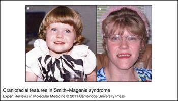
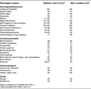
Craniofacial anomalies in SMS include brachycephaly, frontal bossing, hypertelorism, synophrys, upslanting palpebral fissures, midface hypoplasia, a broad square-shaped face, a flat nasal bridge and a tented upper lip (Ref. Reference Allanson, Greenberg and Smith15) (Fig. 1). Dental anomalies include tooth agenesis, especially of premolars, and taurodontism (Ref. Reference Tomona16). A cleft lip or palate is also reported at a higher rate than is seen in the normal population (Ref. Reference Edelman17) (Table 1).
Craniofacial features in Smith–Magenis syndrome. (a) Female child with common Smith–Magenis syndrome deletion, at age 2 years. Note broad, flat face and flat nasal bridge. (b) Same individual as in part a, at age 20 years. Note prognathism, midface hypoplasia and dental anomalies. Parental permission was obtained to use and publish these photos.

Phenotypic features of subjects with deletion 17p11.2 and RAI1 mutation

aData compiled and modified from Ref. Reference Girirajan4.
bData compiled from Refs Reference Girirajan4 and Reference Truong18.
Other skeletal and developmental abnormalities include short stature (<5th percentile in younger children), brachydactyly, scoliosis, fifth-finger clinodactyly, and forearm and elbow limitations. Adult height is typically less than expected based on parental heights, but will generally fall within the normal range, with an average between the 15th and 25th percentile. Early-onset obesity is observed, with >50% of individuals at or above the 85th percentile by age 9 years (Ref. Reference Burns19) and truncal obesity most commonly noted. A wide range of cardiac defects are also present in ~30% of reported cases (Refs Reference Edelman17, Reference Gropman20).
Otolaryngological abnormalities such as hearing loss (Ref. Reference Edelman17), a hoarse deep voice, and vocal cord nodules and polyps are also common (Refs Reference Di Cicco21, Reference Greenberg22, Reference Liburd23) (Table 1). Additionally, ophthalmological features are present in 64% of SMS patients, including myopia, iris anomalies [such as heterochromic irides or Wolfflin spots (iris hamartomas)], strabismus, microcornea and, rarely, retinal detachment (which can result from violent behaviours) (Refs Reference Edelman17, Reference Chen24, Reference Finucane25).
Neurological findings and behaviour
The neurological and behavioural phenotypes seen in SMS consist of stereotypies/tics such as self-hugging and hand twirling, self-abusive behaviours including nail yanking and skin-picking, hyperactivity, and oral or motor dysfunction (Refs Reference Girirajan4, Reference Elsea and Girirajan12, Reference Edelman17, Reference Gropman20, Reference Greenberg22, Reference Smith, Dykens and Greenberg26, Reference Finucane and Jaeger27, Reference Gropman, Duncan and Smith28, Reference Potocki29). Additionally, speech and motor delays are observed, with hypotonia typically present at birth in >90% of cases (Ref. Reference Elsea and Girirajan12). Lesser observed but significant phenotypic manifestations include seizures and polyembolokoilamania (Table 1).
An inverted circadian rhythm, which results in nighttime awakenings and napping periods during the day and even during times of activity, complicates the neurological and behavioural phenotypes observed (Refs Reference Potocki30, Reference De Leersnyder31). Sleep disturbance is reported in up to 100% of SMS cases and is a key diagnostic feature (Refs Reference Girirajan4, Reference Edelman17, Reference Greenberg22, Reference Smith, Dykens and Greenberg26). Interestingly, infants usually experience hypersomnolence during the first year of life, with circadian changes beginning typically between 12 and 18 months of age (Ref. Reference Gropman, Duncan and Smith28). The complicated sleep phenotype occurs in older children and includes reduced 24 h sleep time, reduced REM (rapid eye movement) sleep, and fragmented night sleep with daytime sleepiness (Refs Reference Smith, Dykens and Greenberg26, Reference Potocki30, Reference De Leersnyder32). Until recently, these clinical phenotypes have been attributed to an inverted rhythm of melatonin secretion, but several instances of individuals with normal melatonin secretion and significant sleep disturbance have been reported (Refs Reference Potocki30, Reference Boudreau33). These findings indicate a molecular role for RAI1 in proper sleep cycling, which should be further investigated.
Mechanisms of deletion of the 17p11.2 region
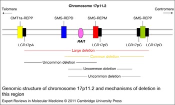
Chromosome 17p11.2 deletion or duplication results from NAHR between LCRs situated along the chromosome. Within the SMS region on chromosome 17p11.2 there are four distinct LCRs, termed LCR17pA, LCR17pB, LCR17pC and LCR17pD (Ref. Reference Park34). Additionally, three large highly homologous LCRs (SMS-REPs) are present in the region, and based on their chromosomal position are designated as SMS-REPP (proximal), SMS-REPM (middle) and SMS-REPD (distal), each represented by a YAC clone (Ref. Reference Chen35) (Fig. 2). These repeats are paralogous repeat gene clusters of ~200 kb. The distance between proximal and distal REPs was estimated to be ~4–5 Mb and is now calculated as 3.7 Mb (Refs Reference Chen35, Reference Shaw, Withers and Lupski37, Reference Stankiewicz38). A common junction fragment was also identified in ~90% of SMS patient samples (Ref. Reference Chen35).
Genomic structure of chromosome 17p11.2 and mechanisms of deletion in this region. The RAI1 (retinoic acid induced 1) gene is represented by the purple oval. SMS-REPs (highly homologous repetitive regions) are shown in blue (SMS-REPD), red (SMS-REPM) and green (SMS-REPP) (Ref. Reference Shaw and Lupski36). Smaller and lesser utilised low-copy repeats are shown in black. The Charcot–Marie–Tooth disease type 1A (CMT1a) proximal REP is shown in yellow. Below the 17p11.2 schematic are examples of large (Ref. Reference Girirajan4), common (Ref. Reference Girirajan4) and uncommon deletions (Ref. Reference Shaw and Lupski36). Additional examples of various deletions can be found in Refs Reference Girirajan4 and Reference Shaw, Withers and Lupski37. All deletions include the RAI1 gene. Additional homologous repeat-rich regions are present but not shown, and details can be found in Refs Reference Shaw and Lupski36 and Reference Shaw, Withers and Lupski37. The figure is not drawn to scale. Abbreviation: SMS, Smith–Magenis syndrome.

Based on these data, it was predicted that SMS-REPP and SMS-REPD would mediate most of the recurrent rearrangements (duplications/deletions) associated with this region (Ref. Reference Potocki39) (Fig. 2). Genetic evidence of NAHR between the distal and proximal SMS-REPs was obtained by genotyping ~24 patients carrying 17p11.2 rearrangements (Refs Reference Shaw, Withers and Lupski37, Reference Bi40, Reference Shaw, Bi and Lupski41). Data show that >75% of SMS deletions are due to recombination involving proximal and distal repeats, causing an ~3.7 Mb common deletion. Large deletions can be seen when recombination between LCR17pA and LCR17pD occurs, giving rise to an ~5 Mb deletion in ~4% of SMS cases (Ref. Reference Shaw, Withers and Lupski37) (Fig. 2). Additionally, uncommon deletions occur in ~16% of cases when alternate LCRs are paired in recombination (Ref. Reference Shaw, Withers and Lupski37). Rearrangements mediated by nonhomologous mechanisms involving repetitive elements such as Alu–Alu recombination have also been reported (Ref. Reference Stankiewicz38).
Mutation of RAI1
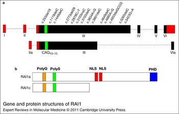
De novo mutations in the RAI1 gene that result in SMS are observed in ~10% of cases (Refs Reference Girirajan4, Reference Slager5, Reference Elsea and Girirajan12, Reference Edelman17, Reference Truong18, Reference Bi42, Reference Girirajan43, Reference Bi44) (Fig. 3). RAI1 contains six exons, four of which are protein coding (Fig. 3). Mutations include small deletions and insertions within the RAI1 coding region, which cause frameshifts and premature stop codons; also observed are nonsense mutations, which again produce a truncated protein product, and missense mutations, which lead to nonfunctional or reduced-function proteins. To date, all mutations observed have been in coding exon 3 of the RAI1 gene, and recently a ‘hotspot’ was identified in this region for frameshift of RAI1 (c.3103delC and c.3103insC) (Ref. Reference Truong18). Thus far, the mutations reported include ten frameshift, one nonsense and three missense changes in the RAI1 gene (Refs Reference Girirajan4, Reference Slager5, Reference Bi42, Reference Girirajan43, Reference Bi44) (Fig. 3). These molecular data combined with clinical information support haploinsufficiency of RAI1 as the primary gene defect responsible for most of the phenotypic load in SMS, accounting for mild intellectual disability, attention-seeking behaviour, self-injurious behaviour, circadian defects, craniofacial features and other minor anomalies (Table 1). To date, major organ anomalies have not been associated with mutation of RAI1, and so other genes are probably contributing to these developmental defects.
Gene and protein structures of RAI1. (a) RAI1 (retinoic acid induced 1) gene structure, including noncoding exons (red) and coding regions (black). Represented are those cDNAs cloned from biological samples and subsequently evaluated, encoding isoform a (top: NP109590, EAW55692.1) and isoform c (bottom: AAH21209.1, BC021209). A CAG repeat region is shown in green. Other isoforms are predicted to exist but have not been confirmed. Shown above the gene structure for isoform a are all reported mutations in the RAI1 gene. (b) RAI1 protein isoforms a and c, including polyglutamine tract (polyQ, orange), polyserine tract (polyS, green), bipartite nuclear localisation signals (NLS, red) and plant homeodomain (PHD) (blue).

Potocki–Lupski syndrome
As described above, RAI1 is a dosage-sensitive gene, and duplication of the 17p11.2 region results in ‘duplication 17p11.2 syndrome’, also known as PTLS. PTLS results in mild to severe intellectual disability, infantile hypotonia, failure to thrive, cardiovascular anomalies, sleep-disordered breathing, developmental delay, hyperactivity and autism (Refs Reference Treadwell-Deering, Powell and Potocki45, Reference Potocki46, Reference Soler-Alfonso47). Craniofacial features include a triangular face, microcephaly, micrognathia, a broad nasal bridge, a highly arched palate, hypertelorism, and dental anomalies in the form of malocclusion of the teeth (Ref. Reference Potocki39). Limb abnormalities, including flexion deformity of the fingers and club foot, have also been described, as well as pharyngeal dysphagia and electroencephalogram abnormalities (Refs Reference Treadwell-Deering, Powell and Potocki45, Reference Potocki46). Less commonly reported features are hearing impairment, structural otolaryngological defects and ophthalmic abnormalities, such as myopia and iris hamartoma. Other rarely reported features are genitourinary or renal anomalies, scoliosis and hypercholesterolaemia (Ref. Reference Potocki46).
As discussed in the introduction, although the predicted incidence of 17p11.2 duplication syndrome is the same as that of 17p11.2 deletion, it is probably underdiagnosed because the phenotype can be much more subtle than that of SMS (Ref. Reference Zhang8). Reported duplications of this 17p11.2 region range between 0.4 and 13.3 Mb; 3.7 Mb is the most common duplication (between distal and proximal repeats flanking RAI1) and 5 Mb is the least common recurrent duplication (located between LCR17pA and LCR17pD), having been recently identified in only two individuals (Ref. Reference Zhang8).
The clinical and molecular findings for deletion and duplication syndromes such as SMS and PTLS that involve the same genomic region, with evidence that the same gene is contributing to the phenotypes in both disorders, illustrate the dosage sensitivity of the key gene in that genomic region (Refs Reference Slager5, Reference Zhang8). SMS and PTLS data, in addition to the mouse models described below, indicate the need for tight regulation of expression of RAI1.
Mouse models for SMS and PTLS
Mouse models that mimic human conditions are invaluable tools to help understand the molecular, developmental and behavioural role of a gene or genes. The sequencing of the mouse genome has allowed for regions of synteny to the human genomic sequence to be mapped and utilised for research (Ref. Reference Waterston48).
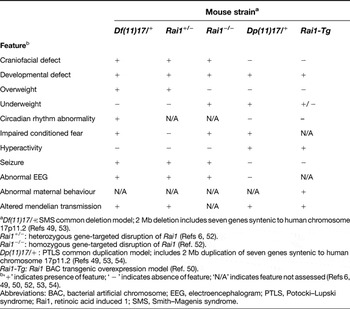
Murine chromosome 11qB1.3–B2 is syntenic to human chromosome 17p11.2. An ~34 cM region surrounding human RAI1 and murine Rai1 is conserved between human and mouse genomic structure, making this region fit for creation of knockout and transgenic mice modelling human conditions arising from changes in gene dosage. Mice lacking [Df(11)17/+] or duplicated [Dp(11)17/+] for a 2 Mb region containing seven genes syntenic to human chromosome 17p11.2, including Rai1, were originally created by Walz et al. (Ref. Reference Walz49), using chromosome engineering technology, to mimic the common deletion or duplication seen in SMS or PTLS, respectively. These mice recapitulate many of the core features observed in PTLS or SMS, including lean body weight, hyperactivity and altered sociability in Dp(11)17/+ mice, and obesity, abnormal behaviour and craniofacial features in Df(11)17/+ mice. Such features are also seen in later gene-targeted models of altered Rai1 expression (Refs Reference Bi6, Reference Burns19, Reference Girirajan and Elsea50, Reference Ricard51, Reference Bi52) (Table 2).
Phenotypes of Rai1-engineered mice

aDf(11)17/+: SMS common deletion model; 2 Mb deletion includes seven genes syntenic to human chromosome 17p11.2 (Refs Reference Walz49, Reference Walz53). Rai1 +/−: heterozygous gene-targeted disruption of Rai1 (Refs Reference Bi6, Reference Bi52).Rai1 −/−: homozygous gene-targeted disruption of Rai1 (Ref. Reference Bi52). Dp(11)17/+ : PTLS common duplication model; includes 2 Mb duplication of seven genes syntenic to human chromosome 17p11.2 (Refs Reference Walz49, Reference Walz53, Reference Walz54). Rai1-Tg: Rai1 BAC transgenic overexpression model (Ref. Reference Girirajan and Elsea50).
b‘+’ indicates presence of feature; ‘ − ’ indicates absence of feature; ‘N/A’ indicates feature not assessed (Refs Reference Bi6, Reference Walz49, Reference Girirajan and Elsea50, Reference Bi52, Reference Walz53, Reference Walz54).Abbreviations: BAC, bacterial artificial chromosome; EEG, electroencephalogram; PTLS, Potocki–Lupski syndrome; Rai1, retinoic acid induced 1; SMS, Smith–Magenis syndrome.
Recently, Girirajan and colleagues (Refs Reference Girirajan and Elsea50, Reference Girirajan55) generated and performed an in-depth behavioural analysis of bacterial artificial chromosome (BAC) transgenic Rai1-overexpression mice (Rai1-Tg) harbouring two additional copies of the Rai1 gene. These mice showed a variety of defects, including neurological deficits and growth retardation, which resolved by 20 weeks of age (Ref. Reference Girirajan55). Abnormal maternal behaviour was observed wherein Rai1-Tg mothers could not properly take care of their pups (1/5 survival average for wild-type or transgenic pups); however, when pups were placed with a wild-type foster mother, all (5/5, average) survived (Ref. Reference Girirajan and Elsea50). Additionally, Rai1-overexpressing mice show a reduction in the levels of the serotonin metabolite 5-HIAA, suggesting a defect in the regulation or expression of monoamine oxidase, which converts serotonin to 5-HIAA. Further, these mice have altered social behaviour, with impaired nesting behaviour, social dominance, aggression, reduced social memory, hyperactivity, anxiety-related behaviour and altered sociability – all 5-HIAA-mediated behaviours (Refs Reference Girirajan and Elsea50, Reference Girirajan55, Reference Molina56).
Later, Burns et al. (Ref. Reference Burns19) showed that Rai1 +/− mice are obese and hyperphagic but lack metabolic disease, such as signs of insulin resistance commonly associated with high fat stores (Ref. Reference Burns19). Female Rai1 +/− mice also have altered fat deposition, with increased deposition of truncal fat when compared with wild-type female mice. However, these mice have increased leptin levels, which might account for their increased food intake after fasting. These data suggest a role for Rai1 in the satiation response or metabolism/synthesis of adipose tissue.
Taken together, Df(11)17/+, Rai1 +/−, Dp(11)17/ + and Rai1-Tg mice have proved to be good models for their respective human syndromes. Rai1 +/− mice have craniofacial, behavioural and developmental abnormalities consistent with those of SMS. Additionally, Dp(11)17/+ duplication and Rai1-Tg mice have behavioural, neurological and developmental abnormalities consistent with those of PTLS. These data reinforce the dosage sensitivity of RAI1. A summary of the phenotypes seen in these mice is provided in Table 2.
Molecular function of RAI1
Although data show that haploinsufficiency of the RAI1 gene results in SMS, little is known about the molecular function of RAI1. Given the phenotypic consequences of RAI1 mutation or deletion, RAI1 must be involved in pathways associated with development, behaviour, neurological function and circadian rhythm.
Mutations in transcription factors have long been associated with human disease, including ATRX syndrome (X-linked α-thalassaemia/mental retardation syndrome), myeloid leukaemia and autoimmune dysfunction (Ref. Reference Capili57). Because transcription factors can regulate many genes, deciphering their global function can be difficult. With regard to RAI1, there is an additional layer of complexity because, other than insights from bioinformatic analysis, very little is known about the true molecular function of RAI1.
Rai1, originally called Gt1, was identified in mouse P19 embryonal carcinoma cells, which can be differentiated into neurons and glial cells when treated with retinoic acid. It was found to be expressed in all tested tissues and highly expressed in neurons (Refs Reference Slager5, Reference Imai58). At least three human isoforms are predicted by bioinformatic analysis. RAI1 contains a plant homeodomain (PHD) and a bipartite nuclear localisation signal, which are commonly seen in transcription factors. PHD domains typically bind zinc and are involved in chromatin remodelling, whereas nuclear localisation signals have the ability to recruit chaperone proteins, thus facilitating transport into the nucleus (Ref. Reference Bienz59) (Fig. 3). Indeed, RAI1/Rai1 protein localises to the nucleus and has transactivational activity in a luciferase reporter system (Refs Reference Bi6, Reference Carmona-Mora7, Reference Burns19). Complicating protein analysis, the three predicted isoforms of RAI1 might serve different functions dependent on spatial and temporal expression.
In a functional network module of SMS, small interfering RNA technology was used to achieve in vitro haploinsufficiency (50% knockdown) of RAI1 (Ref. Reference Girirajan60). Differentially expressed transcripts identified in this study included genes important in (but not limited to) metabolism, the cell cycle, insulin signalling, lipid biosynthesis and cholesterol homeostasis. Additionally, knockdown of RAI1 resulted in reduced expression of NR1D2 (nuclear receptor subfamily 1, group D, member 2), which is an essential component of the circadian clock. It has been well established that disruption of the circadian clock can lead to obesity: the Clock Δ19 mutant mouse model presents with obesity, hyperphagia and metabolic disease, including hyperleptinaemia, hyperlipidaemia, hepatic steatosis, hyperglycaemia and hypoinsulinaemia (Ref. Reference Turek61). CLOCK is a transcriptional regulator of the circadian clock, including NR1D2 as well as PER1/2 (period homologue 2) and CRY1/2 (cryptochrome 1/2). Taken together, these data and the well-established sleep disturbance in SMS suggest a possible role for RAI1 in the circadian loop.
Recent studies in mice showed that haploinsufficiency of Rai1 results in reduced expression of brain-derived neurotrophic factor (Bdnf) (Ref. Reference Burns19). BDNF promotes striatal neurons, and reduced expression has been associated with schizophrenia, depression and obsessive compulsive disorder (Refs Reference Angelucci, Brene and Mathe62, Reference Duncan63, Reference Wendland64). Additionally, tissue-specific knockout of Bdnf in mouse hypothalamus results in obesity and hyperphagia, much like that seen in Rai1 +/− mice. Furthermore, a luciferase reporter system showed that RAI1 overexpression induces BDNF expression (Ref. Reference Burns19), lending support to the idea that these two genes function in a common pathway with RAI1 acting as an enhancer of BDNF. These facts present an interesting opportunity to exploit the RAI1–BDNF relationship by targeting Bdnf expression through ampakine treatment in mice; ampakine compounds have been shown to enhance Bdnf expression in several animal model studies, suggesting a possible pharmacological target for treatment in humans (Refs Reference Kramar65, Reference Lauterborn66, Reference Ogier67, Reference Simmons68). Additionally, given the fact that RAI1 has been shown to upregulate the transcription of the BDNF gene in vitro and that Bdnf expression is reduced in Rai1 +/− mice, it is likely that RAI1 serves as an enhancer that is integral to modulating genes involved in metabolism, and these pathways should be explored in detail in future studies.
RAI1 has also been implicated in metabolism and neurobehaviour in a recent publication that shows that RAI1 expression levels are reduced in subjects with brachydactyly mental retardation syndrome (BDMR) (Ref. Reference Williams69). This syndrome results from a deletion of chromosome 2q37 or a mutation of the histone deacetylase 4 gene (HDAC4) (Ref. Reference Williams69). Individuals with BDMR have a strikingly similar phenotype to individuals with SMS (including obesity, sleep disturbance and self-abusive behaviour) and have been previously misdiagnosed with SMS (Ref. Reference Williams14). All BDMR patient lymphocytes tested for expression of RAI1 had reduced levels, indicating that RAI1 is probably downstream of HDAC4. One major difference between SMS and BDMR is the distinctive brachydactyly type E that BDMR subjects often present with, which is not seen in SMS subjects. These phenotypic differences should be taken into account when considering the differential diagnosis.
Clinical implications/applications
The study of SMS, and RAI1 in particular, provides an interesting opportunity to analyse the molecular aspects of a single-gene disorder involving intellectual disabilities, neurobehavioural problems, sleep disturbance and obesity. Because of the phenotypic overlap with many other syndromes in the diagnostic spectrum, it will be important to analyse the ‘finer details’ of these molecularly distinct syndromes in the clinic. Supporting this, a recent study showed that up to 50% of subjects referred for molecular evaluation of SMS did not have SMS, indicating the need for close evaluation of behaviour and attention to the physical phenotype, including craniofacial and skeletal clues (Ref. Reference Williams14).
Proper diagnosis is key to all downstream treatments. Because of the strong overlap of SMS with so many other syndromes, this is a challenge every day for clinical geneticists. It would be appropriate to devise a detailed panel of phenotypes to aid in distinguishing the clinical differences between SMS and overlapping syndromes, taking into account the following: neurobehavioural features including, but not limited to, stereotypies, self-injurious and aggressive behaviours, and sleep disturbance; craniofacial features, including a broad face, a flat nasal bridge and brachycephaly; and skeletal features including short stature, brachydactyly and displaced toes 3–5. This is a short list, but if specific combinations of features are matched with most likely syndromes, the diagnostic process might be simplified. Development of a tiered molecular testing panel to facilitate correct diagnosis would further support this process and should include aCGH (to detect 17p11.2 and 2q37.3 deletions as well as other phenotypically overlapping syndromes), RAI1 mutation analysis and HDAC4 mutation analysis. Further studies of gene expression changes in RAI1 (and HDAC4) could support diagnosis in complex cases not identified by the above methodologies.
Medical management, behavioural modifications and other pharmacological treatments are probably appropriate for people with SMS, but any treatment should be on a case-by-case basis in order to improve daily life. No single medication works for all people with SMS (Ref. Reference Laje70); thus, additional research into the affected molecular and metabolic pathways is required in order to target appropriate pharmaceuticals to this population. A promising avenue to explore in the future is sleep rhythms and their impact on behaviour and metabolism. Circadian rhythms are present in living organisms ranging from bacteria to mammals. Disrupted circadian rhythms are associated with obesity and increased risk for high blood pressure and heart attack in shift workers (Ref. Reference Di Lorenzo71). Because of the strong circadian phenotype seen in SMS, directly addressing this basic biological problem is key for improved behaviour overall. Very basic treatments, such as light therapy and strictly enforced feeding schedules, might significantly improve the behaviour and metabolic outcomes in this population. Studies have shown that robust and appropriate circadian rhythms of transcription can be restored in mice lacking circadian rhythm (Cry1 −/−/Cry2−/−) (Ref. Reference Vollmers72) by enforcing a restricted feeding schedule, which holds promise that patterned feeding schedules during the day for people with SMS might provide an avenue for more consistent sleep patterns at night. Restoration of a consistent sleep pattern would probably have a positive impact on the behavioural and metabolic aspects of SMS (Ref. Reference De Leersnyder31).
Outstanding research questions
There are many aspects of research that are yet to be uncovered in the field of intellectual disabilities syndromes, especially SMS. Further molecular analysis of the RAI1 gene, its transcriptional targets and the genes regulating RAI1 is important if targeted pharmacological treatments are to be pursued in a meaningful way.
Any proteins that RAI1 might bind in order to exert its gene-regulatory activity, and whether it functions only as an enhancer or can act as a repressor too, are unknown. Molecules that modulate transcription rarely work alone. If binding partners are identified, it will be easier to identify specific points at which RAI1 functions in metabolic, neurological and circadian pathways. Additionally, the exact targets of RAI1 are still undefined. This can be analysed by tissue-specific temporal chromatin immunoprecipitation with sequencing (ChIP-seq) analysis, which will give an insight into the genes that RAI1 regulates during and after development.
To that end, it would be especially useful to create an inducible Rai1-knockout mouse in order to separate the developmental impact of loss of Rai1 from the postnatal influence of Rai1 on metabolism and neurological development and behaviour. This approach would also provide data to indicate whether RAI1 would be a good target for pharmacological intervention in SMS. If the primary impact of Rai1 haploinsufficiency is on the developing fetus, then it is unlikely that simply increasing the expression level of RAI1 would make an effective treatment strategy. However, it is more likely that haploinsufficiency of RAI1 has a major role throughout life and that targeting this gene pharmacologically could be explored using conditional mouse models with the ability to focus on specific affected pathways, such as satiety, control of behaviours and circadian rhythm.
At the very basic level, the specific structure of the RAI1 protein, the composition and function of the various isoforms, and any tissue specificity are not understood. These details are especially of interest to identify the specific role that RAI1 has in critical molecular and metabolic pathways.
Given that proper dosage of RAI1 is crucial to normal development, investigation into the regulation of RAI1 expression is important, and understanding the players involved might lead to the identification of other genes that, when insufficient, might result in phenotypes with significant overlap to SMS. In this same line, we question whether RAI1 might be targeted by microRNAs. Is there an antisense transcript that might be targeting RAI1 in a temporal pattern? These details hold a wealth of information for future work, and if there are microRNAs targeting RAI1, this might be a crucial step in the creation of therapeutics in the future. A screen of known microRNAs and subsequent evaluation of RAI1 expression would provide the first indication of whether this is a possibility. Bioinformatic analysis should also be assessed when seeking these answers.
In summary, RAI1 has a major role in pathways that impact metabolism, neurology, development and circadian rhythm. Although the specific role of RAI1 in these pathways is unknown, the SMS (and PTLS) phenotype illustrates the importance of this gene and proper gene dosage. Additionally, because of the phenotypic overlap and emerging molecular role for RAI1 in other syndromes with intellectual disability and congenital abnormalities, it is essential that RAI1 not be overlooked. A combined, collaborative and focused clinical and research strategy is of the utmost importance and will aid progress in the future.
Acknowledgements and funding
We thank all the clinicians who work towards accurate diagnosis of intellectual disabilities syndromes, the families who live with these syndromes every day, and the scientists, students and laboratory staff who make this research possible and worthwhile. This study was supported, in part, by the Jérôme Lejeune Foundation.