Introduction
Investigation of the stapedius reflex by detecting acoustic admittance changes at the eardrum is one of the main diagnostic tools in audiology. The acoustically evoked stapedius reflex (acoustic reflex) enables exploration of peripheral and central auditory pathways and furnishes valuable information on the mechanics of the middle ear ipsilateral to the recording side.Reference Jerger and Jerger1
Ossicular fixation or discontinuity, facial palsy, or even absence of the stapedius muscle tendon are possible causes of dysfunction of the efferent branch of the stapedius reflex, which usually leads to the disappearance of the response. However, severe to profound bilateral hearing impairment due to inner-ear disorders, with or without middle-ear dysfunction, may also result in the absence of the stapedius reflex, due to attenuation of the acoustic input to the afferent portion of the reflex. In this situation, the acoustic reflex cannot be utilised to detect an underlying disorder in the mechanics of the middle ear, since the reflex is absent independently from the conditions of the middle ear. Diagnostic doubts may therefore occur, for example, when very advanced otosclerosis is suspected.
This drawback may be circumvented by investigating middle-ear admittance changes induced by non-acoustic stimuli which are able to evoke a stapedius reflex. This may be accomplished by stimulating reflexogenic skin areas with tactile or electrical stimuli to the contralateral concha or the ipsilateral face.Reference Klockhoff2, Reference Djupesland and Tvete3 The largest responses are elicited when the skin of the external ear canal is touched or when an air puff is directed towards the orbital region.Reference Djupesland and Tvete3 This stimulation is able to activate both the stapedius and the tensor tympani muscles.Reference Djupesland and Tvete3
However, non-acoustic activation of the stapedius reflex presents some drawbacks, which have precluded widespread clinical use, that is: difficulty in quantifying the eliciting stimuli; discomfort; and lack of reproducibility of the response. The present paper describes an alternative technique for activating middle-ear muscles, as revealed by acoustic admittance modifications at the eardrum – transcranial magnetic stimulation.
Materials and methods
Subjects
Admittance changes induced by transcranial magnetic stimulation were evaluated in eight normal subjects, in two subjects with prelingual deafness, and in 22 patients suffering from other disorders characterised by absence of the acoustic stapedius reflex or by the impossibility of recording this reflex using admittance audiometry.
The normal subjects (four men and four women aged 19 to 40 years) had hearing levels within 15 dB HL in the frequency range 0.25–8 kHz.
The prelingual deaf subjects (one man and one woman aged 23 and 27 years, respectively) had normal external and middle-ear function. Their hearing level was characterised by bilateral profound hearing loss with residual hearing (equal to or higher than 110 dB HL) only at 250, 500 and 1000 Hz. The acoustic stapedius reflex could not be evoked in these patients, even at the highest intensity of stimulation (120 dB HL).
The patients with absent acoustic reflex comprised 12 women and 10 men, with ages ranging from 22 to 42 years. They suffered from otosclerosis (n = 8), subtotal tympanic membrane perforation (n = 3), fibroadhesive otitis media (n = 1), previous first stage tympanoplasty with absence of the malleus and the incus (n = 3), previous stapedotomy with stapedius tendon section (n = 4), or facial palsy due to complete resection of the facial nerve during acoustic tumour surgery (n = 1) or to Bell's palsy (n = 2). Patients with stapedotomy had a stapedial prosthesis devoid of ferromagnetic properties.
None of the subjects had any history of neurological or general diseases.
Both ears were examined in the normal and prelingually deaf subjects, whereas only the involved ears were examined in the remaining patients. All subjects underwent otomicroscopy and pure tone audiometry (PTA), using a clinical audiometer (A319; Amplaid, Milan, Italy).
Admittance audiometry was preliminarily performed in all subjects with intact tympanic membranes. An admittance meter (Amplaid 702) with a probe tone of 220 Hz, connected to an X–Y plotter, was utilised for this purpose. A type A tympanogram with a peak within ±50 daPa was obtained in all ears except those with previous tympanoplasty. A stapedius reflex was present at 500, 1000 and 2000 Hz in all normal subjects and absent in the affected ears of the remaining patients.
All subjects were informed about the experimental procedure and the aim of the study, and gave their free consent.
Magnetic stimulation
Transcranial magnetic stimulation utilised a Magstim 200 (Magstim, Withland, Wales, UK) connected to an 8 cm circular coil. The coil was placed on the scalp surface and centred on the inion. The capacitor was charged up to 90 per cent of its maximal output, with steps of l0 per cent. Four stimulations were performed in all subjects at each intensity, in a randomised order.
Admittance changes were recorded using the same equipment as employed for the preliminary investigation (i.e. an Amplaid 702 admittance meter). The external ear was sealed by the probe, which was stabilised with adhesive tape fixed to the pinna.
Admittance recordings were performed at equipressure between the external and middle ear in subjects with an intact tympanic membrane, and at 0 daPa in the remaining subjects. Downward (negative) deflections indicated decreased admittance and vice versa. During the examination, subjects were in a comfortable sitting position. They were asked to relax and to avoid any voluntary contraction of the facial muscles.
During magnetic activation, in order to rule out any admittance changes originating from air- or bone-conducted acoustic stimuli, the responses were also evaluated in normal subjects with the coil positioned on the vertex and at a distance of 3 cm from the occipital scalp, using a maximal stimulation intensity (90 per cent).
Admittance changes were also recorded in otosclerotic subjects following air jet stimulation directed towards the ipsilateral orbit.
Results
Figure 1 shows typical admittance modifications observed in a normal subject submitted to transcranial magnetic stimulation (90 per cent of maximal output) at the inion. A predominant negative peak (n) characterised the response. Two small positive components were also present, the first one preceding (p1) and the last one following (p2) the negative deflection. The first peak was characterised by a rapid onset and decay, whereas the p2 peak presented greater duration and slower onset and decay.
Typical triphasic admittance changes detected in a normal ear, following transcranial magnetic stimulation at 90 per cent of maximal output. Arrow indicates stimulus onset. Ms = milliseconds

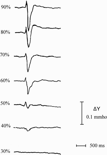
Figure 2 shows representative responses elicited in a normal subject by different intensities of stimulation, i.e. 30 to 90 per cent. The threshold of the response was obtained when the stimulator fired at 40 per cent of the maximal output. The amplitude of the n peak increased with stimulus intensity, whereas this phenomenon was not clearly evident for the p1 and p2 components.
Admittance changes elicited in a normal subject, following transcranial magnetic stimulation at different intensities. Ms = milliseconds

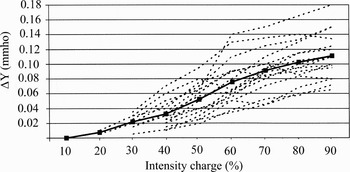
Normal subjects always presented a negative peak, followed by the p2 wave at least at the highest intensities, whereas the p1 peak was only present in five subjects and was quite inconsistent in these same subjects, even between tests. Threshold values for the negative response ranged from 20 to 50 per cent, and the amplitude always increased proportional to the intensity. The individual amplitude/intensity functions of this component, as well as the median values, as expressed by admittance changes (ΔY mmho), are shown in Figure 3.
Individual amplitudes of negative admittance changes as a function of magnetic stimulus intensities observed in all normal ears (n = 16). Bold line represents median values.

Stimulation at 90 per cent intensity, by positioning the coil over the occipital region at a distance of about 3 cm from the scalp, or centred on the vertex in contact with the scalp, was only able to produce inconsistent, small responses with variable polarity, probably attributable to artefacts.
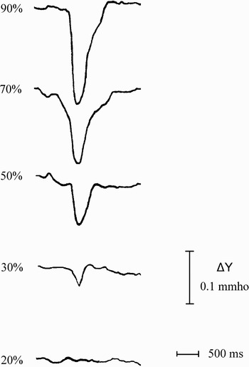
Responses identical to those obtained in normal subjects were elicited in the prelingually deaf subjects, in whom no acoustic stapedius reflex had been obtained (Figure 4).
Admittance changes in a subject with profound hearing loss, following transcranial magnetic stimulation at different intensities. Ms = milliseconds

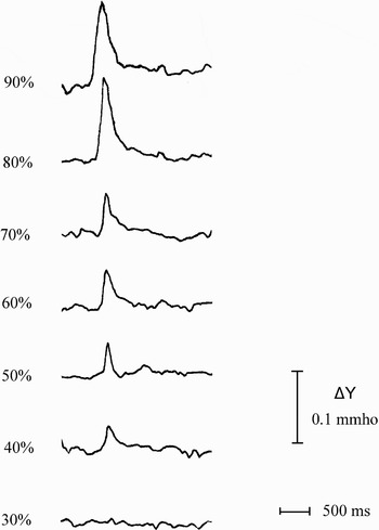
Figure 5 shows representative responses elicited in the right ear in a patient with otosclerosis. His auditory threshold showed an air–bone gap of 32 dB (PTA 0.5–2 kHz) with bone conduction of 7 dB at the same frequencies. No negative deflections were observed, whereas a rapid-onset positive component with a slower decay and amplitude increasing with stimulus intensity was elicited. This positive peak was observed in all otosclerotic patients, with thresholds ranging from 40 to 70 per cent of the maximal output.
Positive admittance changes elicited in an otosclerotic ear. Ms = milliseconds

Findings similar to those achieved in otosclerotic ears were observed in subjects who had undergone stapedotomy with section of the stapedius tendon (Figure 6) and in patients with facial palsy.
Positive admittance changes elicited in an ear which had undergone stapedotomy with stapedius tendon section. Ms = milliseconds

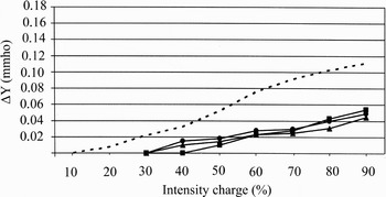
Figure 7 shows the median input-output function of the positive peak amplitude, observed in otosclerotic patients, in patients who had undergone stapedotomy with tendon section and in patients with facial palsy. Positive responses increased slightly with stimulus intensity and showed lower amplitude values, compared with the n component of normal subjects.
Median amplitudes of the positive admittance changes shown by different groups of ears with absent acoustic reflex (triangles = otosclerosis; circles = stapedotomy; squares = facial palsy). Median values of the n wave calculated in normal subjects are presented (dotted line) for comparison.

Stimulation with an air jet directed towards the orbit elicited positive responses very similar to those observed following magnetic stimulation in ears with otosclerosis (Figure 8).
Comparison between a typical response elicited with magnetic stimulation (90 per cent of maximal output) and with tactile stimulation (air jet) in an otosclerotic ear. Ms = milliseconds

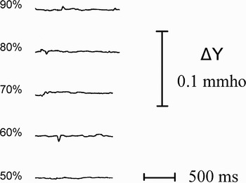
Figure 9 shows the admittance changes recorded from an ear showing chronic otitis media with subtotal tympanic membrane perforation and incus erosion. Inconsistent admittance changes, with variable directions which were not dependent on the stimulus intensity, were observed. Most subjects with chronic otitis media or previous tympanoplasty presented similar findings. These admittance changes were not reproducible, showing large variability among subjects, tests and stimulus intensities. In all cases, they were characterised by very small amplitudes.
Responses observed following magnetic stimulation in a patient with chronic otitis media. Ms = milliseconds

Discussion
Magnetically induced stimulation of the cranial nerves and cerebral cortex is a non-invasive technique for eliciting motor responses.Reference Mills, Murray and Hess4, Reference Bickford, Guidi, Fortesque and Swenson5 Pulsed magnetic fields induce a depolarisation current in the underlying neural tissue, with subsequent generation of action potentials. Since close contact is not required, skin preparation is unnecessary and impedance at the stimulus–tissue interface irrelevant. Magnetic signals are not attenuated by body tissue and are virtually painless.Reference Barker, Freeston, Jalinous and Jarratt6 No side effects have been documented, although caution has been advocated regarding the possible activation of seizures in epilepsy, and the effect of the magnetic field on implanted metallic objects and pacemakers.
Transcranial magnetic stimulation is used for investigating the conduction properties of the facial nerve, as an alternative to electric stimulation at the stylomastoid foramen.Reference Macabee, Amassian, Cracco, Cracco and Anziska7–Reference Rosler, Hess and Schmid10 Transcranial stimulation activates the facial nerve in its intracranial portion,Reference Macabee, Amassian, Cracco, Cracco and Anziska7–Reference Rosler, Hess and Schmid10 or, as shown by Schmid et al. Reference Schmid, Moller and Schmid11 during posterior fossa surgery, at the entrance of the petrous bone. Schmid et al. Reference Schmid, Moller and Schmid12 showed that magnetic stimulation is also able to activate the motor portion of the trigeminal nerve, either proximal or distal to the foramen ovale. Whatever the precise site of neural activation, magnetic pulses stimulate the Vth and VIIth cranial nerves proximal to the neural branches directed to the tensor tympani and stapedius muscles.
In the present investigation, the responses obtained with magnetic stimulation in normal subjects were characterised by a main negative component (n), which may be attributed to stapedius muscle contraction. In fact, total absence of this wave was observed in subjects with facial palsy, otosclerosis, previous stapedotomy with stapedius tendon section, and generally in ears with middle-ear disorders. The response amplitude was proportional to the stimulus intensity and was also elicited in totally deaf subjects, ruling out the possibility that it was a result of air- or bone-conducted acoustic stimulation of the ear. This interpretation is further supported by the absence of any response in normal subjects when positioning the coil in contact with the vertex, a situation theoretically generating bone-conducted acoustic stimulation, or when stimulating at 3 cm over the scalp, a situation which precludes magnetic stimulation but is theoretically able to furnish an air-conducted acoustic stimulation.
• Acoustic admittance testing, with evaluation of the acoustic reflex, may help diagnose impairment of the middle-ear mechanism, e.g. ossicular fixation or ossicular discontinuity
• Non-acoustic stimuli are also able to evoke a stapedius or tensor tympani reflex, when stimulating reflexogenic skin areas with tactile or electrical stimuli
• The present investigation found that transcranial magnetic stimulation at the inion was a reliable method of activating both the stapedius and the tensor tympani muscles
• This technique may be particularly useful in confirming stapes fixation when severe to profound hearing loss is present
• The findings observed in the present investigation support the hypothesis that activation of the tensor tympani occurs with generation of the ‘inverted’ ipsilateral response in otosclerosis
In addition, two positive peaks, one preceding and one following the n wave, were observed. The p1 peak was an inconsistent, rapid-onset, positive deflection possibly caused by an initial overshoot of the stapedius response, or by an artefact.
The increased admittance change characterising the second positive component (p2) was more consistently detected, and might be attributable to activation of the tensor tympani muscle. A positive response was in fact obtained in ears with absent acoustic stapedius reflex, for example in otosclerotic patients, whether operated upon or not, and in patients with facial palsy. In the absence of ossicular movements due to stapedius muscle contraction, the only possible interpretation of such modification is tensor tympani activation. This response increased with stimulus intensity and was not elicited when detection of admittance changes due to tensor tympani contraction was not possible, as in ears with tympanic membrane perforation. Similar responses were elicited in otosclerotic patients following tactile stimulation of the orbital region.
The positive response observed in our patients with absent stapedius reflexes resembles the ‘inverted’ (positive) waveform which is often detected ipsilaterally in ears with otosclerosis or facial palsy.Reference Brask13–Reference Ochi, Ohashi and Kinoshita15 This abnormal morphology has been attributed to a true tensor tympani reflex in response to very intense acoustic stimulation, or to vibration of the eardrum activating the ipsilateral trigeminal nerve.Reference Ochi, Ohashi and Kinoshita15 The findings observed in the present investigation support the hypothesis of an activation of the tensor tympani in the generation of the ‘inverted’ response.
Acoustic admittance, with evaluation of the acoustic reflex, has been a cornerstone of diagnostic audiology since the 1950s.Reference Metz16 Detection of abnormalities of the afferent portion of the reflex has been particularly utilised in the differential diagnosis between cochlear and retrocochlear disorders, whereas impairment of the efferent section has been useful in detecting abnormalities of middle-ear mechanics, e.g. ossicular fixation or ossicular discontinuity.
The most common of these disorders is probably stapes fixation due to otosclerosis. The diagnosis of otosclerosis is usually straightforward, being characterised by the presence of normal otoscopy, conductive or mixed hearing loss, type A tympanogram and absent stapedius reflex. However, the air–bone gap is not always clearly evident in subjects suffering from severe forms of bilateral otosclerosis, i.e. so called ‘far-advanced’ otosclerosis.Reference Sheehy17 The difficulty encountered in assessing cochlear reserve in these patients depends upon a series of factors, the most important of which is probably represented by the problem of masking.Reference Sanders and Rintelmann18 The limited output of the audiometers and the crossover of the masker noise can lead to an overestimation of bone-conduction level. In these cases, the differential diagnosis between stapedo-ovalar fixation with poor cochlear reserve vs pure sensorineural hearing loss may be problematic. The demonstration of an otosclerotic process may be an indication to stapedoplasty, in an attempt to improve hearing level and restore serviceable aided hearing. Various clinical and audiometric tools are employed in order to diagnose far-advanced otosclerosis, including: positive family history, gradual progressive hearing loss, Schwartz sign and computed tomography. Non-acoustic stimulation of the middle-ear muscles may help to confirm a disorder in the middle-ear mechanics. Transcranial magnetic stimulation, in conjunction with admittance audiometry, may represent a reliable method of investigating middle-ear function in the absence of reliable acoustic reflex testing.