INTRODUCTION
Leishmaniasis is still an important tropical disease in the world, affecting people in countries located principally in tropical areas from Latin America, Africa, the Mediterranean basin and Asia. The World Health Organization (WHO) estimates that around 2 million people are infected every year (Desjeux, Reference Desjeux2001). The disease is known for exhibiting a wide spectrum of clinical symptoms, ranging from self-healing cutaneous lesions to fatal visceral infections. More than 20 Leishmania species have been reported as aetiological agents, and taxonomic diversity is underlying this clinical pleomorphism. For instance, Indian Kala-Azar is caused by L. (L.) donovani and muco-cutaneous leishmaniasis is essentially produced by L. (V.) braziliensis. The variation in clinical manifestation between species is complicated by intra-species variability. For instance, L. (V.) braziliensis will cause a cutaneous lesion with no further progression, but in 10% of the cases, this will be followed by metastasis and severe mucosal mutilations known as Espundia (Llanos-Cuentas, Reference Llanos-Cuentas1993).
The clinical outcome of a Leishmania infection results from complex host-parasite relationships. These involve, among others, parasite factors and host factors like the immune status and the genetic background of humans (Liew and O'Donnell, Reference Liew and O'Donnell1993) or the immunomodulatory effect of sandfly saliva (de Almeida et al. Reference de Almeida, Vilhena, Barral and Barral-Netto2003). Obviously, a multi-disciplinary approach is needed for a comprehensive understanding of this phenomenon.
Identification of the parasite factors involved in virulence (defined by Chang (Reference Chang2003) as the degree of pathogenicity of a microorganism genetically endowed with that capacity, as manifested against a host with an intact immune system under normal conditions) is not an easy task. It requires the understanding of the Leishmania life-cycle, with particular attention to the infective stages. During their life-cycle, Leishmania spp. pass through 3 major stages, only 2 being infective for vertebrates: the metacyclic promastigotes (the final insect vector stage, programmed for survival in the host and infection of the macrophages) and the amastigotes (vertebrate host stage, proliferating in the macrophages).
Gene expression is known to vary significantly during the Leishmania life-cycle (Almeida et al. Reference Almeida, Gilmartin, McCann, Norrish, Ivens, Lawson, Levick, Smith, Dyall, Vetrie, Freeman, Coulson, Sampaio, Schneider and Blackwell2004) and its monitoring will allow identification of molecular changes associated with the infective stages. Studies on gene expression can be separated in 2 categories, (i) the specific ones, in which expression of a given gene is analysed, and (ii) the random ones in which the expression of global populations of genes is surveyed. Several PCR-based methods have been developed for studies of complex populations of RNAs without knowledge of sequence information (among others, differential display (Liang and Pardee, Reference Liang and Pardee1992), subtractive hybridization (Diatchenko et al. Reference Diatchenko, Lau, Campbell, Chenchik, Moqadam, Huang, Lukyanov, Lukyanov, Gurskaya, Sverdlov and Siebert1996) and microarrays (Schena et al. Reference Schena, Shalon, Davis and Brown1995)). Differential Display analysis (DD) (Liang and Pardee, Reference Liang and Pardee1992) provides a fingerprint of cDNA of particular cells and allows the subsequent isolation and further characterization of cDNAs that are potentially of interest. DD analysis was recently used to identify genes preferentially expressed in amastigotes of L. (L.) major (Ouakad et al. Reference Ouakad, Chenik, Achour, Louzir and Dellagi2007).
In the present study we focused on L. (V.) braziliensis, a species so far poorly studied in terms of virulence and gene expression, despite being the most pathogenic species in the subgenus Viannia. We aimed to identify transcripts that might be (i) candidate markers of metacyclics and intracellular amastigotes of L. (V.) braziliensis or (ii) potential controls, i.e. constitutively expressed; these markers might serve as a reference for future gene expression profiling studies in L. (V.) braziliensis. To that end, we used a parasite strain originating from a patient with a severe mucosal compromise (Espundia), isolated mRNA from procyclic promastigotes, enriched metacyclics and amastigotes and submitted them to DD analysis. After validation by quantitative real-time PCR, we encountered 2 candidate markers of the infective stages and 2 potential controls.
MATERIALS AND METHODS
Parasites
The strain MHOM/PE/91/LC2043 was isolated in Peru from a patient with a severe mucosal compromise. After typing as L. (V.) braziliensis by the isoenzyme analysis (13 enzymes; Bañuls, Reference Bañuls1998), the strain was cloned by the micro-drop method (Van Meirvenne et al. Reference Van Meirvenne, Janssens and Magnus1975). Parasites were cultured at 26°C in a biphasic medium containing base agar (GIBCO) supplemented with 15% defibrinated rabbit blood, overlaid with Locke's solution. Four bottles containing 100 ml of HO-MEM medium supplemented with 10% heat-inactivated fetal bovine serum (56°C for 30 min), were inoculated with early stationary phase promastigotes at the starting density of 1×10⁶ parasites/ml. Parasites were counted daily using a Neubauer brightline haemocytometer.
Promastigotes were grown at pH7·4 and were collected at the logarithmic-growth phase of culture (second day according to the kinetics of the growth curve, further called pH7·4/day2 promastigotes) from 2 of these culture bottles.
An enriched preparation of metacyclic promastigotes was attempted by cultivation of promastigotes at pH 5·5 (Almeida et al. Reference Almeida, Cuba, de Sa, Pharoah, Howard and Miles1993; Bates and Tetley, Reference Bates and Tetley1993; Zakai et al. Reference Zakai, Chance and Bates1998) and harvesting at the early stationary phase (fourth day of growth according to the growth curve, further called pH5·5/day4 promastigotes). Enrichment in metacyclics was monitored by morphological and biological parameters. The size of the flagellum was measured following the protocol described by Zakai et al. (Reference Zakai, Chance and Bates1998), but using an ocular micrometer with 100 rulings corresponding to a spacing of 1·29 μm between rulings in the object plane. The resistance to complement-mediated lysis was measured with the modified protocol from Zakai et al. (Reference Zakai, Chance and Bates1998) where we used human serum instead of guinea-pig serum. The in vitro infectivity at early stationary phase (day 4, according to the growth curve) was quantified by measuring the percentage of infected macrophages and the average number of amastigotes per infected macrophage.
Amastigotes were obtained from infected Raw 264 macrophages. These cells were infected with pH5·5/day4 promastigotes resuspended in HO-MEM medium supplemented with 10% heat-inactivated fetal bovine serum at 34°C with a parasite to macrophage ratio of 30:1. After 2 h of incubation, the infected cells were washed with the same medium to remove the unattached extracellular parasites and the cultures were incubated for an additional 3 days at 34°C in 5% CO2. Amastigotes were released from macrophages after 72 h post-infection by SDS lysis (0·0125%) and further purified by Percoll gradient centrifugation (Hart et al. Reference Hart, Vickerman and Coombs1981). An additional culture plate, performed under the same conditions described above, was used as a control to monitor the rate of infection.
RNA extraction
For DD analysis total RNA was isolated from 10e7 parasite pellets harvested at the different parasite stages (pH7·4/day2 and pH5·5/day4 promastigotes, and amastigotes) using TRIzol reagent (Life Technologies, Gaithersburg, MD) according to the manufacturer's instructions. Any possible genomic DNA contamination was eliminated by treatment with RNAse-free DNase I (GIBCO). RNA isolated in this manner was resuspended in DEPC-treated water, stored at −70°C and its integrity was confirmed by formamide gel electrophoresis. For quantitative real-time PCR, RNA was extracted using the RNAqueous kit (Ambion), which includes a DNase I treatment to remove any possible genomic DNA contamination. Quality and quantity of the extracted RNA was determined using RNA 6000 Nano Labchip kit on the Bioanalyzer 2100 (Agilent Technologies) and the Nanodrop (ND-1000, Isogen-Life Science) respectively.
Differential display analysis
Total mRNA was reversed transcribed with oligo-dT12CX anchor primers (where X=A, C, G, or T), that anneals to the poly(A) tail of all mRNA, in 4 separated cDNA reactions. Theoretically, by using these primers together with 3 random primers one would generate 12 subfractions of cDNA that should represent almost equally one-twelfth mRNA pools. Total mRNA along with 1X first strand buffer (GIBCO), dNTP's (20 μm final concentration), 5 mm dithiothreitol (DTT; GIBCO-BRL), RNAse inhibitor (RNAsin, Promega) and 200 U of Superscript II RNAase H reverse transcriptase (GIBCO-BRL) was added to each tube in a final volume of 20 μl, mixed and incubated for 1 h at 37°C for cDNA synthesis. Finally the reaction was incubated for 5 min at 94°C to inactivate the reverse transcriptase. The resulting cDNA (2 μl) was used directly for the radioactive PCR reaction mix (20 μl final volume) containing the respective oligo-dT12CX anchor primer (1 μm final concentration), an arbitrary 12mer oligonucleotide at a final concentration of 0·3 μm, dNTP's, 2·5U Taq polymerase (Eurogentec), 1X Taq reaction buffer (Eurogentec) and 0·2 μl [10 μCi α32P] dCTP (Amersham). The sequences of the primers used in this study are listed in Table 1. PCR cycling conditions for all reactions were 1 cycle at 94°C for 5 min, 32 cycles of 94°C 45 s, 38°C 45 s and 72°C 1 min 30 s, followed by a final extension at 72°C for 5 min, and then chilled at 4°C. The PCR was carried out in a Thermal cycler PTC-100 (MJ Research, Inc.). RNA samples were checked for DNA contaminants by including a reverse transcriptase-free control in all Differential Display PCR experiments (DD-PCR). The total volume of the PCR products was analysed on an 8% denaturing polyacrylamide sequencing gel. After electrophoresis, the gel was carefully removed, transferred to a 3MM Whatman paper and exposed to Amersham Hyperfilm MP overnight at −70°C. Finally the gel was aligned with the film and the stage-specific bands and some controls were excised with a clean scalpel, eluted and re-amplified in the absence of the labelled nucleotide. To that end, the same primers and reaction conditions were applied as mentioned above, except for an increase in the dNTP concentration from 2 to 20 μm, and differences in the PCR cycling conditions: 30 cycles at 94°C 30 s, 40°C 2 min and 72°C 30 s and a final extension at 72°C for 5 min. The PCR products were analysed in 2% agarose gel and transferred to Nylon N+ membranes (Amersham) for Southern blotting.
Oligonucleotide primers used in RNA differential display analysis

Southern blot analysis
The PCR blots were incubated overnight at 65°C in hybridization solution (6X SSC, 5X Denhardt's solution, 0·5X SDS) containing genomic DNA of L. (V.) braziliensis (LC2043cl8) that had been randomly 32P-labelled (DecaLabel DNA labelling kit, Fermentas). Filters were washed twice with 2X SSC at 65°C for 15 min and once with 2X SSC, 0·1% sodium dodecyl sulfate (SDS) at 65°C for 30 min. After air drying, the filters were exposed to X-ray film at −70°C.
Cloning and sequencing
All the positive bands after the Southern analysis were re-amplified and purified from the PCR reaction mix using the High pure PCR product purification kit (Boehringer Mannheim) and a restriction reaction was set for each sample according to the specific restriction site of the primers used on the DD-PCR. The purified PCR amplified DNAs were cloned in the pUC19 vector (BioLabs) with T4 ligase (GIBCO-BRL). Sequencing was carried out in a total volume of 20 μl by automated cycle sequencing method on an ABI 3100 DNA sequencer (Perkin Elmer), using universal forward and reverse primers in conjunction with the ABI PRISM Dye Deoxy Terminator Cycle Sequencing kit (PE Applied Biosystems) according to the manufacturer's recommendations.
Bioinformatics analysis
After removal of vector and primer sequences (using Chromas 2.23 software when it was necessary), all the inserts were compared to those in the GenBank sequence database using the BLAST (Basic local alignment search tool) program. BLASTN and BLASTX searchers, from the National Center for Biotechnology Information (NCBI), (http://www.ncbi.nlm.nih.gov), were used for the nucleotide sequences obtained. Generally, hits with BLASTN E values of <1e-05 and hits with BLASTX E values of <1e-04 were considered significant, although some exceptions were made upon inspection of the alignment. Additionaly GeneDB Data Base (http://www.genedb.org/) for L. (V.) braziliensis and L. (L.) major was also used; we selected the sequences with high Score and low P(N) value for further analysis.
Quantitative real-time PCR analysis
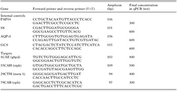
Real-time quantitative PCR (qPCR) using SYBR Green Supermix (Bio-Rad) was used to confirm the differential expression of 4 of the identified clones (further called targets). The cDNA synthesis and qPCR was performed following the protocol described by Decuypere et al. (Reference Decuypere, Rijal, Yardley, De Doncker, Laurent, Khanal, Chappuis and Dujardin2005a), and using an I-cycler (Bio-Rad). The following genes were analysed in parallel with the targets and used as internal expression controls (further called internal controls) for normalization: S8 (internal control used by Coulson et al. Reference Coulson, Connor, Chen and Ajioka1996), PAP14, AQP1, GCS (genes used by Decuypere et al. Reference Decuypere, Rijal, Yardley, De Doncker, Laurent, Khanal, Chappuis and Dujardin2005a, for L. (L.) donovani); primers used for their amplification and for the amplification of the transcripts selected in the present study are listed in Table 2. All the reactions were done in triplicate using a negative control of cDNA synthesis (i.e. without reverse transcriptase) and non-template controls. The analysis of the quantity data was performed using the geNorm VBA applet for MS Excel developed by Vandesompele et al. (Reference Vandesompele, De Preter, Pattyn, Poppe, Van Roy, De Paepe and Spelemen2002) and applied by Decuypere et al. (Reference Decuypere, Rijal, Yardley, De Doncker, Laurent, Khanal, Chappuis and Dujardin2005a) for quantification of gene expression in L. (L.) donovani. The raw (non-normalized) expression levels were determined with the delta Ct method, more specifically the Ct value of a gene for a sample was related to the Ct value of the same gene in the sample with the highest expression (or lowest Ct value), taking the amplification efficiency of the PCR for that gene into account. The geNorm VBA applet for MS Excel was used to determine the most stably expressed genes from the set of 8 tested genes (4 targets and 4 internal controls) in a given sample panel and were subsequently used to determine the normalization factor for each sample as described by Vandesompele et al. (Reference Vandesompele, De Preter, Pattyn, Poppe, Van Roy, De Paepe and Spelemen2002). The relative (normalized) expression levels were obtained by dividing the raw expression levels with the given normalization factor. The 95% confidence intervals (CI) were based on quantitative data collected from 3 repeated experiments that included cDNA-synthesis and quantitative PCR and were calculated on log transformed normalized expression levels, using the T.INV function in MS Excel to calculate the critical t value [CI=mean DPF +/− s.e.m.*t, with s.e.m.=standard error of the mean, and t=critical t value=T.INV (0·05, degrees of freedom)] (Motulsky, Reference Motulsky1995).
Primers used in qRT-PCR

RESULTS
Features of the biological material used for DD analysis
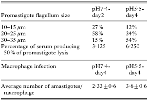
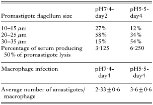
Procyclic and metacyclic promastigotes are reported to differ in the size of their flagellum, their susceptibility to complement lysis and infectivity in vitro and in vivo (Bates and Tetley, Reference Bates and Tetley1993; Zakai et al. Reference Zakai, Chance and Bates1998). These parameters were measured during the in vitro growth of L. (V.) braziliensis LC2043 cl8 in order to monitor the enrichment in metacyclics in pH5·5/day4 promastigotes (Table 3). First, at the morphological level we observed a clear shift of the flagellum distribution size between pH7·4/day-2 and pH5·5/day-4 promastigotes: from a higher percentage around 20–25 μm to 30–35 μm. Secondly, pH5·5/day-4 promastigotes showed a 2-fold increase in their resistance to complement lysis. Thirdly, pH5·5/day-4 promastigotes were found to be significantly (P<0·05) more infective to macrophages than pH7·4/day-4 ones: a higher percentage of infected macrophages and a higher average number of amastigotes per macrophage (72 h post-infection). Both preparations obviously remained a mixture of stages (promastigotes at stationary growth phase: procyclics and metacyclics). However, the pH5·5/day-4 promastigotes were clearly more enriched in metacyclics and suited to screen potential markers of this life stage.
Biological features of the parasite material used in DD analysis
Differential display analysis
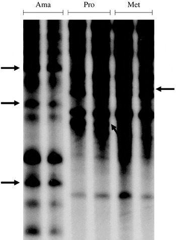
One of the most critical aspects of DD analysis is the reproducibility during cDNA synthesis and subsequent PCR amplifications. It was necessary to ensure that the observed differences in cDNA pattern between samples from different life stages were due to differences in gene expression and not to variations in the RNA preparation or to PCR artifacts. Therefore, we worked with at least 4 different RNA extractions from different biological batches and tested different RNA concentrations for each sample. Fragments were selected for analysis, only if differential expression was observed in all the repeats of a given life stage. One example of differential cDNA patterns between procyclics, metacyclics and amastigotes is shown in Fig. 1. The product specificity was demonstrated by changing 1 of the random primers, which resulted in a different pattern (data not shown). To verify the efficiency of the DNAse treatment of the RNA, non-reverse transcribed RNA samples from the different life stages were incorporated. As expected, no products were observed following the electrophoresis and autoradiography (data not shown).
DD analysis of Leishmania (Viannia) braziliensis: example of stage-specific fragments with primer ApoT12CC-EcoRI (arrows). Ama, amastigotes; Pro, procyclics; Met, metacyclics.

Exploitation of the DD analysis
Comparative analysis of the DD patterns revealed that pH7·4/day2 and pH5·5/day4 promastigotes shared about 90% of amplified cDNA fragments mRNAs that were expressed at similar levels. In contrast, our amastigote preparation shared only about 50% of their cDNA fragments with promastigotes. Of course, the latter percentage has to be taken with caution because of the presence of macrophage contaminants (see below).
We isolated a total of 214 DNA fragments (i) 178 differentially expressed between the 3 biological samples (isolated from pH7·4/day2, pH5·5/day4 promastigotes or amastigotes) and (ii) 36 similarly expressed in all the life stages as negative controls. After the hybridization screening with genomic DNA of L. (V.) braziliensis LC2043cl8, 120 fragments were marked as Leishmania-specific and selected for further analysis (56%). Of these, 48 DNA fragments gave good sequencing data: 7 isolated from pH7·4/day2 promastigotes, 6 from pH5·5/day4 promastigotes, 23 from amastigotes and 12 controls.
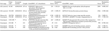
After the analysis of the 48 samples using the GeneDB Data Base for L. (V.) braziliensis, we found only 11 sequences with high Score and low Probability P(N) value (<10e-15), 4 of them corresponding to known proteins. When we performed the analysis using GeneDB from L. (L.) major, 40 sequences showed high scores and low P(N) values (<10e-5), from this group only 7 corresponded to known proteins, 3 of them being similar to the ones found for L.(V.) braziliensis. Interestingly, 1 fragment was identified as a portion of the ribosomal RNA gene of L. (V.) naiffi (Rotureau et al. Reference Rotureau, Ravel, Couppie, Pratlong, Nacher, Dedet and Carme2006). Among the 8 remaining sequences, 5 were identified by BlastN analysis as mouse sequences (all coming from amastigote preparations) and were likely to be contaminants from macrophages. The last 3 sequences did not show significant match. Table 4 presents only the hits corresponding to identified genes, but the complete list is available on request to the authors.
Identification of Leishmania-specific fragments isolated in L. (V.) braziliensis by DD analysis and showing highest score values

* In annotation process.
Validation of the gene expression profile by qPCR
Four DNA fragments were selected for qPCR verification of their expression profile in the 3 biological samples (Table 5): they showed a high score and low P(N) value after the GeneDB analysis for L. (V.) braziliensis and/or L. (L.) major, corresponded to known genes and had an appropriate size (more than 200 nt) for primer design for the qPCR analysis. These fragments corresponded to (i) the infective insect stage-specific protein (meta1), isolated from pH5·5/day4 promastigotes (thus putative marker of metacyclics), (ii) the oligopeptidase b (opb), isolated from amastigotes and (iii) the glucose-6-phosphate dehydrogenase (g6pd) and serine/threonine protein kinase (stpk), both identified in all life stages. In the case of the meta1 gene, expression was shown by qPCR to be highest in pH5·5/day4 promastigotes: up to 8 and 12 times higher than in pH7·4/day2 promastigotes (depending on the preparation, which was likely reflecting the proportion of metacyclics) and amastigotes respectively. With respect to the opd gene, highest expression was encountered in amastigotes: up to 1·6 and 2·5 times higher than in pH7·4/day2 and pH5·5/day4 promastigotes, respectively. The g6pd and stpk transcripts showed a similar expression in all samples.
Q-RT-PCR normalized expression level of the candidate markers (with standard-deviations) obtained from DD analysis
(Four genes were selected: 2 controls (g6pd, glucose-6-phosphate dehydrogenase and stpk, a serine/threonine protein kinase) and 2 differentially expressed (meta1, candidate marker of metacyclics and opb, oligopeptidase b, candidate marker of amastigotes); in bold, higher expression levels observed in pH5·5/day4 promastigotes and amastigotes respectively.)

DISCUSSION
In the present work, we applied differential display analysis (DD analysis) for the identification and characterization of mRNA transcripts differentially expressed in different life stages of a L. (V.) braziliensis strain, with particular attention to the infective stages, metacyclics and amastigotes. Fourty-eight fragments gave good sequencing data: among them, (i) 6 were isolated from pH5·5/day4 promastigotes and thus constituted candidate markers of metacyclics and (ii) 23 were candidate markers of amastigotes. Eight sequences only were identified to known genes.
DD analysis is one of the methods that can be used for comparing large numbers of mRNA between 2 samples. It has been validated in different organisms and, among others, allowed identification of genes preferentially expressed in amastigotes of L. (L.) major (Ouakad et al. Reference Ouakad, Chenik, Achour, Louzir and Dellagi2007) as well as a putative virulence factor (Ben Achour et al. Reference Ben Achour, Chenik, Louzir and Dellagi2002). This approach is complementary to other gene expression methodological approaches like suppression subtractive hybridization (SSH, Diatchenko et al. Reference Diatchenko, Lau, Campbell, Chenchik, Moqadam, Huang, Lukyanov, Lukyanov, Gurskaya, Sverdlov and Siebert1996) or micro-arrays (Saxena et al. Reference Saxena, Worthey, Yan, Leland, Stuart and Myler2003; Almeida et al. Reference Almeida, Gilmartin, McCann, Norrish, Ivens, Lawson, Levick, Smith, Dyall, Vetrie, Freeman, Coulson, Sampaio, Schneider and Blackwell2004). In our hands, the use of SSH in Leishmania was seriously hampered by background caused by the presence of large amounts of poly-adenylated ribosomal RNA (Decuypere et al. Reference Decuypere, Vandesompele, Yardley, De Doncker, Laurent, Rijal, Llanos-Cuentas, Chappuis, Arevalo and Dujardin2005b). This was likely not the problem with DD analysis, as out of 48 sequenced fragments, only 1 corresponded to rRNA. With respect to micro-arrays, this method allowed, for instance, identification of differentially expressed genes in L. (L.) major (Almeida et al. Reference Almeida, Gilmartin, McCann, Norrish, Ivens, Lawson, Levick, Smith, Dyall, Vetrie, Freeman, Coulson, Sampaio, Schneider and Blackwell2004; Leifso et al. Reference Leifso, Cohen-Freue, Dogra, Murria and McMaster2007), in L. (L.) mexicana (Holzer et al. Reference Holzer, McMaster and Forney2006) and in L. (L.) donovani (Saxena et al. Reference Saxena, Lahav, Holland, Aggarwal, Anupama, Huang, Volpin, Myler and Zilberstein2007). However, this method requires sophisticated equipment and is therefore not as readily available as DD analysis. In addition, micro-arrays currently exist only for L. (L.) major, L. (L.) donovani and L. (L.) infantum and it is likely that a different micro-array would ideally need to be developed for each species.
Whatever the method used for differential expression analysis, the crucial issue is not only the molecular method, but also the biological quality of the material used for mRNA extraction. In the present study, pH5·5/day-4 promastigotes were used as proxy of metacyclics on the base of morphological and functional changes compared with pH7·4/day-2 promastigotes, but we are aware that pH7·4/day-2 and pH5·5/day-4 samples both contain metacyclics, the second one being relatively enriched in this infective stage. This does not disqualify the method used here, nor the candidate markers of metacyclics found using this method, but a qualitatively and quantitatively more precise gene expression profiling would require the use of purified stages. With respect to the amastigotes, our experimental approach was based on in vitro intracellular stages and we observed a 50% difference in DD-patterns by comparison with promastigotes. After correction for macrophage contaminants, we estimate that this value should drop by up to 23% (0·5 multiplied by 0·56 – proportion of L. (V.) braziliensis specific sequences as shown by hybridization – multiplied by 0·83 – proportion of Leishmania-specific DNA as shown by sequencing). This value is higher than that reported using micro-arrays and axenic amastigotes (3% differential gene, Saxena et al. Reference Saxena, Lahav, Holland, Aggarwal, Anupama, Huang, Volpin, Myler and Zilberstein2007) and the discrepancy could be due to the nature of the biological material. This is supported by micro-array studies that revealed 17 times more differences in gene expression between intracellular amastigotes and promastigotes than between axenics and promastigotes (Holzer et al. Reference Holzer, McMaster and Forney2006). However, the values observed in our study are also higher than those observed after micro-array analysis of lesion-derived L. (L.) major amastigotes (1·4%, Leifso et al. Reference Leifso, Cohen-Freue, Dogra, Murria and McMaster2007). We cannot exclude Leishmania species-related differences, but these discrepancies also point out the fundamental differences between DD-analysis and micro-array, such as the extent of genome coverage, the sensitivity and the risk with DD-analysis to detect several fragments corresponding to a same transcript. Altogether, particular care should be taken in the interpretation of our DD-analysis results as well as the comparison with other reports made on other species, in biologically different experimental conditions and with other molecular methods.
With regard to the high frequency of false positive results with DD analysis, confirmation of the expression profiles needed to be performed with a more robust technique for relative quantification of transcripts. This was done here by quantitative reverse transcription PCR, for 4 candidate markers: 2 differentially expressed in DD analysis and 2 controls. (1) The transcripts corresponding to the infective-insect stage protein (also known as the Meta 1 protein) were identified as candidate markers of metacylics. This gene was first identified by Coulson and Smith (Reference Coulson and Smith1990) in L. (L.) major, and then in L. (L.) donovani and L. (L.) amazonensis by Uliana et al. (Reference Uliana, Goyal, Freymüller and Smith1999). This gene is predominantly expressed in infective metacyclics of L. (L.) major (Nourbakhsh et al. Reference Nourbakhsh, Uliana and Smith1996) and over-expression of corresponding Meta 1 protein in L. (L.) amazonensis generates parasites that are more virulent than wild-type organisms in vivo (Uliana et al. Reference Uliana, Goyal, Freymüller and Smith1999). Our results support the hypothesis that the product of this gene could be an important virulence factor in parasites of the subgenus Viannia. (2) The transcripts corresponding to the oligopeptidase B were identified as candidate markers of amastigotes. This is a serine oligopeptidase, a member of the prolyl oligopeptidase family (Morty et al. Reference Morty, Lonsdale-Eccles, Morehead, Caler, Mentele, Auerswald, Coetzer, Andrews and Burleigh1999), involved in (i) Ca(2+)-signalling during host cell invasion by Trypanosoma cruzi (Burleigh et al. Reference Burleigh, Caler, Webster and Andrews1997; Caler et al. Reference Caler, Vaena, Haynes, Andrews and Burleigh1998) and (ii) in degradation of regulatory peptide hormones in the blood of infected hosts in African tryponosomiasis (Troeber et al. Reference Troeber, Pike, Morty, Berry, Coetzer and Lonsdale-Eccles1996). So far, it has not been reported as a potential virulence factor in leishmania. (3) Transcripts corresponding to a serine/threonine protein kinase were likely expressed at the same level in the different biological preparations. Protein kinases constitute a large family of important regulators of many different cellular processes such as transcriptional control, cell cycle progression and differentiation (Naula et al. Reference Naula, Parsons and Mottram2005). A large number of protein kinases (179) were identified in Leishmania (Naula et al. Reference Naula, Parsons and Mottram2005), some of them being exclusively expressed in a given stage (f.i. LmxMKK in the promastigote stage, Wiese et al. Reference Wiese, Kuhn and Grunfelder2003). For others (like CRK3), mRNA was found to be constitutively expressed throughout the parasite life-cycle (Wang et al. Reference Wang, Dimitrov, Garrity, Sazer and Beverly1998). (4) The transcripts corresponding to the glucose-6-phosphate dehydrogenase were also found to be constitutively expressed in the strain here analysed. A proteomic study of L. (V.) panamensis revealed that this protein was more abundant in amastigotes (Walker et al. Reference Walker, Vasquez, Gomez, Drummelsmith, Burchmore, Girard and Ouellette2006). This protein could be another example of post-transcriptional regulation mechanisms which are common in Leishmania and other Trypanosomatids (Stiles et al. Reference Stiles, Hicock, Shah and Meade1999). Noteworthy, this is likely not the case for all genes of the parasite: there was for instance 53% correlation between L. infantum amastigote-specific protein isoforms and mRNA expression trends (McNicoll et al. Reference McNicoll, Drummelsmith, Müller, Madore, Boilard, Ouellette and Papadopoulou2006). In a screening phase, transcript levels would be enough to identify potential markers, but particular care should be taken when interpreting their biological meaning and this should be completed by functional studies, including analyses at the protein level.
The present study demonstrates the feasibility of DD screening for transcripts coming from parasites obtained at different life stages and provided a set of potential markers of the infective stages of L. (V.) braziliensis that might be used as references when monitoring gene expression in that species. Further work is needed to validate our markers in other strains of that species and in related less-pathogenic species like L. (V.) peruviana. Last but not least, it should be tested whether some of them are indeed virulence factors in L. (V.) braziliensis.
This study was funded by WHO-TDR, Research Training Grants (M8/181/4/G.261, ID 990815), UNESCO-L'OREAL fellowship for Women in Science 2003, EC (INCO-CT2005-015407), and by the Directorate-General for Development Cooperation (DGDC) of the Belgian Government (framework agreement 02, project 95501.