Introduction
Glomus jugulare tumours originate from paraganglionic chemoreceptor cells. They can occur in the middle ear or at other sites such as the temporal bone, neck and jugular vein. Most patients present with conductive deafness or pulsatile tinnitus, but there have been isolated reports of cases presenting with raised intracranial pressure leading to papilloedema.Reference Beck, Kassell and Drake1, Reference Angeli, Sato and Gantz2 Magnetic resonance venography was not reported in these cases.
We present the case of a man who became ophthalmically symptomatic just three weeks after embolisation and surgical resection of a left-sided glomus jugulare tumour. He was found to have established, haemorrhagic optic disc oedema. An urgent magnetic resonance venography scan confirmed the presence of cerebral venous sinus thrombosis in the left lateral sinus. It is feasible that this was affecting the drainage of the venous sinus network, leading to raised intracranial pressure and papilloedema.
Post-operative hydrocephalus and benign intracranial hypertension have been documented after skull base surgery. To the best of our knowledge, the present case represents the first report of magnetic resonance venography confirmed cerebral venous sinus thrombosis occurring post-operatively in a symptomatic patient following glomus jugulare tumour removal.
Patients undergoing surgery involving resection or manipulation of the internal jugular vein may be at higher risk of developing thrombosis superior to the level of resection. If this affects drainage of the venous sinus network, it may cause raised intracranial pressure and papilloedema. Our case highlights the importance of careful post-operative management, and the diagnostic benefits of magnetic resonance venography, in such cases.
Case
A forty-two-year-old heavy goods vehicle driver was referred to the ophthalmology casualty clinic with blurred vision in both eyes. At the time, he was under the care of the otolaryngologists, having been diagnosed with a left-sided glomus jugulare tumour following a 12-month history of pulsatile tinnitus. He had undergone embolisation and surgical resection of the tumour (infratemporal type A approach) three weeks prior to referral. Carotid angiography prior to surgery had demonstrated a 2.2 × 2.0 × 2.7 cm, left skull base tumour with arterial supply from the retroauricular, ascending pharyngeal and occipital arteries. No major complications had been documented at the time of the procedure. However, within three days of discharge he had become aware of blurred vision, particularly in the left eye, associated with a mild headache. There was no history of visual obscuration, transient visual loss or other neurological features.
There was no relevant medical history of note, apart from an episode of right orbital cellulitis 15 years previously which had required surgical intervention, resulting in a right superior altitudinal field defect.
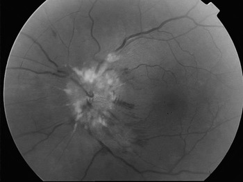
On examination, systemic blood pressure was recorded as 124/89 mmHg. Snellen visual acuity was recorded as 6/5 + 3 in the right eye and 6/18 in the left, improving to 6/9 with pinhole. No relative afferent pupillary defect was demonstrable. Colour vision testing with Ishihara plates revealed a reduction in the left eye (10/17 plates detected slowly, compared with 17/17 in the right eye). Cranial nerve assessment showed a left lower motor neurone facial nerve palsy and left sensorineural deafness. Corneal sensation was normal. The anterior segment examination was within normal limits. Fundoscopy revealed extensive bilateral optic disc oedema with surrounding exudates and haemorrhages (Figure 1). Humphrey visual field analysis (24–2) confirmed the presence of a superior altitudinal defect in the right eye and a few central defects in the left eye which did not align to any recognisable pattern.
Fundus photograph of left optic nerve, showing haemorrhagic optic disc oedema and exudates.

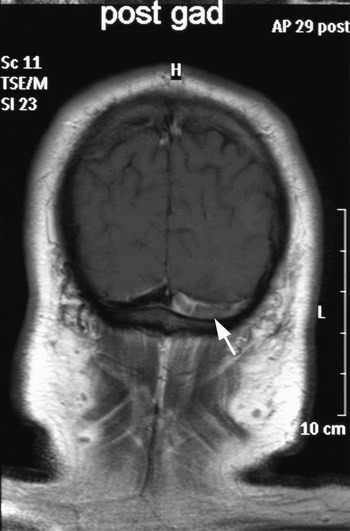
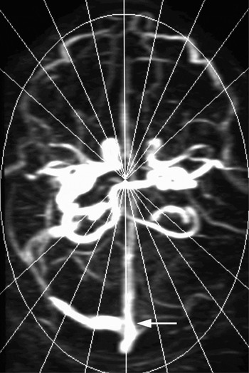
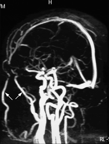
A computed tomography scan of the brain failed to show any ventriculomegaly or signs of raised intracranial pressure. Further imaging included magnetic resonance imaging (Figure 2) and magnetic resonance venography (Figure 3) of the dural sinuses, and revealed the presence of fresh thrombus within the left lateral sinus, which was occluded, and slight narrowing of the right proximal lateral sinus. Magnetic resonance venography showed the facial veins to be significantly dilated due to the obstruction (Figure 4). The superior sagittal sinus and the cavernous sinus appeared normal. It was proposed that the occluded lateral sinus was responsible for inadequate cerebrospinal fluid (CSF) drainage, leading to raised intracranial pressure and papilloedema.
Coronal magnetic resonance imaging scan; arrow highlights fresh thrombus within the left lateral sinus.

Axial magnetic resonance venography image. Arrow indicates obstruction of venous drainage through the left lateral sinus, which is occluded by thrombus.

MRV (saggital section). Arrows highlight dilated facial veins secondary to an obstruction within the venous sinus network.

The patient was promptly commenced on intravenous anticoagulation. He was later discharged home on oral anticoagulants (warfarin) for six months. A thrombophilia screen was not performed for our patient prior to commencing warfarin, but should have been considered to exclude any thrombophilic tendency. At the time of writing, he remained well and was being followed up regularly.
Discussion
The presence of glomus jugulare tumours is known to be associated with cerebral venous sinus thrombosis due to obstruction of the venous sinuses.Reference Bousser3 Treatment options include surgical excision and radiotherapy. Post-operative surgical complications include infection, hearing loss, new cranial nerve palsies and CSF leak. However, to the best of our knowledge there have been no previous reports of symptomatic cerebral venous sinus thrombosis occurring post-operatively following surgical excision of a glomus jugulare tumour.
• Glomus jugulare tumours originate from paraganglionic chemoreceptor cells. They can occur in the middle ear or at other sites such as the temporal bone, neck or jugular vein
• This paper describes the case of a male patient who became ophthalmically symptomatic just three weeks after embolisation and surgical resection of a left-sided glomus jugulare tumour
• Post-operative hydrocephalus and benign intracranial hypertension have been documented after skull base surgery
• Patients undergoing surgery involving resection or manipulation of the internal jugular vein may be at higher risk of developing thrombosis superior to the level of resection
A case of benign intracranial hypertension, also known as idiopathic intracranial hypertension, has been reported as a complication of glomus jugulare tumour surgery.Reference Brookes and Graham4 However, neither post-operative cerebral angiography nor magnetic resonance venography was reported in this case, and it is possible that there may have been undetected cerebral venous sinus thrombosis present.
A recent study by Lin et al. considered the overlap between the clinical presentation of idiopathic intracranial hypertension and cerebral venous sinus thrombosis.Reference Lin, Foroozan, Danesh-Meyer, De Salvo, Savino and Sergott5 In these authors' study of 106 patients with presumed idiopathic intracranial hypertension, 9.4 per cent had cerebral venous sinus thrombosis observed via magnetic resonance venography.
Our patient had no visual symptoms prior to undergoing surgical resection of a glomus jugulare tumour. He developed extensive papilloedema and visual deterioration within three weeks of surgery, secondary to extensive cerebral venous sinus thrombosis. This was diagnosed on magnetic resonance venography and treated with warfarin. It is possible that the reported cases of glomus jugulare tumours presenting with raised intracranial pressure, and the reported case of post-surgical benign intracranial hypertension, may have represented undiagnosed, coexisting cerebral venous sinus thrombosis.Reference Beck, Kassell and Drake1, Reference Angeli, Sato and Gantz2, Reference Brookes and Graham4
Patients undergoing surgery involving resection or manipulation of the internal jugular vein, as often occurs in glomus jugulare tumour surgery, are at an increased risk of developing thromboses superior to the level of resection.Reference Wustenberg, Offergeld, Zahnert, Huttenbrink and Kittner6 In the case of glomus jugulare tumours, in which both the presence of the tumour and the process of surgical resection predisposes to cerebral venous sinus thrombosis, particular attention must be given to early signs and symptoms of raised intracranial pressure. There should be a high index of clinical suspicion for cerebral venous sinus thrombosis, and a low threshold for performing magnetic resonance venography to diagnose this potentially life-threatening, yet treatable, condition.