
Introduction
Lanthanides possess long luminescent lifetimes with narrow emission bands in the visible and near-IR regions.[Reference Dang, Yu, Wang, Guo, Sun, Deng, Feng, Fan and Zhang1–Reference Ji, Wu, Song, Han, Wang, Liu, Guo and Zhao4] In contrast to organic fluorophores and quantum dots, lanthanides are known for their emission stability without either photobleaching or blinking. However, the luminescence intensities of lanthanides are relatively low. To enhance their luminescence, lanthanides are complexed with organic conjugate ligands, such as 2-thenoyl trifluoroacetone (tta) and 1,10-phenanthroline (phen). These ligands serve as “antenna” molecules which improve luminescence intensity.[Reference Liu, Wang, Yu and You5–Reference Yang and Feng8] By this strategy, lanthanide complexes can be prepared with outstanding optical, electronic, and catalytic properties. In particular, the optical properties of luminescent lanthanide complexes make them excellent candidates for hybrid functional materials for artificial lighting, lasers, electro-luminescence devices, biosensors, and large flat panel displays.[Reference Binnemans9] However, these organic complexes usually have poor water solubility and are incompatible with aqueous biologic environments. Therefore, there is interest in improving the chemical characteristics by modifying the coordination compounds used to form luminescent lanthanide complexes.[Reference Boissé, Rieger, Belal, Dicicco, Beaunier, Li and Charleux10–Reference Fang, Wang, Hu, Wang, Zhou, Gao and Liu12]
To take advantage of the excellent luminescence properties of lanthanide complexes for biomedical applications, such as fluorescent labels and biosensors, the complexes must be made compatible with aqueous environments.[Reference Jha and Chandra13] Polyelectrolytes with opposite charges can combine to form stable, water-soluble complexes.[Reference Tsuchida, Sou, Nakagawa, Sakai, Komatsu and Kobayashi14] When a polyanion and polycation are combined under favorable conditions (at non-stoichiometric charge ratios) colloidal nanoparticles can be formed. These nanoparticles which can improve the efficiency of lanthanide complex stabilization, are stabilized by surface charges from the polyelectrolyte in excess. This results in the enhancement of Eu3+ complex luminescence as the chitosan (CHI) content is increased. Biocompatible polyelectrolytes can be combined to form polyelectrolyte complexes that stabilize and deliver fragile bioactive moieties to cells.[Reference Volpato, Almodóvar, Erickson, Popat, Migliaresi and Kipper15, Reference Place, Sekyi and Kipper16]
In this study, our goal is to demonstrate that polyelectrolyte complex nanoparticles can be formed from biocompatible polysaccharides, to compatibilize lanthanide complexes with aqueous environments. The photoluminescent Eu3+–ligand complexes using the ligands 2-tta and 1,10-phen, are dispersed homogeneously in aqueous solution by the association with water-soluble hyaluronic acid (HA). Furthermore, crystallization of the Eu3+–tta–phen aggregates is observed in HA–Eu3+–tta–phen complexes. CHI–HA–Eu3+–tta–phen luminescent nanoparticles are obtained by further complexing the HA–Eu3+–tta–phen with CHI. We report the initial characterization of the structural and luminescent properties of these lanthanide-induced polysaccharide aggregates (LIPAs) containing Eu3+–tta–phen complexes. The Eu3+ can serve as the central multivalent ion to induce the negatively charged carboxylic acid groups in HA to form into nanoparticles. The water solubility of HA enables the nanoparticles to be homogeneously dispersed in aqueous solution. Moreover, if positively charged CHI is co-added into HA–Eu3+–tta–phen solution, nanoparticles can be formed via charge reactions between the two polyelectrolytes. The size of nanoparticles and their luminescence properties are reported here as a function of the mixing ratio of CHI-to-HA. This comprehensive structural and fluorescence characterization supports the eventual development of functional nanobiomaterials with biologic and blood compatibility.[Reference Liu, Yuan, Li, Tang, Wang, Chen, Chen and Brash17, Reference Chanphai and Tajmir-Riahi18]
Experimental
Materials
Chitosan and hyaluronan (HA) were purchased from Aladdin Company in Shanghai. Europium oxide, hydrochloric acid, tta, and phen were bought from Darui Company (Shanghai, China). The solvents including the acetate buffer and alcohol were analytical grade and were used without purification.
Characterization
Transmission electron microscopy (TEM) was performed with a JEM-1200EX (JEOL Ltd, Japan). The samples of LIPAs were ultrasonicated for 5–6 min before testing. The solution was dropped onto a copper grid, and the solvent (ethanol) was evaporated at room temperature for 1 or 2 s.
The dispersion of Eu3+ ions in LIPAs and the nanostructures were characterized by high-resolution transmission electronic microscope (JEM 2100F). The sample preparation was the same as TEM test.
The zeta potentials of the Eu3+ complexes, HA–Eu3+ complexes, and CHI–HA–Eu3+ polyelectrolyte complex nanoparticles were measured by a Malvern Zetasizer NanoZS90. Each sample was measured three times at room temperature and mean zeta potentials are reported for each sample.
The size distribution images of CHI–HA–Eu3+ polyelectrolyte nanoparticles were obtained by using Dynamic light scattering (Dynapro Tian TC). The measurement of 2 mL sample was conducted for 60 s. Each sample was tested for three times.
The photoluminescence spectra were obtained at room temperature using a Cary Eclipse fluorescence spectrophotometer (Varian, USA) equipped with a 75 kW xenon lamp as the excitation source. The excitation slit width and emission slit width were both 5 nm.
Preparation of CHI and HA solution
The CHI and HA solutions were obtained via dissolving the polyelectrolytes in acetate buffer. Polymer solutions (0.9 mg/mL for CHI and 1 mg/mL for HA) were prepared using 0.1 M, pH 5.0 acetate buffer. (sodium acetate and acetic acid). At pH 5.0 HA has sufficient carboxylate groups to form electrostatic complexes with Eu3+ ions.
Preparation of Eu3+ chloride solution and Eu3+–tta–phen complexes
Europium oxide was reacted with hydrochloric acid to form europium chloride. Europium oxide (0.88 g) and excess hydrochloric acid (6 mol/L, 2 mL) aqueous solution were mixed together by stirring until the solid was completely dissolved, the solutions were distilled at 50 °C for about 10 h for form transparent crystals of EuCl3■6H2O. Then the crystals were dissolved in ethanol to obtain a 0.01 mol/L solution.
Charge reaction between HA–Eu3+ complexes
The amounts of the reagents used to prepare different samples are shown in Table I. The complexation of Eu3+–tta–phen with HA was obtained as follows. Luminescent Eu3+–tta–phen complexes were formed by previously reported methods.[Reference Rajamouli and Sivakumar19] Then HA–Eu3+–tta–phen samples were prepared at room temperature by dropwise titration method using Eu3+ complexes as the starting solution and dropping the negatively charged HA dissolved in the acetate buffer while stirring constantly at 1000 rpm, for 2 h. Eu3+ and HA complexed in solution to produce uniform HA–Eu3+–tta–phen complexes.
The amounts of reagents and the concentration of Eu3+ in each charge reaction.

a The amounts of europium chloride, tta, and phen added to the reaction.
b The final concentration of Eu3+ that was calculated as follows:.
The volume of Eu3+, tta, phen HA, or ethanol added to the reaction was 300 µl, 300 µl, 300 µl, 2 mL, respectively. The concentration of each Eu3+, tta, and phen was 0.01 mol/L. The mount of Eu3+ was 3 × 10⁻⁶ mol, Total solution volume was (300 µl × 3 + 2 mL). So, the final concentration of Eu3+ (3 × 10⁻⁶ mol/300 µl × 3 + 2 mL) was 1.03 × 10⁻³ mol/L.
Formation of Eu-containing polyelectrolyte complexes
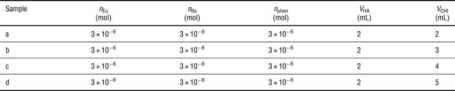
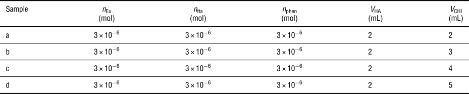
HA–Eu3+–tta–phen complexes were added to the CHI solution, to form polyelectrolyte complex nanoparticles. The solution amounts used to prepare different samples are shown in Table II. The nanoparticles form by complexation between the positive charges in CHI and the residual negative charges on HA. The morphology of the polyelectrolyte complex nanoparticles formed from different proportions of the HA and CHI are evaluated.
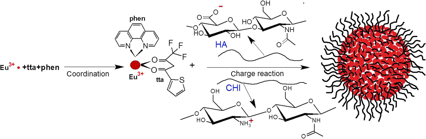
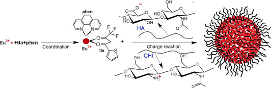
The compound principle of polyelectrolyte complex formation, containing Eu3+ complexes is shown in Scheme 1.
Formation of polyelectrolyte complexes containing Eu–tta–phen, HA, and CHI.

Results and discussion
Zeta potential measurement
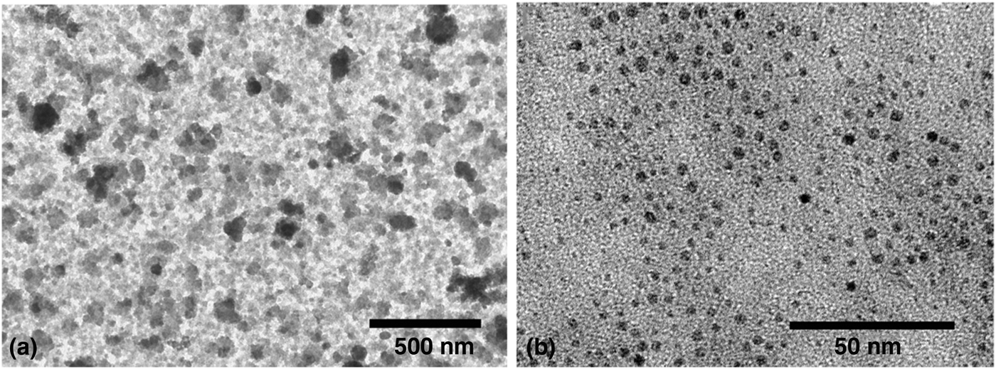
The zeta potential values of each sample verify that the HA–Eu3+–tta–phen complexes, and CHI–HA–Eu3+–tta–phen nanoparticles are formed by electrostatic complexation. The zeta potential values are 2.43 mV, −13.2 mV, 11.5 mV, corresponding to the Eu3+–tta–phen complexes, HA–Eu3+–tta–phen complexes, and CHI–HA–Eu3+–tta–phen polyelectrolyte complexes, respectively. The zeta potential value of Eu3+–tta–phen complexes suggests that the Eu3+–tta–phen complex is prone to aggregate. This is also confirmed by the TEM image of Eu3+–tta–phen complexes adsorbed onto the copper TEM grids, as seen in Fig. 1(a). The aggregation of coordinated Eu3+–tta–phen complexes reduced when these are complexed to HA.
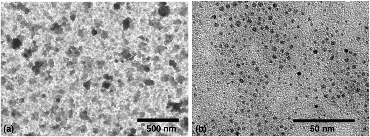
Morphological structures of Eu3+–tta–phen complexes and HA–Eu3+–tta–phen complexes
Transmission electron micrographs are shown in Fig. 1. The Eu3+–tta–phen complexes adhere to each other tightly by forming irregularly shaped aggregates, observed as cloudy suspensions in Fig. 1(a). HA–Eu3+–tta–phen complexes are colloidally stable particles with diameters of about 3–6 nm in Fig. 1(b). The high contrast of these particles in electron microscopy can be attributed to the formation of a densely packed structure consisting of Eu3+–tta–phen complexes. Eu3+–tta–phen complexes serve as a multivalent cross-linker to coordinate the charged carboxylic acid groups between macromolecular chains of HA. The Eu3+–tta–phen complexes that are dispersed homogeneously is a prerequisite for the formation of HA–Eu3+–tta–phen complexes with uniform distribution.[Reference Zhao, Huang, Wang, Tang, Wang, Liu, Belfiore and Kipper20]
Structure of CHI–HA–Eu3+-tta-phen complex nanoparticles
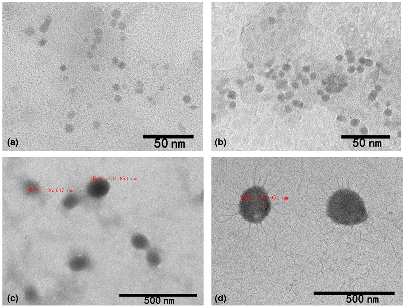
Solutions of Eu3+–tta–phen–HA complexes were added to the CHI solution to form polyelectrolyte complex nanoparticles. The morphology of the polyelectrolyte complexes can be seen in Fig. 2. The diameter of the nanoparticles ranges from tens to hundreds of nanometers. The complexation results in spherical core-corona particles containing collapsed HA–CHI complexes in the core, surrounded by a corona of excess CHI.
TEM images of CHI–HA–Eu3+–tta–phen polyelectrolyte complex nanoparticles (sample a, b, c, d in Table II).
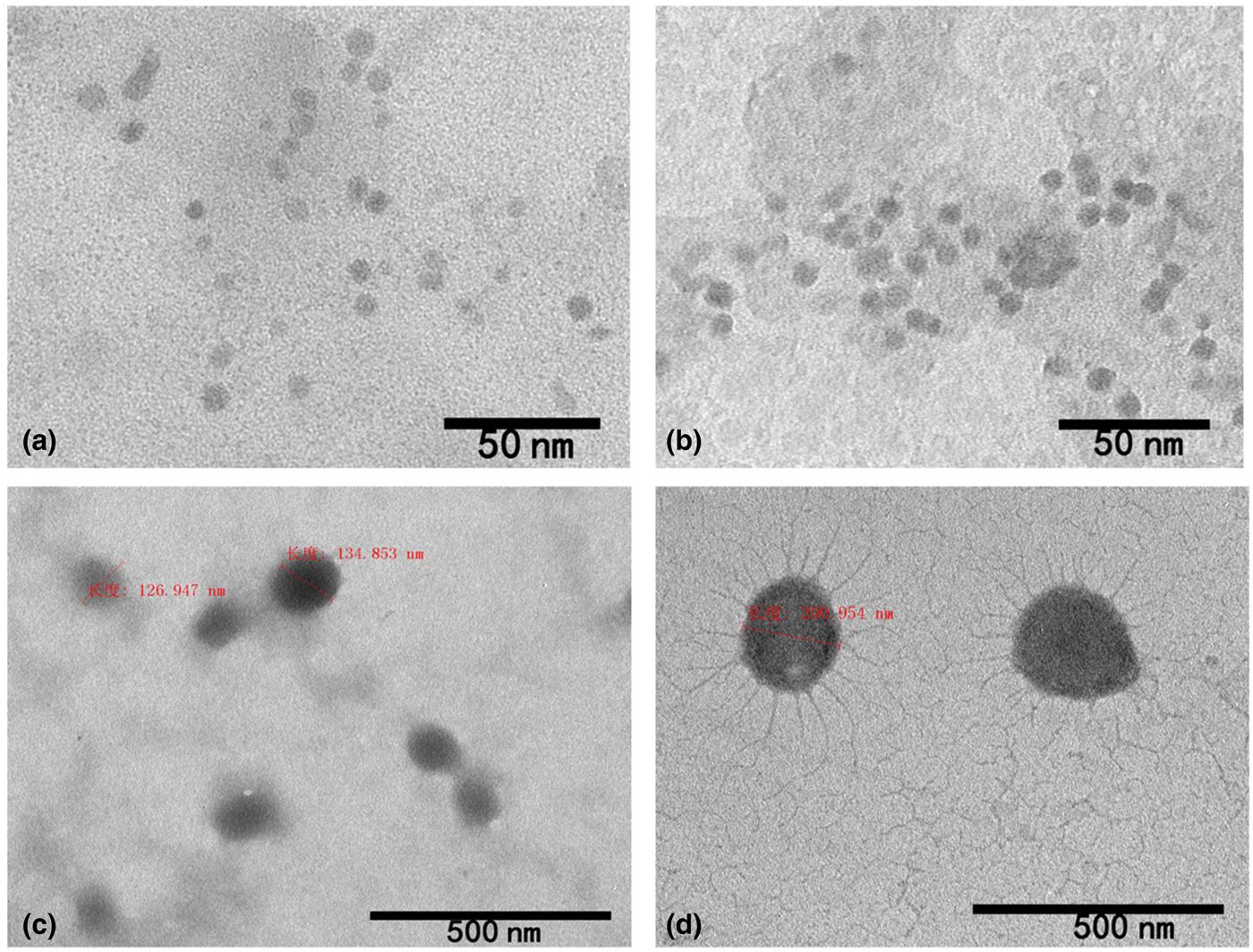
Figure 2(a) shows the nanoparticles formed from with the composition a in Table II. Which are the smallest among the four different compositions. There are residual Eu3+ complexes around the nanoparticles. As the amount of CHI is increased (samples b, c, and d), the size of the nanoparticles is correspondingly increased, and at the same time, the core of the nanoparticles becomes gradually darker. This can be attributed to the higher segregation of Eu3+–tta–phen complexes in the particle cores as the amount of CHI is increased. Finally in the case of sample d, after reaction with negatively charged HA, the CHI polymer chains with excess positive charges dangle outside of the particles under the interfacial tension between the particles and the solvent, as seen in Fig. 2(d). The size of CHI chains depends on the charge density as well as ionic strength.
The amounts of reagents and the mixing ratios of HA and CHI in the charge reaction.
There are several factors that determine the morphology of polyelectrolyte complex nanoparticles. The size can be affected by the stretched polymer segments that participated in the core, and also the interfacial tension between the core and the solvent phases.[Reference Xu, Tang, Wang, Liu, Wang, Huang, Huang, Wang, Shen and Belfiore21–Reference Müller-Buschbaum, Hermsdorf, Roth, Wiedersich, Cunis and Gehrke24] Size of polyelectrolyte complex nanoparticles can also be tuned through control of charge mixing ratio, charge density, and the concentration of small molecular weight counter ions in the solution systems. Boddohi et al. also have demonstrated that polyelectrolyte complexes of CHI and HA become larger when the amount of CHI is increased, as observed here.[Reference Boddohi, Moore, Johnson and Kipper25]
Photoluminescent properties of Eu3+–tta–phen complexes, HA–Eu3+–tta–phen complexes, and CHI–HA–Eu3+–tta–phen polyelectrolyte complex nanoparticles
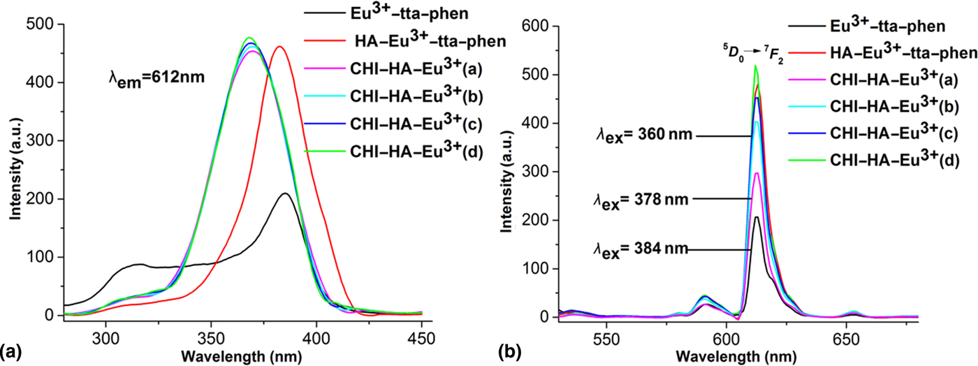
The optical properties of the three complexes containing Eu3+ were measured by fluorescence spectrophotometry. The photoluminescence intensity is a function of the CHI–HA composition, as shown in Fig. 3.
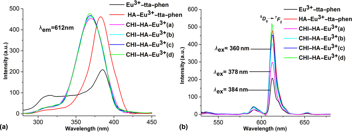
The excitation peaks of six different compositions are at 384 nm, 378 nm, 360 nm, for samples a, b, from Table I (shown in Fig. 1), and samples a–d from Table II (shown in Fig. 2), respectively. The characteristic emission band of Eu3+–tta–phen complexes, corresponding to the 5D 0→7F 2 transition is observed at 612 nm for all four samples [Fig. 3(b)].[Reference Lapina, Pavich and Pershukevich26] The intensity of this band increases along with amounts of CHI in the nanoparticles. This result can be attributed to the favorable circumstance of the chemical coordination for Eu3+–tta–phen complexes,[Reference Gupta, Parbhakar, Behera and Hussain27] in which the coordination of the Eu3+–tta–phen complexes increases with increasing CHI composition. This result is consistent with the TEM images (in Fig. 2) showing more electron density contrast in the particles as the CHI content increases. Oppositely, a large amount of Eu3+ exists outside of the polyelectrolyte complexes as residuals in sample a, which display relatively weaker luminescence. This could be attributed to the coordination number of ligands to Eu3+ in the Eu3+–tta–phen complexes being not fully satisfied.[Reference Wang, Tang, Wang, Shen, Wang, Liu, Huang, Jiao and Song28] As the CHI composition increases from samples a to d, the uncomplexed residual Eu3+ is reduced. The increased association of the Eu3+–tta–phen with the polysaccharides results in the enhancement of the lanthanide photoluminescence.
Luminescent lifetime and fluorescence quantum yield
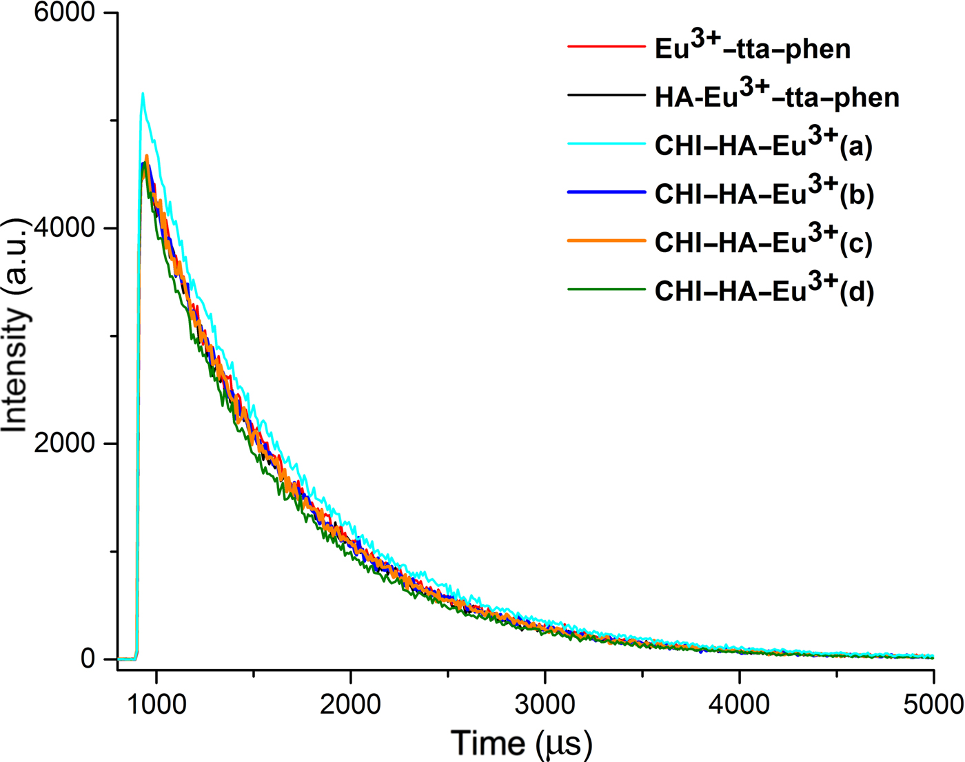
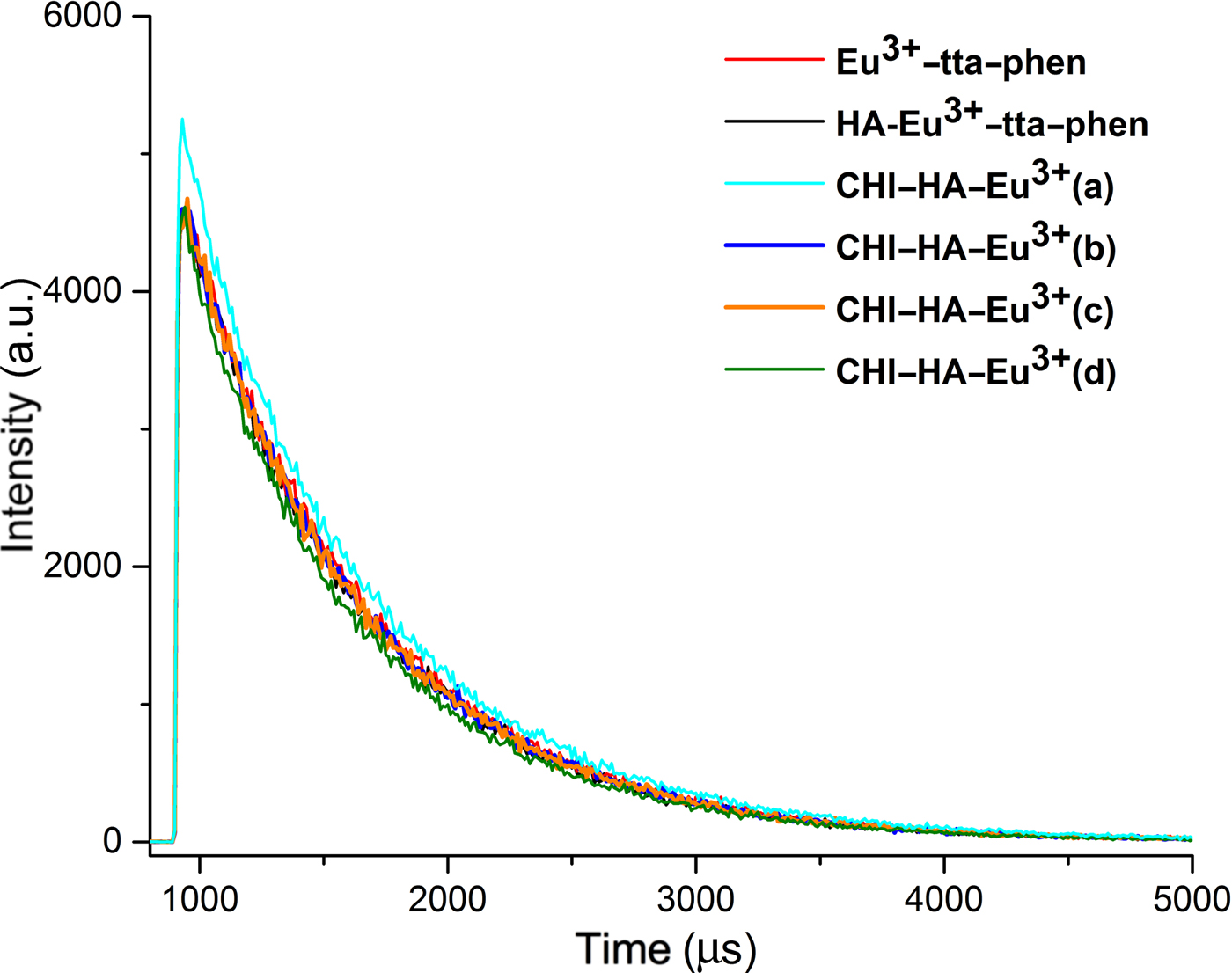
The luminescent lifetime is an important parameter to characterize the fluorescent nanoparticles. The luminescent lifetime of Eu3+–tta–phen, HA–Eu3+–tta–phen, and CHI–HA–Eu3+–tta–phen complexes were obtained at room temperature as shown in Fig. 4. The luminescent lifetimes of six different compositions are at 754.24, 735.46, 789.04, 756.84, 749.92, and 724.42 µs, corresponding to the samples of a, b, from Table I (shown in Fig. 1) and a–d from Table II (shown in Fig. 2), respectively. This demonstrates the luminescent lifetime declined as the nanoparticles increased. The longest luminescent lifetime of CHI–HA–Eu3+ (789.04 µs) is longer than the pure Eu3+–tta–phen complexes (754.24 µs).
The fluorescence quantum yields of six different compositions are 71.27, 83.88, 69.81, 79.89, 80.88, and 87.28%, regarding the six samples mentioned above. We can find that the fluorescence quantum yields ascended as the nanoparticles increased. The maximum increased ratio of quantum yield is 22.46%.
Conclusion
In this work, photoluminescent Eu3+–tta–phen complexes were dispersed homogeneously in aqueous solution by association with the polyanionic polysaccharide, HA. Polyelectrolyte complex nanoparticles are obtained by further complexing the HA–Eu3+–tta–phen with the polycationic polysaccharide CHI. Here, we demonstrate a novel series of luminescent polyelectrolyte complex nanoparticles and investigate how the luminescence, the luminescent lifetime and quantum yield, are affected by the composition and particle morphology. The CHI–HA–Eu3+–tta–phen nanoparticles become larger and more stable as the ratio of CHI-to-HA is increased. For the largest particles with highest charge density (formed with the greatest excess of CHI). CHI chains dangled outside of the nanoparticles. The lattice arises from crystallization of the Eu3+–tta–phen in HA–Eu3+–tta–phen complexes, which stabilize the lanthanide complex in the aqueous environment. These results can be extended to other biologically active polyelectrolytes and other lanthanide ions with different photoluminescence spectra such as Tb3+ (which emits green light) and La3+. The stable, water-soluble LIPA complexes will lead to advances in biologic applications, such as biosensing and bioimaging, where the luminescent rare-earth ion is protected or even enhanced through polyelectrolyte complexation in the aqueous biologic environment.
Supplementary material
The supplementary material for this article can be found at https://doi.org/10.1557/mrc.2018.107
Acknowledgments
This work was supported by (1) The Natural Scientific Foundation of China (Grant No. 51473082); (2) State Key Project of International Cooperation Research (2017YFE0108300, 2016YFE0110800); (3) The Program for Introducing Talents of Discipline to Universities (“111” plan); (4) The 1st class disciplines of Shandong Province; (5) The National One-Thousand Foreign Expert Program (Grant No. WQ20123700111); (6) Key Research and Development Plan of Shandong Province (Grant No. 2017GGX20112).