Neonates with hypoplastic left heart syndrome and an intact atrial septum are at the highest risk for mortality, and occasionally require emergent transcatheter atrial septoplasty after birth.Reference Rychik, Rome, Collins, DeCampli and Spray 1 , Reference Bernstein, Moore, Stanger and Silverman 2 The levoatriocardinal vein provides an alternative egress and plays an important role as a natural decompression pathway. We present two cases of neonates with unique levoatriocardinal veins that arose from the top of the left atrium and directly drained into the right atrium.
Case reports
Case 1
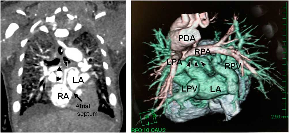
A 2.6 kg baby girl was born at 39 weeks’ gestation. Shortly after birth, she developed tachyponea and cyanosis, with systemic oxygen saturation level of 75%. Echocardiography revealed aortic atresia, mitral atresia, and a diminutive left ventricle and intact atrial septum; however, color Doppler imaging demonstrated that a vessel from the left pulmonary vein drained into the right atrium, which differed from the foramen ovale. Contrast-enhanced CT revealed that a communicating vessel arose from the top of the left atrium along the atrial roof in the transverse sinus and drained into the right atrium (Fig 1). We considered that this communication was not enough to maintain the pulmonary circulation and thus attempted an urgent transcatheter atrial septoplasty.
CT images of Case 1. The coronal image (left side) and volume-rendering image from the back side (right side) show a communicating vessel arising from the top of the left atrium along the atrial roof in the transverse sinus and draining into the right atrium. LA=left atrium; LPA=left pulmonary artery; LPV, left pulmonary vein; PDA=patent ductus arteriosus; RA=right atrium; RPA=right pulmonary artery; RPV=right pulmonary vein.

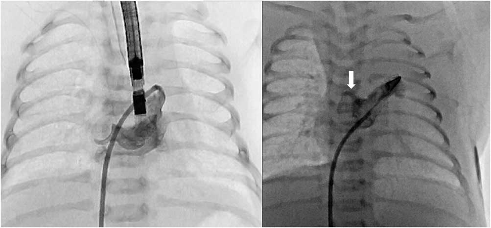
During the procedure, a 5-French multipurpose catheter was unintentionally advanced into the left atrium, not through the foramen but through the communication route that was identified in the previous CT (Fig 2, left). The measurement at the tip of the catheter showed an increase in the left atrial pressure to 20 mmHg. We exchanged to only the dilator of an 8-French long sheath, and subsequently a trans-septal radiofrequency needle was advanced through the dilator. After repetitive confirmation of the catheter position, several applications of energy resulted in a successful perforation of the septum. After removal of the radiofrequency needle and exchanging it for a 0.014-inch high-torque wire, balloon atrial septoplasty was performed using a 5-mm coronary angioplasty balloon catheter, and subsequently using a 10-mm angioplasty balloon catheter. Finally, we successfully obtained sufficient atrial communication, and the left and right atrial pressures were 8 and 6 mmHg, respectively. The patient experienced immediate improvement in her systemic saturation level to 85%. At 5 days of age, the patient underwent bilateral pulmonary arterial bandings. Her systemic oxygen saturation level was 85%, and the interatrial communication was sufficient to maintain appropriate pulmonary circulation. The patient underwent the Norwood stage I procedure with Sano shunt at 2 months of age. Unfortunately, she died at 5 months of age because of septic shock.
Fluoroscopic images obtained during transcatheter atrial septoplasty in Case 1 (left side) and Case 2 (right side). The catheter was advanced into the left atrium through the levoatriocardinal vein (left side). An angiogram of the left atrium after trans-septal puncture in Case 2 (right side), showing a levoatriocardinal communication arising from the top of the left atrium along the atrial roof in the transverse sinus and draining into the right atrium (arrow).

Case 2
A 2.1 kg baby girl was born at 36 weeks’ gestation. She was transferred to the referred hospital because of cyanosis and not doing well shortly after birth. Her systemic oxygen saturation level was 50%, and respiratory support was started. Echocardiography revealed hypoplastic left heart syndrome and an intact atrial septum; however, a color Doppler image demonstrated that a vessel from the left pulmonary vein drained into the right atrium, which was similar to the communicating vessel in Case 1. The patient was transferred to our institution. Her leg blood pressure was 42/15 mmHg and her femoral pulsation was weak because of the narrowing ductus. After the immediate intravenous administration of prostaglandin, the ductus was opened wide. We considered that the communicating vessel was not sufficient to maintain pulmonary circulation and thus performed urgent transcatheter balloon atrial septoplasty as described in Case 1.
The left atrial pressure was increased to as high as 31 mmHg. An angiogram of the left atrium showed that a communicating vessel arose from the top of the left atrium along the atrial roof in the transverse sinus and drained into the right atrium, similar to that in Case 1 (Fig 2, right). We successfully obtained sufficient atrial communication; however, the patient developed pericardial effusion, hypotension, and renal failure after the procedure, following transfusion and peritoneal dialysis for several days. At 21 days of age, the patient underwent bilateral pulmonary arterial bandings via median thoracotomy. Her systemic oxygen saturation level was 75%, and the interatrial communication was enough to maintain appropriate pulmonary circulation. The patient is currently awaiting the Norwood procedure.
Discussions
An intact atrial septum is present in 10% of neonates with hypoplastic left heart syndrome, which is associated with fetal hydrops, arterialisation of the pulmonary veins, and pulmonary lymphangiectasis.Reference Pesonen, Haavisto, Ammala and Teramo 3 , Reference Moerman, Van Dijck and Lauweryns 4 Usually, the levoatriocardial vein arises from the left atrium or one of the pulmonary veins and drains into the innominate or superior caval vein.Reference Bernstein, Moore, Stanger and Silverman 2 We demonstrated two cases of neonates with unique levoatriocardinal veins that arose from the top of the left atrium and drained into the right atrium.
In the development of the pulmonary venous system, the plexus of the intrapulmonary veins initially connects to the systemic cardinal venous system. In fetuses with hypoplastic left heart syndrome and an intact atrial septum, an inadequate connection between the pulmonary and systemic cardinal venous systems provides the only route and egress for pulmonary venous drainage, which becomes the levoatriocardinal vein.Reference Tweddell, Hoffman, Ghanayem, Frommelt, Mussatto and Berger 5 When a channel to the anterior cardinal systemic venous system persists, the levoatriocardinal vein drains into the innominate or superior caval vein. The levoatriocardinal vein originated from the left atrium in 68% and from the pulmonary vein in 32% of cases, and drained into the innominate vein in 48%, the superior caval vein in 36%, and the jugular vein in 8% of cases.Reference Bernstein, Moore, Stanger and Silverman 2 As the incidence of the primordial connection between the plexus of the foregut and the cardinal venous system is expected to become more widespread, we assume that the levoatriocardinal vein can directly drain into the right atrium in the present cases.
Conclusions
We demonstrated two cases of neonates with hypoplastic left heart syndrome and an intact atrial septum who had unique levoatriocardinal veins. We emphasise that transcatheter atrial septostomy should be performed not through the communicating vessel but using another trans-septal approach in neonates with hypoplastic left heart syndrome and an intact atrial septum.
Acknowledgements
None.
Financial Support
This research received no specific grant from any funding agency, or commercial or not-for-profit sectors.
Conflicts of Interest
None.