Coexistence of both right and left heart obstruction in the same patient is exceptional. Over the past 40 years, four infants with tetralogy of Fallot associated with coarctation of the aorta underwent surgery in our department. To the best of our knowledge, only 14 patients have been described to date in the literature, including two of our patients.Reference Rey, Coeurderoy and Dupuis 1
Cases presentation
Patient characteristics are summarised in Table 1.
Main information about the cases.

BTT shunt=Blalock–Taussig–Thomas Shunt; LSCA=left subclavian artery; TOF=tetralogy of Fallot; TOF-PA=tetralogy of Fallot with pulmonary atresia; RCCA=right common carotid artery; RSCA=right subclavian artery
First case
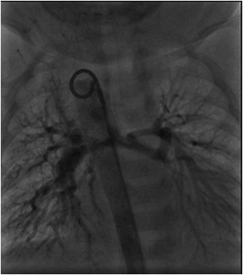
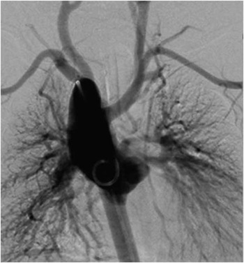
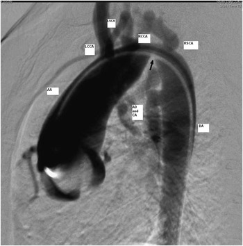
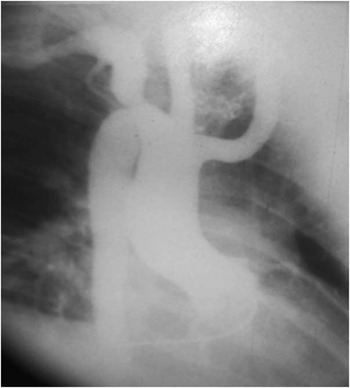
A 1-month-old infant with 22q11 microdeletion was referred for tetralogy of Fallot with pulmonary atresia diagnosed at birth. He had no signs of cardiac failure and normal femoral pulses. Cardiac catheterisation showed confluent hypoplastic pulmonary branches and two major aortopulmonary collateral arteries from the descending aorta (Fig 1). There was a right aortic arch (Fig 2) and a mild coarctation located between the right carotid artery and the right subclavian artery. The systolic pressure gradient across the coarctation was 15 mmHg (Fig 3). A left modified Blalock–Taussig–Thomas shunt was performed. The coarctation was considered to be mild, and thus not repaired.
Aortic frontal angiogram. Right-sided aortic arch. Pulmonary atresia. Major aortopulmonary collateral arteries (MAPCA) (Case 1).

Aortic frontal angiogram. Right-sided aortic arch. Major aortopulmonary collateral arteries (MAPCA) (Case 1).

Aortic lateral angiogram. Coarctation of the aorta located between the right carotid and the right subclavian arteries. Major aortopulmonary collateral arteries (MAPCA) (Case 1). The arrow shows the coarctation of the aorta. AA=ascending aorta; AD=arterial duct; CA=collateral arteries; DA=descending aorta; LCCA=left common coronary artery; LSCA=left subclavian artery; RCCA=right common carotid artery; RSCA=right subclavian artery.

Second case
In the 1990s, a 3-month-old infant was referred with a diagnosis of tetralogy of Fallot and aortic coarctation. Cardiac catheterisation showed a right aortic arch with a hypoplastic horizontal segment located before the right subclavian artery. The left subclavian artery was retro-oesophageal. The ascending to descending aorta gradient was 75 mmHg. Coarctation repair using a modified Crafoord technique was performed at the age of 15 months. Complete repair of tetralogy of Fallot was performed at 21 months of age.
Third case
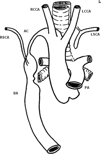
In the 1980s, a 3-month-old child was admitted for tetralogy of Fallot with right aortic arch and coarctation of the aorta.Reference Rey, Coeurderoy and Dupuis 1 Transthoracic echocardiography and cardiac catheterisation showed a cervical right aortic arch with a long narrowed segment located between the right carotid artery and the right subclavian artery. The left subclavian artery issued from the left vertebral artery (Fig 4). An 8 mm Goretex conduit was inserted between the ascending and descending aorta. Complete repair of tetralogy of Fallot was performed 2 years later. After 12 years, the child presented a residual aortic coarctation with a systolic pressure gradient of 40 mmHg. A Crafoord anastomosis was then performed.
Drawing of the congenital heart disease by the surgeon. Cervical right aortic arch, long narrowed segment between the right carotid artery and the right subclavian artery. Left subclavian artery issued from the left vertebral artery (Case 3).

Fourth case
In the 1970s, a 4-month-old child was admitted for severe cyanosis and faintness.Reference Rey, Coeurderoy and Dupuis 1 Angiography revealed tetralogy of Fallot with right aortic arch and mild aortic narrowing. A Waterston shunt was performed and complete repair was postponed until 5 years later. During the follow-up, a second angiography revealed a tight coarctation of the aorta with a systolic pressure gradient of 40 mmHg, located between the right carotid artery and the right subclavian artery (Fig 5). A Crafoord anastomosis was performed.
Frontal aortography. Right aortic arch with coarctation of the aorta between the right carotid artery and the right subclavian artery (Case 4).

Discussion
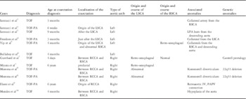
Coarctation of the aorta in the setting of tetralogy of Fallot with or without pulmonary atresia is exceedingly rare. In a recent study on about 2235 patients with tetralogy of Fallot, aortic coarctation was described in only one patient.Reference Changela, John and Maheshwari 2 We report here four cases of this exceptional association, two of them having already been published in 1984.Reference Rey, Coeurderoy and Dupuis 1 To the best of our knowledge, only 12 similar cases, apart from our two first cases, have been described to date in the literature.Reference Iannucci, Mahle and Clabby 3 – Reference Morales, Dibardino, Vick, Fraser and McKenzie 10 Their characteristics are summarised in Table 2. Aortic arch and brachiocephalic branching abnormalities are frequently associated. Right-sided aortic arch is particularly frequent, occurring in our four patients and in seven of the 12 patients described in the literature. A total of four patients had a left-sided aortic arch and the pattern of the arch was unknown in one (Table 2). When the aortic arch was left-sided, the coarctation was situated in the most common location for coarctation in a left aortic arch, distal to the left subclavian artery. In contrast, in all patients reported with right-sided aortic arch, as well as in our four patients, coarctation was located between the right carotid artery and the right subclavian artery, and was associated with anomalies of insertion of the left subclavian artery (Table 2). Only one of our patients had proven or suspected DiGeorge syndrome, whereas two patients described in the literature had 22q11 microdeletion. Associated lesions were present in eight patients, including two diverticulums of Kommerell. Although this diverticulum can be considered as part of the malformation, it is far from being constant and was therefore included in the associated lesions.
Review of the literature.

AD=arterial duct; IV=innominate vein; LPA=left pulmonary artery; LSCA=left subclavian artery; PAPVC=partial anomalous pulmonary venous connection; RCCA=right common carotid artery; RSCA=right subclavian artery; TOF=tetralogy of Fallot; TOF-PA=tetralogy of Fallot with pulmonary atresia
Tetralogy of Fallot with or without pulmonary atresia belongs to the group of conotruncal defects, which we define as all the congenital heart diseases presumably caused by abnormal migration of neural crest cells, and subsequent failure of the anterior part of the second heart field to add myocytes to the developing outflow tract, including also common arterial trunk, malalignment ventricular septal defects with overriding aorta, certain forms of interrupted aortic arch, and certain forms of double outlet right ventricle. However, there is now experimental evidence that neural crest cells are also involved in both arterial valves malformations and double outlet right ventricle in mice, and also in the development of the intrapericardial arterial trunks.Reference Phillips, Mahendran, Singh, Anderson, Chaudhry and Henderson 11 , Reference Anderson, Chaudhry and Mohun 12 For this reason, we chose to group all these malformations under the term “outflow tract defects”.
Right-sided aortic arch is frequently associated with outflow tract defects, such as tetralogy of Fallot and common arterial trunk.Reference Rauch, Rauch and Koch 13 It can also occur without any intracardiac anomaly, the incidence of 22q11 microdeletion being as high as 24% in such cases.Reference McElhinney, Clark and Weinberg 14 The branching of the brachiocephalic arteries can be either in mirror-image – left brachiocephalic artery followed by right carotid artery then right subclavian artery – or it can be more abnormal, including aberrant left subclavian artery with retro-oesophageal course, or isolated left subclavian artery. The ductus can be right-sided, left-sided, or bilateral. Congenital obstructions of a right aortic arch – coarctation, interruption or obstruction of a cervical arch – although very uncommon, have already been reported, but no patient in this series had tetralogy of Fallot.Reference McElhinney, Tworetzky, Hanley and Rudolph 15 Interestingly, in patients of this series with an interrupted right aortic arch, the interruption was always located between the right common carotid and the right subclavian artery, mirror-image of interrupted left aortic arch type B; a ventricular septal defect was always present; and a majority of patients had 22q11 microdeletion.Reference McElhinney, Tworetzky, Hanley and Rudolph 15 , Reference Celoria and Patton 16 Interrupted left aortic arch type B and type A have a very different etiologic background.Reference Van Mierop and Kutsche 17 Type B is strongly associated with 22q11 microdeletion and may be due to an abnormal development of the fourth aortic arch, whereas type A may be considered as an extreme form of aortic coarctation.Reference Van Mierop and Kutsche 17
The pathogenesis of coarctation of the aorta in the setting of tetralogy of Fallot could then be different according to the pattern of the aortic arch. In all patients reported with a left aortic arch, aortic coarctation had a usual location, at the level of the isthmus, without aortic arch hypoplasia and without subaortic obstruction. This type of coarctation might then be due to an abnormal expansion of the ductal tissue into the aortic wall, and its association with tetralogy of Fallot might be then fortuitous. In contrast, in patients with right aortic arch, the very unusual location of the coarctation, which is in the same site as mirror-image interrupted left aortic arch type B, almost always associated with anomalies of the left subclavian artery, could be considered to be due to an anomaly of the fourth aortic arch, itself related with abnormal migration of cardiac neural crest cells.Reference Rauch, Rauch and Koch 13 We hypothesise that this abnormal migration may explain the association of an outflow tract defect such as tetralogy of Fallot with or without pulmonary atresia with a right aortic arch, anomalies of the branching of the brachiocephalic arteries, and an unusual coarctation of the aorta, located between the right common carotid artery and the right subclavian artery.Reference Momma 8 Indeed, combination of both right and left-sided outflow tract obstruction cannot be explained by Rudolph’s theory about flow-related development of the great arteries in the foetus, in which coarctation of the aorta is a consequence of a reduced blood flow through the left ventricular outflow tract, and the right aortic arch is a consequence of a reduced flow through a narrowed right ventricular outflow tract.Reference Rudolph, Heymann and Spitznas 18 However, a recent study reconciles the genetic and the hamodynamic hypothesis by demonstrating that laterality genes such as Pitx2c, inducing, by their action on the second heart field, the rotation of the outflow tract resulting in the normal position of the great arteries, determine differential blood flow in the aortic arches.Reference Yashiro, Shiratori and Hamada 19 Normally, blood flow is favoured within the left fourth aortic arch and reduced in the right fourth aortic arch, leading to normal left aortic arch. If the rotation is impaired because of abnormal migration of the neural crest cells,Reference Hutson and Kirby 20 the right fourth aortic arch development is favoured, which explains its frequent association with outflow tract defects.Reference Yashiro, Shiratori and Hamada 19 Experiments on mice found some genes and pathways implied in the patterning of the great arteries and outflow tract of the heart. One of them, Hox gene, is expressed in precursors of cardiac neural crest cells. A mutation in this gene in the mouse is associated with aortic arch anomalies, including interrupted aortic arch type B and aberrant subclavian artery, and occasionally tetralogy of Fallot but without association between these different anomalies.Reference Makki and Capecchi 21
Clinical implications
Although rarely seen in the setting of tetralogy of Fallot, screening for aortic coarctation should be performed in all such patients because of the major potential clinical implications. Diagnosis can be challenging as the clinical presentation is not always the same.Reference Changela, John and Maheshwari 2 Coarctation may be discovered at birth or during the follow-up in asymptomatic patients. Diagnosis is also crucial because of its impact on both mortality and morbidity in the post-operative course after repair of tetralogy of Fallot.Reference Morales, Dibardino, Vick, Fraser and McKenzie 10 Knowing where the arch obstructions are typically found in right- and left-sided aortic arch in association with tetralogy of Fallot should help the preoperative analysis of anatomy of the aortic arch on echocardiography, in helping focus the assessment of the arch. This is of particular importance for surgery. If the coarctation is not clinically significant, primary repair of tetralogy of Fallot can be performed without repairing the aortic arch.Reference Changela, John and Maheshwari 2 Other options include complete repair of both tetralogy of Fallot and coarctation or coarctation repair with a systemic to pulmonary artery shunt followed by later complete repair of the tetralogy of Fallot.Reference Changela, John and Maheshwari 2
Conclusion
The rare association between tetralogy of Fallot with or without pulmonary atresia and coarctation of the aorta should not be considered as haemodynamically illogical coexistence of left and right heart obstruction but rather, at least when the aortic arch is right-sided, as a complex anomaly of the aortic arches in the setting of a cardiac neural crest defect. Screening for this unusual anatomic and developmental syndrome is critical in the initial assessment of all patients with tetralogy of Fallot.
Acknowledgements
We thank Pr Richard Van Praagh for his critical revision of the manuscript.
Financial Support
This research received no specific grant from any funding agency or commercial or not-for-profit sectors.
Conflicts of Interest
None.