Case report
A six-year-old male with the cardiac diagnosis of unbalanced complete atrioventricular septal defect, pulmonary atresia, pulmonary venous drainage to the right side of a common atrium, left-sided inferior caval vein, as well as heterotaxia with asplenia presented two years after completion of an extracardiac fenestrated Fontan with progressive desaturations into the low 80’s, even more pronounced and noticeable with activities. He was otherwise clinically well and asymptomatic. Echocardiography documented normal ventricular function with the fenestration not being visualised.
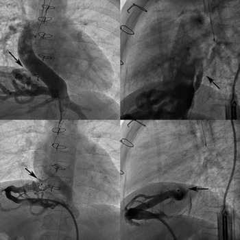
He was taken to the catheterisation laboratory to evaluate haemodynamics and to assess for any source of right-to-left shunting that would be amenable to transcatheter therapy. Baseline haemodynamics documented mean Fontan pressures of 13 mmHg. Angiography documented no identifiable residual fenestration, but a large hepatic collateral meshwork draining to what appeared to be the right side of the common atrium (Fig 1). Balloon test occlusion at the atrial entry site resulted in an increase in systemic saturation to 92% (from 81% at baseline) with no change in Fontan pressures. Attempts were made to occlude the communication at the entry to the common atrium using a 16 mm Amplatzer Vascular Plug II but resulted in the device also occluding hepatic side branches that entered close to the common atrium (Fig 1). The procedure was therefore abandoned and discussed at case management conference. Surgical reimplantation of what was considered to likely be a hepatic vein that had not been incorporated into the Fontan (and drained directly to the atrium) was considered. To further aid surgical planning, a CT scan was obtained, which suggested the hepatic channel was curving posterior to the atrium, with some length that may in fact allow device placement with sufficient distance from any hepatic vein entry. It was therefore felt that another attempt at transcatheter closure should be undertaken.
6-year-old male after Fontan completion. Top left and right: Inferior caval vein injection documenting a hepatic venous collateral meshwork draining towards the common atrium (arrow). Bottom left and right: An Amplatzer Vascular Plug II (arrow) sized to occlude the atrial entry, obstructing several hepatic venous side branches while elongating in a smaller diameter component of the hepatic vein.

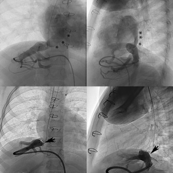
The patient was again taken to the catheterisation laboratory, and this time a rotational angiography was obtained (Fig 2). This documented that what was thus far considered to be a hepatic vein draining to the common atrium was in fact a direct communication to the coronary sinus, which itself opened towards the left side of the common atrium. While positioning a device into the coronary sinus would have been feasible to avoid obstructing a hepatic venous side branch, it was felt that this may potentially incur a higher risk of thrombus formation and impact coronary venous flow, and as such was considered a less desirable option. Given that placement of a device within the coronary sinus was not a suitable option, this significantly limited the available length for device placement without obstructing hepatic side branches. Eventually, it was decided to place a 6 mm Amplatzer Muscular Ventricular Septal Defect Occluder, which was felt to have the best occlusive properties and be preferrable to a vascular plug. This was performed successfully without obstructing any hepatic side branches (Fig 2). There was only trivial foaming through the device with improved systemic saturation to 93%. The patient was discharged the following day.
6-year-old male after Fontan completion, undergoing a repeat cardiac catheterization. Top left and right: Still frames from a rotational angiography, delineating drainage of a hepatic vein to the coronary sinus, with the coronary sinus (marked by stars) then draining towards the left atrium. Bottom left and right: Occlusion of the hepatic vein proximal to the coronary sinus using a 6 mm Amplatzer Muscular Ventricular Septal Defect Occluder (arrows). The arrows point to hepatic side branches that were not occluded by this device.

On follow-up, saturations have remained between 89% and 93%, with a trivial residual shunt having developed around the device. The patient has been placed on aspirin, and placement of additional devices/coils will be considered whenever clinically needed.
Brief discussion
Drainage of a (usually left-sided) hepatic vein to the coronary sinus is an extremely rare abnormality with very few cases described in the literature. Reference Karolczak, Mądry and Zacharska-Kokot1–Reference Lee and Saremi3 Embryologically, it results when the left horn of the sinus venosus (which forms the coronary sinus) forms a persistent connection with the left vitelline vein system, thereby creating the left hepatic vein to coronary sinus communication. Reference Karolczak, Mądry and Zacharska-Kokot1,Reference Morshuis, de Lind van Wijngaarden, Kik and Bogers4 In most cases, this anomaly is of no clinical consequence. However, several reports suggest the importance of this finding during cardiac surgery using extracorporeal bypass, use of cardioplegia via the coronary sinus, and in cases of Fontan completion. Reference Morshuis, de Lind van Wijngaarden, Kik and Bogers4,Reference de Oliveira, Horvat and de Azambuja5
In our patient, the abnormal connection existed in the context of visceral heterotaxia with asplenia and anomalous pulmonary venous drainage to a common atrium, which is the first time such a connection has been described in the literature. This had a clinically important impact in this patient, resulting in significant desaturations after Fontan completion. The anomaly was only detected after multiple angiographic and CT evaluations. Patients with heterotaxia often have pulmonary and/or systemic venous abnormalities, not all of which are readily identified on standard pre-Fontan imaging. Not fully incorporating the entire hepatic venous drainage into a Fontan circulation can lead to intrahepatic collateralisation and “pop-off” from a higher-pressure Fontan circulation to a lower pressure atrial chamber, irrespective of whether a solitary hepatic vein draining directly to the atrium was missed, or an extremely rare communication between a hepatic vein and coronary sinus as seen in this patient. This patient did have an angiography performed inside the inferior caval vein during the pre-Fontan cardiac catheterisation (in anterior–posterior and lateral projection), but with venous pressures at that stage being a lot lower than post Fontan, it only documented reflux of contrast into left- and right-sided hepatic veins, but not a connection from hepatic vein to coronary sinus. As such, if there is doubt about hepatic venous drainage, then detailed pre-Fontan axial imaging should be performed (even though in this patient, the intracardiac connection to the coronary sinus was missed even by axial imaging).
Acknowledgements
None.
Financial support
This research received no specific grant from any funding agency, commercial, or not-for-profit sectors.
Conflicts of interest
The authors have no conflicts of interest to disclose.
Ethical standards
Not applicable.