Background
Patients with pulmonary valve disease can often be treated with hybrid approach, especially in cases with oversized right ventricular outflow tracts and high surgical risks.Reference Sosnowski, Matella and Fogg1,Reference Güzeltaş, Tanıdır, Gökalp and Haydin2 These patients are often in need of additional interventional procedure which is not typically performed in the same encounter. Here, we reported a successful combined hybrid pulmonary valve replacement and atrial septal defect device closure.
Case report
A 15-year-old female patient with a history of congenital pulmonary stenosis status-post pulmonary valvectomy as a neonate following failed pulmonary valvuloplasty presented with progressive dyspnea on exertion and decreased exercise tolerance. She was also known to have secundum atrial septal defect. Her echocardiogram revealed moderate sized secundum atrial septal defect with adequate rims. The right atrium and the right ventricle were dilated secondary to volume overload. Doppler echocardiography showed severe pulmonary valve regurgitation with holodiastolic reversal of flow in the proximal branch pulmonary arteries. Subsequently, cardiac MRI confirmed severe pulmonary regurgitation with a regurgitant fraction of 50%, as well as severely dilated right ventricle (indexed end diastolic right ventricular volume 162 ml/m2). The right and left ventricular systolic function was preserved with normal ejection fraction on MRI scan. Her right ventricular outflow tract measurements were too large to accommodate a transcatheter pulmonary valve. Patient was referred to our cardiac catheterisation laboratory for elective hybrid pulmonary valve replacement and atrial septal defect closure.
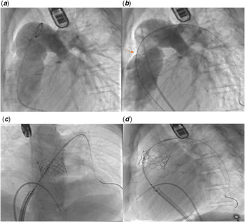
The procedure was performed in our hybrid catheterisation laboratory under general anaesthesia. Right ventricular outflow tract angiogram was performed and confirmed main pulmonary artery dilation, which measured 29 mm at the narrowest diameter without balloon compliance testing (Fig 1a). Given the large main pulmonary artery size, we decided to proceed with a hybrid approach to reduce the main pulmonary artery size and establish adequate landing zone for transcatheter pulmonary valve. Through a small left anterior thoracotomy incision, the right ventricular outflow tract and the main pulmonary artery were exposed. Two 2-0 Tevdek pledgeted sutures were used to plicate the main pulmonary artery to approximately 25 mm. After plication, repeat right ventricular outflow angiogram demonstrated a waist of roughly 25 mm (Fig 1b). Compliant testing of the main pulmonary artery with 30 mm × 40 mm sizing balloon (NuMED, Hopkinton, NY) was performed, which demonstrated a waist of 25 mm in the main pulmonary artery and no evidence of coronary compression. A 29 mm Edwards SAPIEN S3 valve was prepared on the Edwards Commander delivery system (Edwards Lifesciences, Irvine, CA). The valve was deployed uniformly under fluoroscopic guidance at the site of plicated main pulmonary artery (Fig 1c and d). Repeat main pulmonary artery angiograms showed well-positioned valve without significant insufficiency.
Angiograms and fluoroscopy images. (a) Initial right ventricular outflow angiogram demonstrating dilation of the main pulmonary artery measuring 29 mm. (b) Right ventricular outflow tract angiogram following main pulmonary artery plication demonstrating a waist (red arrow) of 25 mm. (c) and (d) Fluoroscopy images after deployment of the SAPIEN valve showing well-positioned valve with final diameter of 28 mm.

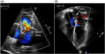
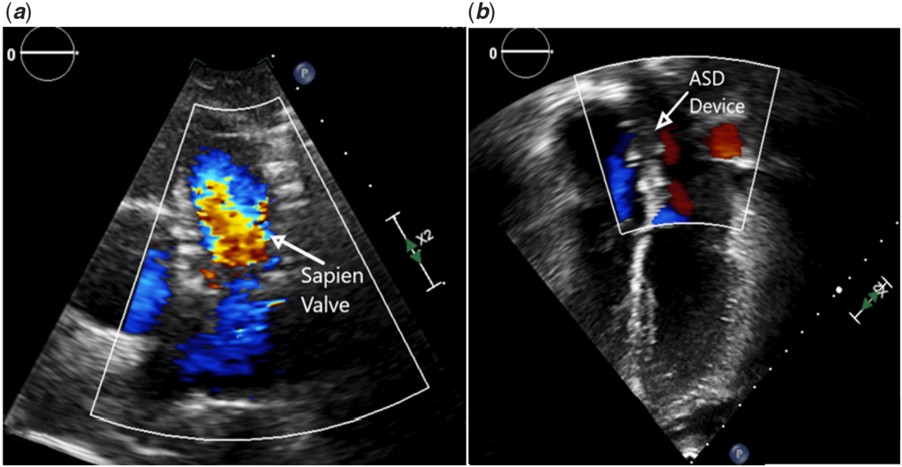
Following valve placement, we proceeded with transcatheter atrial septal defect closure from the right femoral vein. Transesophageal echocardiography demonstrated a defect measuring 11 mm with a total septal length of 36 mm and adequate surrounding rims. A 30 mm Gore Cardioform Septal Occluder (GSO, W.L. Gore & Associates, Flagstaff, AZ) was chosen for transcatheter closure. The delivery sheath was advanced across the atrial septum into the left atrium. The device was deployed successfully across the atrial septum under echocardiography and fluoroscopy guidance. The procedure was uneventful, and patient was discharged the next day on aspirin therapy for 6 months. Pre-discharge echocardiogram showed well-functioning pulmonary valve with no pulmonary insufficiency and well-seated atrial septal defect device without any residual shunt. At 6-month follow-up, the patient was well and her echocardiogram showed no significant pulmonary insufficiency and no residual atrial level shunt (Fig 2a and b).
(a) Transthoracic echocardiography with colour Doppler showing SAPIEN valve in pulmonary position with no regurgitation. (b) Four-chamber view with colour Doppler showing ASD device with no residual shunt. ASD = atrial septal defect.
Discussion
Patients with CHD require multiple cardiac interventions over their lifetime. These interventions are not routinely performed in the same setting. Recent studies have shown that combining procedures in patients with CHD is feasible, safe, and can reduce costs.Reference Bansal, Molian, Maldonado, Aldoss, Ochoa and Law3,Reference Asaki, Orcutt and Miyake4
Transcatheter pulmonary valve implantation has revolutionised the non-surgical treatment of dysfunction right ventricular outflow tracts in the past two decades.Reference Boudjemline, Brugada and Van-Aerschot5,Reference McElhinney, Hellenbrand and Zahn6 There are two valves currently approved by the U.S. Food and Drug Administration for use only in previously placed surgical conduits, the Melody valve (Medtronic, Minneapolis, MN), and the Edwards SAPIEN valve (Edwards Lifesciences, Irvine, CA). The Medtronic Melody valve consists of a 16 mm or 18 mm bovine jugular valve hand-sewn onto a bare metal Cheatham platinum stent (NuMED, Hopkinton, NY) and can be expanded from 16 to ~24 mm. The Edwards SAPIEN valve is a trileaflet bovine pericardial tissue valve hand-sutured in a balloon-expandable cobalt-chromium stent. It is available in 23, 26, or 29 mm diameters that require 22- or 24-F delivery sheaths.
Hybrid approach in pulmonary valve replacement is an increasingly more utilised option in oversized right ventricular outflow tracts as an alternative to surgical replacement.Reference Sosnowski, Matella and Fogg1,Reference Güzeltaş, Tanıdır, Gökalp and Haydin2 It can also be used in certain situations such as complex patients who are considered high-risk surgical candidates, small size patients, or absent venous access. Potential benefits using this method include the avoidance of full sternotomy, cardiopulmonary bypass, and shorter hospital stay. Pre-procedural comprehensive planning and coordination between the different services is a key in hybrid procedures. Currently, there are no randomised controlled trials to compare the long-term outcomes of the hybrid approach with the standard surgical method.
Atrial septal defects are one of the most common CHDs accounting for 25–30% of CHDs diagnosed in adulthood.Reference Lindsey and Hillis7 Atrial septal defects are further classified based on the location in the interatrial septum to ostium secundum, ostium primum, sinus venosus, and coronary sinus defect. Ostium secundum defect occurs due to excess resorption of septum primum, or inadequate development of septum secundum.Reference Geva, Martins and Wald8 Percutaneous closure has become the treatment of choice in most child and adult patients with secundum atrial septal defect due to lower rate of complications and shorter recovery time.Reference Villablanca, Briston and Rodés-Cabau9
Conclusion
Hybrid approach in patients with dilated right ventricular outflow tracts allows for pulmonary valve placement without the need for cardiopulmonary bypass. Hybrid pulmonary valve replacement and percutaneous atrial septal defect closure in one interventional procedure can be safe, feasible, and effective.
Acknowledgements
None.
Financial Support
This research received no specific grant from any funding agency, or from commercial or not-for-profit sectors.
Conflict of Interest
None.