Introduction
Gastrointestinal (GI) parasites cause great harm to commercial agriculture, costing the livestock industry £1000 million per annum in resources to control parasitism (Newton & Munn, Reference Newton and Munn1999). Haemonchus contortus, which parasitizes the abomasums of cattle and sheep, is a major GI parasite with a worldwide distribution. Parasitism by this blood-feeding nematode causes serious effects, ranging from mild anaemia to mortality in young animals (Nikolaou & Gasser, Reference Nikolaou and Gasser2006). The current means of control, which are dependent on excessive treatment with anthelmintic chemicals, have led to severe drug resistance (Newton & Munn, Reference Newton and Munn1999). Together with the growing concern over residual chemicals in the environment and food chain, this has led to the development of alternative or supplementary means of control, including the development of molecular vaccines and new anthelmintic chemicals that leave little residue.
Parasitic nematodes utilize glucose for energy production, and glycogen is the major substrate for energy conservation (Kohler, Reference Kohler1985). Glyceraldehyde-3-phosphate dehydrogenase (GAPDH) is an important enzyme in the glycolytic pathway. It catalyses the reversible oxidative phosphorylation of d-glyceraldehyde-3-phosphate to 1,3-diphosphoglycerate in the presence of inorganic phosphate and nicotinamide adenine dinucleotide (NAD+) (Kasuga-Aoki et al., Reference Kasuga-Aoki, Tsuji, Suzuki, Matsumoto, Arakawa and Isobe2002). This NAD+-dependent glycolytic enzyme is widely distributed and conserved in organisms from bacteria to humans (Fothergill-Gilmore & Michels, Reference Fothergill-Gilmore and Michels1993) and is approximately 35–37 kDa in size (Fothergill-Gilmore & Michels, Reference Fothergill-Gilmore and Michels1993). In some species, four such proteins may combine to form a tetramer structure with a native molecular weight in the range of 140–150 kDa (Oukhattar et al., Reference Oukhattar, Baibai, Moutaouakkil, Assobhei and Soukri2008).
GAPDH has frequently been regarded as a housekeeping gene (Kasuga-Aoki et al., Reference Kasuga-Aoki, Tsuji, Suzuki, Matsumoto, Arakawa and Isobe2002) because of the role of the enzyme in the glycolytic pathway. However, increasing evidence indicates that GAPDH also has several other functions. For example, it plays a central role in the Embden–Meyerhoff pathway in widely differing organisms, not only in glycolysis but also in glyconeogenesis (Fothergill-Gilmore & Michels, Reference Fothergill-Gilmore and Michels1993). In addition, a variety of other biological activities of the mammalian GAPDH protein have been reported, including membrane, cytoplasmic and nuclear endocytosis (Sirover, Reference Sirover1999), mRNA regulation, tRNA export (Singh & Green, Reference Singh and Green1993), DNA replication and DNA repair (Baxi & Vishwanatha, Reference Baxi and Vishwanatha1995). Recently, it has been reported that the GAPDH of Schistosomiasis mansoni can be a protective antigen (El Ridi et al., Reference El Ridi, Farouk, Sherif, Al-Sherbiny, Osman, El Gengehi and Shoemaker1998). It was also reported that this enzyme can be utilized as a useful allozyme in population genetic research (Maltagliati et al., Reference Maltagliati, Belcari, Casu, Casu, Sartor, Vargiu and Castelli2002).
To date, only an expressed sequence tag (EST) of H. contortus GAPDH has been published (GenBank accession no. AW670737). In the current study, we report the complete coding sequence of the GAPDH of this species (HcGAPDH) and the enzymatic activity of its recombinant protein.
Methods and materials
Parasite preparation and mRNA extraction
Adult H. contortus worms were collected from the abomasums of the donor goats as described previously (Muleke et al., Reference Muleke, Ruofeng, Lixin, Yanming and Xiangrui2006). The nematodes were washed three times with normal saline, then stored in liquid nitrogen. Total RNA was prepared from pooled parasite samples utilizing the single-step protocol (Chomczynski & Sacchi, Reference Chomczynski and Sacchi1987).
3′- and 5′-rapid amplification of cDNA ends
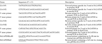
A 3′-end of cDNA was amplified by a 3′-full rapid amplification of cDNA ends (RACE) kit (TaKaRa Biotech, Dalian, P.R. China) using the forward gene specific primers GA-3-F1 and GA-3-F2 (table 1), designed using H. contortus EST (GenBank accession no. AW670737) in combination with the 3′ outer primer and 3′ inner primers provided in the RACE kit (table 1). The primary polymerase chain reaction (PCR) system (final volume, 25 μl) contained 2 μl of 3′-RACE-Ready cDNA, 1.0 U LA Taq ® polymerase (TaKaRa Biotech), 40 μm 1 × cDNA Dilution Buffer II, 50 μm 10 × LA PCR Buffer II (Mg2+ free), 3.0 mm MgCl2 and 400 nm of each primer. The primary PCR was performed using GA-3-F1 and 3′ outer primer followed by treatment at 94°C (3 min), 30 cycles at 94°C (30 s), 55°C (30 s) and 72°C (1 min), and a final extension at 72°C for 10 min. The product of the reaction was used as a template in a second PCR with primers GA-3-F2 and 3′ inner primer. This second PCR system contained the same ingredients as the first except that it lacked the 40 μm 1 × cDNA Dilution Buffer II and contained 400 μm dNTP mixture. The temperatures and durations used in the second PCR were the same as those in the first. The HcGAPDH 3′-end fragment was then obtained and sequenced.
Primers used in this study.

The name, sequence and description of the primers used in this study are shown. The restriction endonuclease sites are underlined.
The 5′-end of the cDNA was amplified by 5′-RACE PCR using the same method as the 3′-RACE PCR. The primary PCR was performed using GA-5-R1 and 5′ outer primer, with GA-5-R2 and 5′ inner primer in the second PCR.
Both of the products of the second PCRs were cloned into the pMD18-T vector (TaKaRa Biotech) and sequenced by Invitrogen Biotech Co., Ltd (Shanghai, P.R. China). The complete sequence of the HcGAPDH cDNA was deduced from the overlapping sequences of both amplification products using BioEdit Version 7.0.1 (T.A. Hall, North Carolina State University, USA). All oligonucleotides used in this research (table 1) were synthesized by Invitrogen Biotechnology Co., Ltd.
Sequence analysis
Sequence similarity was studied using BLASTP and BLASTX (http://blast.ncbi.nlm.nih.gov/Blast.cgi). GAPDH protein sequences were aligned using CLUSTALW1.82 (http://www.ebi.ac.uk/Tools/msa/clustalw2/). The signal peptide, secondary structure and protein motifs were predicted using approaches accessible on the Internet: SignalP (http://www.cbs.dtu.dk/services/SignalP/), PSIpred (http://bioinf4.cs.ucl.ac.uk:3000/psipred/) and Motifscan (http://myhits.isb-sib.ch/cgi-bin/motif_scan), respectively. Phylogenetic analyses among GAPDHs were inferred by the neighbour- joining (NJ) method.
Expression and purification of recombinant HcGAPDH
Primers were designed to subclone the complete H. contortus GAPDH open reading frame (ORF) according to the predicted ORF. The forward primer (HcGAPDHEcoRI 5′-GAATTCATGGTAAAACCAAAGGTTGG-3′) contained an EcoRI site (underlined) and the reverse primer (HcGAPDHSalI 5′-GTCGACTTAGGCCTTGCTTGCAATG-3′) contained a SalI site (underlined). The PCR reactions (final volume, 25 μl) contained 2 μl cDNA, 1.0 U Taq DNA polymerase (TaKaRa Biotech), 3.0 mm MgCl2, 400 μm dNTP mixture, 50 μm 10 × PCR Buffer (Mg2+ free) and 400 nm of each primer. Cycling conditions were: 94°C for 3 min followed by 30 cycles of 94°C (30 s), 55°C (30 s), 72°C (1 min), and a final extension at 72°C for 10 min.
The amplicon was digested with EcoRI and SalI and cloned in a frame into the bacterial expression vector pET28a(+) (Novagen, Merck, Darmstadt, Germany) according to the standard method. The recombinant plasmid was sequenced to confirm that the HcGAPDH insert was in the proper reading frame and to exclude PCR errors. The correct recombinant pET28a(+) plasmid was transferred into competent Escherichia coli BL21 (DE3) cells and the recombinant protein was induced and expressed by addition of 0.8 mm isopropyl thiogalactoside (IPTG; Sigma-Aldrich, St. Louis, Missouri, USA) to the cell culture after the A600 of the culture reached 0.6 at 37°C. The cells were incubated at 37°C for 5 h after the addition of IPTG. The cell lysates were prepared by sonication and analysed by sodium dodecyl sulphate–polyacrylamide gel electrophoresis (SDS–PAGE) using 12% gels to confirm the distribution of the expressed recombinant protein.
To purify the recombinant protein, induced E. coli cells were harvested by centrifugation and sonicated for 15 min on ice. After centrifugation at 10,000 × g, the supernatant was added to a Ni2+-nitrilotriacetic acid (Ni-NTA) column (GE Healthcare, Bio-sciences Corp., USA) and purified according to the manufacturer's instructions. An elution buffer (300 mm NaCl, 40 mm NaH2PO4, pH 8.0) containing 400 mm of imidazole was used to wash the His-tagged proteins from the Ni-NTA column. Purity of the protein was detected by 12% SDS-PAGE and the concentration of purified protein was determined according to the Bradford procedure (Bradford, Reference Bradford1976), using bovine serum albumin (BSA) as a standard. The protein was stored at − 20°C in 50% glycerol for later use.
Generation of polyclonal antibodies
To generate polyclonal antibodies, around 0.3 mg of the purified HcGAPDH was mixed with Freund's complete adjuvant, and then injected into Sprague–Dawley (SD) rats (Qualitied Certificate: SCXK 2008-0004; Experimental Animal Center of Jiangsu, P.R. China) subcutaneously in multiple places as described previously (Yanming et al., Reference Yanming, Ruofeng, Muleke, Guangwei, Lixin and Xiangrui2007). After the first injection, rats were boosted four times at 2-week intervals with the same dose. The serum, containing specific anti-HcGAPDH polyclonal antibodies, was harvested 10 days following the last injection and then stored at − 20°C. The goat anti-sera used in this study was collected from native goats infected with H. contortus.
Western blot analysis
After electrophoresis in SDS-PAGE (12% w/v), Western blotting was performed according to the method reported by Yi et al. (Reference Yi, Xu, Yan and Li2010). Separated protein bands were transferred electrophoretically from the gel to a nitrocellulose filter (Sigma). The blots were then incubated for 1 h in blocking solution containing 5% (w/v) fat-free dry milk, 0.1 m phosphate-buffered saline Tween (PBST) (pH 8.0) and 0.05% (v/v) Tween-20, and incubated with the primary antibodies (rat anti-sera and goat anti-sera, respectively) for 1 h at 37°C (dilutions 1:1000 to rat sera in 5% (w/v) fat-free milk/PBST, but 1:100 to goat sera). The membrane was washed four times for 10 min each in PBST, and then incubated with the second antibody (HRP-conjugated goat anti-rat IgG or HRP-conjugated rabbit anti-goat IgG, Sigma Immuno-Chemicals) in 1:5000 dilution of 5% milk powder/PBST for 1 h at 37°C. After four 10 min rounds of washing with PBST, the immunoreaction was visualized using freshly prepared 3,3′-diaminobenzidine (DAB) (Sigma) as a chromogenic substrate after 2–5 min.
Determination of the recombinant protein GAPDH activity and the optimal pH and temperature for the enzyme reaction
The GAPDH activity of the purified recombinant protein was measured according to the method of Ferdinand (Reference Ferdinand1964) with some modifications. The 1 ml reaction mixture contained 0.1 m triethanolamine-HCl buffer, 2 mm EDTA, 50 mm Na2HPO4, 1 mm NAD+ and 2 mmd-glyceraldehyde-3-phosphate solution (d-G3P). To determine the apparent optimal temperature, reactions were carried out at temperatures ranging between 10 and 75°C using a thermostated cuvette holder connected to a refrigerated bath circulator. To determine the optimal pH, 0.1 m triethanolamine-HCl buffer of pH 5.0–9.0, was used and the reaction was carried out for 2 min after the addition of 30 μg recombinant protein. The changes in absorbency were monitored at 340 nm before the addition of the recombinant protein and at the end of the reaction. One unit of enzyme activity was defined as the amount of enzyme that catalysed the reduction of 1 mol of NAD+ per min under the conditions used. Meanwhile, the commercial GAPDH (Sigma) and BSA were used as positive and negative controls, respectively.
Results
Cloning and sequence analysis of HcGAPDH
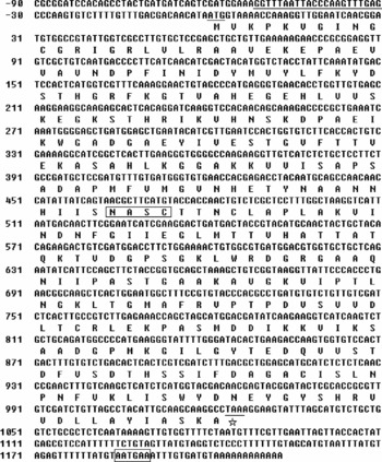
By overlapping the 3′- and 5′-RACE fragments, a 1303 bp transcript was obtained. A 22 bp spliced leader sequence (SL1) was identified at the 5′-end. Following the SL sequence, a 30 bp 5′-untranslated region (5′-UTR) was detected before the ATG initiation codon and a 1026 bp ORF was found that terminated with the TAA stop codon. On the 3′-end, the cDNA had a 175 bp 3′-UTR with a potential functional polyadenylation signal (AATGAA) followed by a 12 bp poly(A) tail (fig. 1). By analysis with SignalP, the ORF was found to encode a protein of 341 amino acids with a molecular mass of 36,446 Da. The theoretical pI of the protein was 8.15. No signal peptide was found in the deduced protein. Motifscan analysis showed that a NAD+ binding domain (residues 4–158), one possible N-glycosylation site (155NASC158) and an enzymatic activity site of GAPDH (156ASCTTNCL163) were present in this putative protein.
Complete cDNA sequence and deduced amino acid sequence of the HcGAPDH. The coding region starts at nucleotide (nt) 1 and ends at nt 1026; the SL1 sequence at the 5′-end, the ATG start and the TAA stop codons are underlined; and the putative N-glycosylation site 155NASC158 and one potential functional polyadenylation signal AATGAA at the 3′-end are boxed.

Alignment of multiple GAPDH sequences
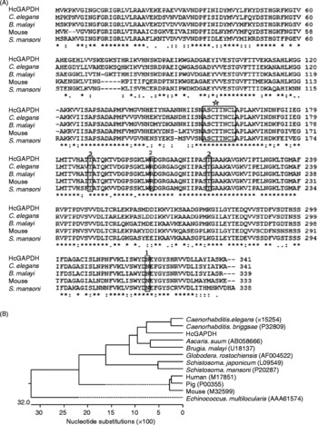
Alignment of the predicted amino acid sequence of H. contortus with those of the GAPDHs from other animals showed that this protein had a significant similarity (74–91% identical at the amino acid level) with GAPDH in parasites and mammals (fig. 2A). The similarity of this protein to GAPDH of Dictyocaulus viviparus was 91%. The residues involved in the NAD+, inorganic phosphate and GAP phosphate binding sites were fully conserved in all examined organisms (fig. 2A). Phylogenetic analysis of the various GAPDH was performed by the neighbour joining (NJ) method (fig. 2B). HcGAPDH was phylogenetically close to the GAPDHs from filarial parasites and remotely related to that from mice.
(A) Alignment of the deduced amino acid sequence of HcGAPDH with those of GAPDHs from other species. The deduced amino acid sequences and the previously reported sequences in the GenBank database were analysed with CLUSTALW version 1.8. Sequences shown in panel (A) are from B. malayi (U18137), C. elegans (X15254), mouse (M32499) and S. mansoni (P20287). Identical residues are marked with asterisks. The dashes demonstrate gaps introduced between the sequences. Amino acid residues essential for the enzymatic activity of GAPDH are indicated in the boxes. These signatures are listed as follows: (1) NAD binding site; (2) inorganic phosphate binding site; (3) GAP phosphate binding site; (⋆) putative enzymatic activity site. (B) Phylogenic trees inferred from homologies of GAPDH amino acid sequences utilizing the neighbour-joining (NJ) method. The scale bar represents one amino acid substitution/100 amino acid residues. The GAPDHs are represented by the name of their species. In parentheses, amino acid database accession numbers are listed. NJ analysis was carried out using the program CLUSTALW version 1.8.

Expression and purification of recombinant protein
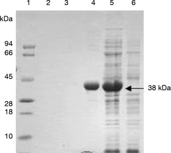
SDS–PAGE showed that the recombinant protein was mostly found in the sonicated bacterial supernatant. After purification from the supernatant by chromatography on the Ni-NTA, the protein was seen as a single band with a molecular mass of 38 kDa on the SDS–PAGE gel (fig. 3, lane 4). Because of the 2 kDa fused protein in the vector, the molecular weight of the recombinant protein was more than the value of 36,446 Da calculated from the deduced amino acid sequence.
Ni2+-nitrilotriacetic acid purification of HcGAPDH produced in E. coli BL21 (DE3). Proteins were resolved by SDS–PAGE on 12% polyacrylamide gel and stained with Coomassie brilliant blue R250. Lanes: 1, molecular standards; 2–4, different concentrations of soluble GAPDH fractions after Ni-NTA affinity chromatography; 5, crude extract from induced cells; 6, crude extract from cells with pET28a(+) expression vector.

Western blotting
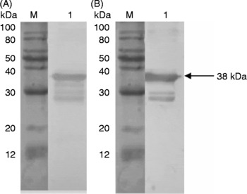
The immunoblotting results showed that the HcGAPDH was recognized by sera from goats naturally infected with H. contortus (fig. 4A). Western blot analysis also showed that mouse anti-HcGAPDH antiserum bound to a band at 38 kDa in the somatic extract of H. contortus adult worms (fig. 4B). This result indicated that natural HcGAPDH protein was distinguished by rat anti-rHcGAPDH antisera. The natural HcGAPDH appeared to have the same molecular mass as the recombinant HcGAPDH expressed in E. coli.
(A) Western blotting analysis of recombinant HcGAPDH protein with sera from goat naturally infected with H. contortus (lane 1); (B) Western blot analysis of crude somatic extracts from H. contortus with the antibody against recombinant HcGAPDH. Bound antibody was located by coupled immunoreactions with peroxidase-conjugated rabbit anti-goat IgG and goat anti-rat IgG, respectively. The arrow indicates the band corresponding to the 38 kDa GAPDH.

Enzymatic activity assay
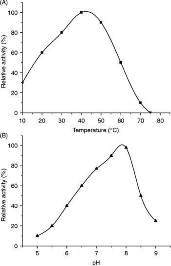
In the enzymatic activity assay, the purified protein yielded a specific activity of 46 U/mg. Without the addition of d-G3P to the reaction mixtures, there was no detectable spontaneous conversion of NAD+ to NADH. The assay showed that temperatures varying between 20 and 45°C did not greatly affect enzymatic activity. However, thermal inactivation occurred above 45°C and resulted in a total loss of activity at 75°C (fig. 5A). Studies on the effect of assay temperature on enzymatic activity revealed an optimal value of about 40°C. The optimal pH assay indicated that this recombinant protein exhibited the highest GAPDH activity at about pH 8.0 (fig. 5B). When the pH was higher above 8.0, the HcGAPDH displayed a sharp decrease in catalytic activity. However, when the pH was below 8.0, the enzymatic activity did not decrease so sharply.
Influence of temperature and pH variations on the relative activity of purified H. contortus GAPDH. (A) Relative enzymatic activity of purified GAPDH from adult H. contortus was tested at a temperature range from 10 to 75°C, revealing the effects of the most suitable temperature. (B) Relative enzymatic activity of purified HcGAPDH was measured in the triethanolamine–HCl buffer using the pH range 5.0–9.0.

Discussion
Several glyceraldehyde-3-phosphate dehydrogenases have been isolated or recombined from a wide range of nematodes (Erttmann et al., Reference Erttmann, Kleensang, Schneider, Hammerschmidt, Buttner and Gallin2005). However, there are only limited reports on Haemonchus contortus GAPDH. In the present study, we reported, for the first time, the cloning, expression and characterization of a H. contortus cDNA sequence that encodes a 38 kDa protein.
Based on multiple sequence alignment of GAPDHs, the predicted amino acid sequence of the HcGAPDH had an 86% identity with GAPDH of Caenorhabditis elegans and comparable similarity to other species: Caenorhabditis briggsae (85%), Brugia malayi (84%) and Onchocerca volvulus (83%). All of these indicated a high level of identity to members of the GAPDH protein family. The primary sequence of HcGAPDH revealed several structural features characteristic of GAPDHs, including an NAD+-binding domain (residues 4–158) and conserved amino acid residues Cys-158, Thr-188 and Arg-203, which were thought to be involved in catalysis. The residues surrounding the active site residue Cys-158, namely 156ASCTTNCL163, exactly matched the consensus sequence that was conserved in both prokaryotic and eukaryotic GAPDHs. Finally, these results were further strengthened by the in vitro test showing that the recombinant HcGAPDH protein had the enzymatic activity of glyceraldehyde-3-phosphate dehydrogenase. All of the above results suggested that it was a member of the GAPDH family.
In our investigation, the mRNA coding for GAPDH in H. contortus was shown to be preceded by a spliced leader. The 22 nucleotides at the 5′-end of mature mRNA of GAPDH were identical to the trans-spliced leader SL1 of C. elegans. In C. elegans, trans-splicing of mRNAs is a common phenomenon with at least 70% of mRNAs having an SL1 sequence (Zorio et al., Reference Zorio, Cheng, Blumenthal and Spieth1994). Additionally, SL1 tends to be spliced very close to the translation start site. It plays major roles in the formation of the optimal length of the 5′-UTR in mature mRNA and in the formation of nucleotide constructs that benefit the translation of genes (Williams et al., Reference Williams, Xu and Blumenthal1999; Stover & Steele, Reference Stover and Steele2001). In this paper, we have shown that the presence of SL1 genes in H. contortus results in mRNA containing an SL1 sequence at its 5′-end. At the 3′-end, the canonical polyadenylation signal AATAAA was not observed in this sequence, but another polyadenylation signal AATGAA was found 11 bases upstream of the poly(A) addition site.
No signal sequence was found in this deduced protein, indicating that it might be an intracellular protein. However, recombinant HcGAPDH could be detected by the sera of infected goats in Western blot assays (fig. 4A). These observations appear to be contradictory. One possible explanation is that the GAPDH enzyme could be a target for several covalent ions, such as nitrated fatty acids and nitric oxide. When nitrated fatty acids bind to GAPDH, the combination increases GAPDH hydrophobicity, and induces its translocation to membranes (Batthyany et al., Reference Batthyany, Schopfer, Baker, Duran, Baker, Huang, Cervenansky, Branchaud and Freeman2006). The nitric oxide bound to GAPDH allows its transport in platelets (McDonald et al., Reference McDonald, Reep, Lapetina and Molina y Vedia1993). Thus, the intracellular GAPDH protein was exposed to the host immune system and resulted in an antibody response. Another possibility is that the GAPDH of other stages of the parasite could be secreted into the host tissue fluid or the GAPDH from the dead adult parasite could be absorbed by the host and recognized by the host immune system.
The special antibody against recombinant HcGAPDH could recognize the somatic extract of adult H. contortus worms. The size of the native HcGAPDH detected by the special antibody was similar to that of the recombinant HcGAPDH protein. This indicated that native GAPDH did not undergo post-translational modification or that only a small modification occurred that was not detected by SDS–PAGE. This phenomenon has also been reported in Leishmania mexicana by Quinones et al. (Reference Quinones, Pena, Domingo-Sananes, Caceres, Michels, Avilan and Concepcion2007).
Analysis of enzymatic activity demonstrated that the recombinant protein showed the activity of glyceraldehyde-3-phosphate dehydrogenase. The activity of the HcGAPDH was dependent on NAD+ as a cofactor, because without NAD+ the reaction was ineffective. Storage in 50% (v/v) glycerol at − 20°C did not alter the enzymatic activity of the recombinant protein. It has been reported that pH can modulate the activities of bacterial and parasitic GAPDH enzymes (Villamon et al., Reference Villamon, Villalba, Nogueras, Tomas, Gozalbo and Gil2003). The GAPDH enzymes of Trypanosoma brucei, Trypanosoma cruzi and bacteria prefer a neutral/mildly alkaline pH (Iddar et al., Reference Iddar, Valverde, Serrano and Soukri2002). In the present study, the optimal pH for GAPDH enzymes was also in this range. We observed that the theoretical pI of recombinant HcGAPDH was 8.15, which approximated to the optimal pH. This may be related to the components of the amino acids which form the protein. Meanwhile, recombinant HcGAPDH displayed high levels of activity within a broad temperature range (20–50°C). Assays of the effect of temperature on enzymatic activity revealed an optimal value of about 40°C, which is similar to that reported in other organisms. The optimal temperature of enzymatic activity of Streptococcus pyogenes was about 40°C (Iddar et al., Reference Iddar, Valverde, Serrano and Soukri2003), and 45°C in Oryza sativa (Arumugam Pillai et al., Reference Arumugam Pillai, Lihuang and Akiyama2002). Although these species survive in different surroundings with different living temperatures, each possesses GAPDH, suggesting that GAPDH was both conserved and could be adapted to the environmental circumstances.
Acknowledgements
This work was supported by the college postgraduate research and innovation project in Jiangsu province (No. CX09B-245Z).