Introduction
Turbellaria, a traditional subdivision of the Platyhelminthes, comprises mostly free-living species, as well as a few parasitic ones. The group Turbellaria has been shown to be paraphyletic, and now is represented by several taxa (see Egger et al., Reference Egger, Lapraz and Tomiczek2015; Laumer et al., Reference Laumer, Hejnol and Giribet2015; Jondelius et al., Reference Jondelius, Raikova, Martinez and Pontarotti2019), but the name is still in use, being a convenient designation of a flatworm lacking neodermis. In this paper, we refer to the members of this group as ‘turbellarians’ in inverted commas.
Though most ‘turbellarians’ are free-living predators occurring in freshwater and marine habitats, soils and wetlands (Cannon & Francis, Reference Cannon and Francis1986; Westheide & Rieger, Reference Westheide and Rieger1996; Jondelius et al., Reference Jondelius, Raikova, Martinez and Pontarotti2019), quite a few form associations with echinoderms, crustaceans, molluscs, annelids and some other animals (Jennings, Reference Jennings1971; Sudo et al., Reference Sudo, Hirano and Hirano2011). The relationships in these associations are diverse, ranging from commensalism to parasitism (Jennings, Reference Jennings1971, Reference Jennings1989, Reference Jennings1997).
The ‘turbellarian’ order Fecampiida comprises 15 species (Tyler et al., Reference Tyler, Artois, Schilling, Hooge and Bush2006–2022) unevenly distributed over five families. Fecampiids are parasites of various animals such as crustaceans (Christensen, Reference Christensen1981; Shinn & Christensen, Reference Shinn and Christensen1985; Williams, Reference Williams1988; Hyra, Reference Hyra1993; Kuris et al., Reference Kuris, Torchin and Lafferty2002), bivalve molluscs (Westblad, Reference Westblad1955; Robledo et al., Reference Robledo, Caceres-Martinez, Sluys and Figueras Huerta1994), polychaetes (Christensen, Reference Christensen1981) and fish (Syromyatnikova, Reference Syromyatnikova1949). The largest families are Fecampiidae (ten species) and the Genostomatidae (four species), while the other three families are monotypic. One of them, the Notenteridae Joffe, Selivanova & Kornakova, Reference Joffe, Selivanova and Kornakova1997, is represented by Notentera ivanovi, reported from the gut of the polychaete Micronephthys minuta at the White Sea.
Cephalopods are ancient marine animals (Kröger et al., Reference Kröger, Vinther and Fuchs2011). In the course of their long-term coevolution with other invertebrates, diverse host–parasites’ associations have formed. Cephalopods are involved in the life cycles of parasitic crustaceans, nematodes, acanthocephalans, cestodes, monogeneans, digeneans (Roumbedakis et al., Reference Roumbedakis, Drábková, Tyml and Di Cristo2018; Tedesco et al., Reference Tedesco, Bevilacqua, Fiorito and Terlizzi2020) and dicyemids (Rhombozoa) (Hochberg, Reference Hochberg1982; Westheide & Rieger, Reference Westheide and Rieger1996). However, to the best of our knowledge, no ‘turbellarians’ have ever been found in association with cephalopods.
During our parasitic survey of the deep-sea fauna in the Antarctic we dissected two specimens of the giant Antarctic octopus Megaleledone setebos and found numerous flatworms in their intestine and liver. They were identified as ‘turbellarians’ from the family Notenteridae (Fecampiida) but could not be assigned to any known species or genus.
In this paper we describe a new ‘turbellarian’ genus and species, Octopoxenus antarcticus gen. nov., sp. nov. (Fecampiida: Notenteridae), on the basis of morphological and molecular data. This is the first report of a parasitic ‘turbellarian’ from a cephalopod mollusc.
Material and methods
Collection data and morphological analysis
The first specimen of giant Antarctic octopus M. setebos (Robson, 1932) (Octopoda: Megaleledonidae) was caught from the fishing vessel Yantar-31 on 30 January 2012 when fishing for toothfish in the Ross Sea (76°30′S; 170°18′E) at a depth of 707 m. Its weight was 8.64 kg; its tentacle length is unknown. The second specimen of M. setebos was caught from the fishing vessel Yantar-35 on 4 January 2015 when fishing for toothfish in the Ross Sea (77°35′S; 179°42′W) at a depth of 657 m (Petrov et al., Reference Petrov, Kuznetsova and Gordeev2015). Its weight and tentacle length are unknown. The specimens were dissected straight after capture using standard methods (Byhovskaja-Pavlovskaja, Reference Byhovskaja-Pavlovskaja1985; Klimpel et al., Reference Klimpel, Kuhn, Münster, Dörge, Klapper and Kochmann2019).
For the histological analysis, the worms were fixed in 70% ethanol and then transferred to 4% formalin. The samples were dehydrated, cleared with xylol, embedded in paraffin, cut into sections 5 μm thick and stained with haematoxylin and Ehrlich's eosin. Histological sections were viewed under a light microscope (Olympus BX45) equipped with a digital camera (Leica DC 100). Infection indices were calculated following Bush et al. (Reference Bush, Lafferty, Lotz and Shostak1997). For the genetic analysis the worms were fixed in 96% ethanol.
DNA extraction, amplification, sequencing, alignment and phylogenetic analysis
Total DNA was extracted from one adult worm fixed in 96% ethanol using the Wizard SV Genomic DNA Purification System (Promega), as recommended by the manufacturer. The nuclear 28S rRNA gene was amplified using the polymerase chain reaction (PCR) with the primers ZX-1 (5′-ACCCGCTGAATTTAAGCATAT-3′), 1500R (5′-GCTATCCTGAGGGAAACTTCG-3′), LSU_300F (5′-CAAGTACCGTGAGGGAAAGTTG-3′), 1090F (5′-TGAAACACGGACCAAGG-3′), LSU_1200F (5′-CCCGAAAGATGGTGAACTATGC-3′), ECD2 (5′-CTTGGTCCGTGTTTCAAGACGGG-3′), which were described earlier (Waeschenbach & Littlewood, Reference Waeschenbach, Littlewood, Caira and Jensen2017). The initial PCR was performed in a total volume of 20 μl that contained 0.25 mm of each primer pair: 1 μl DNA in water; 1× Taq buffer; 1.25 mm dinucleotide triphosphates; 1.5 mm magnesium chloride; and 1 unit of Taq polymerase. The amplification was carried out by CJSC Eurogen (Moscow) with a 3-min denaturation hold at 94°C, 40 cycles of 30 s at 94°C, 30 s at 55°C and 2 min at 72°C, and a 10-min extension hold at 72°C. Negative and positive controls were amplified using all primers. The PCR products were directly sequenced using the ABI Big Dye Terminator v.3.1 Cycle Sequencing Kit, as recommended by the manufacturer, with the PCR primers. The PCR products were analysed by CJSC Eurogen (Moscow). The obtained sequences were submitted to GenBank (NSBI) with accession numbers MZ262534 and MZ330691.
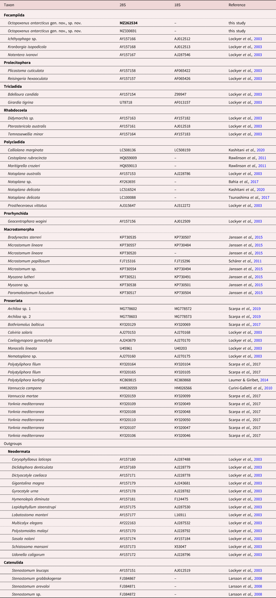
A partial sequence of the 28S rRNA gene (~1100 base pairs) was used to evaluate the phylogenetic relationships of our specimen. Raw reads were assembled using Geneious ver. 10.0.5 software (Kearse et al., Reference Kearse, Moir and Wilson2012). To place this species into the phylogenetic framework, we used publicly available sequences, which showed most similarities to the newly obtained sequence under the Basic Local Alignment Search Tooln algorithm run over the GenBank nr/nt database (Altschul et al., Reference Altschul, Gish, Miller, Myers and Lipman1990). In addition, this dataset was accompanied by the available 28S and 18S data obtained in recent phylogenetic studies; only sequences with good coverage in 28S data were used in the phylogeny (table 1). Catenulida and Neodermata were used as outgroups following Bacon et al. (Reference Bacon, Dallas and Piertney1999) and Lockyer et al. (Reference Lockyer, Olson and Littlewood2003). Original data and publicly available sequences were aligned with the MUSCLE (Edgar, Reference Edgar2004) algorithm in MEGA7 (Kumar et al., Reference Kumar, Stecher and Tamura2016). Indel-rich regions of the 18S and 28S alignments were identified and removed in Gblocks (Talavera & Castresana, Reference Talavera and Castresana2007) with least stringent settings. The best-fitting nucleotide evolution model was tested in the MEGA7 toolkit based on the Bayesian information criterion for each partition. For both markers the general time-reversible model GTR + G + I was chosen. The single-gene datasets (28S + 18S) were concatenated by a simple biopython script following Chaban et al. (Reference Chaban, Ekimova, Schepetov and Chernyshev2019). The concatenated analysis was performed applying evolutionary models separately. The Bayesian inference (BI) was performed in MrBayes 3.2 (Ronquist & Huelsenbeck, Reference Ronquist and Huelsenbeck2003). Markov chains were sampled at intervals of 500 generations. The analysis was started with a random starting tree and run for 107 generations. The burn-in values were 2,500,000 for the ‘sump’ and ‘sumt’ options. The robustness of the phylogenetic relationship was estimated using posterior probabilities (PP) estimated for BI (Ronquist & Huelsenbeck, Reference Ronquist and Huelsenbeck2003). The convergence of parameters and topologies was checked using TRACER 1.7 (Rambaut et al., Reference Rambaut, Drummond, Xie, Baele and Suchard2018). Maximum likelihood (ML) phylogeny inference was performed in the HPC-PTHREADS-AVX option of RaxML HPC-PTHREADS 8.2.12 (Stamatakis, Reference Stamatakis2014) with 1000 pseudoreplicates. Bootstrap values (BS) were placed on the best tree found with SumTrees 3.3.1 from DendroPy Phylogenetic Computing Library 3.12.0 (Sukumaran & Holder, Reference Sukumaran and Holder2010). Final phylogenetic tree images were rendered in FigTree 1.4.0 and their visual components were further modified in Adobe Illustrator CS 2015.
List of species involved in the present molecular analysis based on 18S and 28S rRNA gene sequences.

Results
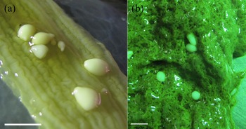
Spherical or slightly oblong worms were found in the intestine (fig. 1a) and in the liver tissue (fig. 1b). The worms in the intestine were sometimes twice as large, or more, than the ones in the liver. A total of 56 specimens of parasitic worms were found in the first octopus (dissected in 2012): 12 in the intestine; and 44 in the liver. The second octopus (dissected in 2015) contained 60 individuals, distributed almost equally between the intestine and the liver.
Octopoxenus antarcticus gen. nov., sp. nov. in Megaleledone setebos: (a) in the intestine; and (b) in the liver tissue. Scale bar: 1 cm.

Taxonomy
Phylum: Platyhelminthes
Order: Fecampiida
Family: Notenteridae Joffe & Kornakova, Reference Joffe and Kornakova1998
Genus: Octopoxenus gen. nov.
Diagnosis: the worms were spherical or oblong and had two morphologically different poles. The frontal pole bears a small conical protrusion containing large elongated pear-shaped frontal glands and large polygonal cells. The ducts of the frontal glands open terminally to form the frontal organ. The caudal pole has an opening shaped as a folded tube connected by the genital pore with a common genital atrium, which continues into a canal with a muscular sheath. Parasite of the digestive system of the cephalopods.
Distribution: Antarctic (Ross Sea).
Etymology: genus name is derived from the order name of the host, Octopoda, and the Ancient Greek word xenos meaning ‘guest’. Thus, Octopoxenus means ‘a guest of the octopodes’.
Type species: Octopoxenus antarcticus sp. nov.
Octopoxenus antarcticus sp. nov. (figs 1–4)
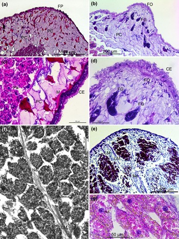
Morphology of the frontal pole and parenchymal body of Octopoxenus antarcticus gen. nov., sp. nov.: (a) frontal pole with light polygonal large cells; (b) frontal glands; (c) integument with ciliary epithelium, mucoid glands and musculature; (d) the frontal organ with large cells of the frontal glands; (e) longitudinal body musculature at the frontal end of the body; (f) longitudinal nerve and longitudinal muscle fibre in the body parenchyma; and (g) the structure of vitelline cells with lipid droplets. CE, ciliary epithelium; FG, frontal glands; FO, frontal organ; FP, frontal pole; LM, longitudinal muscles; M, muscle fibre; Ne, nerves; nu, nucleus; SC, secretory cells; PC, polygonal cells; and Pe, parenchymal cells.

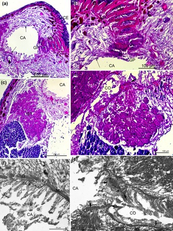
Caudal pole of Octopoxenus antarcticus gen. nov., sp. nov. with genital pore, common genital atrium and copulatory organ: (a, b) morphology of the common genital atrium and genital pore, long arrows note the wall of the atrium, short arrow notes the copulatory organ; (c) two types of the reproductive glands (rg1 and rg2), associating with the common genital atrium and copulatory organ; (d) the copulatory organ and reproductive glands – arrow denotes a sphincter position; (e) the sphincter muscle (arrows) between the common genital atrium and copulatory organ; (f) the epithelium, basal lamina and a musculature of the atrium wall. BL, basal lamina; CA, common genital atrium; CE, ciliary epithelium; CO, copulatory organ; CM, circular muscle; E, epithelium cells; GP, genital pore; LM, longitudinal muscle; M, muscle fibre; MF, muscle folds; and rg1 and rg2, reproductive glands.

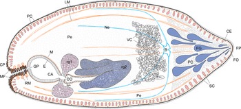
Scheme of Octopoxenus antarcticus gen. nov., sp. nov. B, brain; CA, common genital atrium; CE, ciliary epithelium; CO, copulatory organ; CP, caudal pole; E, epithelium cells; GP, genital pore; FG, frontal glands; FO, frontal organ; FP, frontal pole; LM, longitudinal muscles; M, muscle fibre; MF, muscle folds; Ne, nerves; SC, secretory cells; Sph, sphincter; PC, polygonal cells; Pe, parenchymal cells; RM, ring muscles; and VC, vitelline cells.

Description
The worms were spherical or slightly oblong and had two morphologically different poles, which could be clearly distinguished in histological sections (figs 2 and 3). We refer to them as the frontal and the caudal pole. At the caudal pole there is an opening shaped as a folded tube connected by the genital pore with a spherical cavity (common genital atrium), continuing into a narrowed canal with a muscular sheath (copulatory organ). The frontal pole bears a small conical protrusion filled with large light polygonal cells, which are also present in the area of the body adjacent to the protrusion, occupying up to a quarter of the worm's body volume. Large frontal glands can be seen at the frontal pole. They have an elongated pyriform shape and their ducts open terminally to form the frontal organ (fig. 2b, d).
Integument
The integument of the worms is represented by a ciliary epithelium underlain by an easily visible basal lamina (figs 2 and 3). The cilia cover the body in a dense layer. The cilia at the poles are longer than elsewhere on the body. The epithelium has approximately the same height throughout the body of the worm. The nuclei of the epithelial cells are not submerged, being located above the basal plate.
The cells of the ciliary epithelium are interspersed with intensely stained secretory cells. The nuclei of secretory cells lie under the basal lamina and under the layers of the subepidermal muscles. The cell bodies are elongated and the long ducts are intensely stained. The epidermal secretory cells are more developed at the frontal pole (fig. 2c).
In addition to the intensely stained secretory cells, very large lightly stained polygonal cells are located at the frontal pole under the basal lamina. They lie in several layers and occupy much of the anterior part of the body (fig. 2b). Their layer gradually narrows laterally and is completely absent at the posterior body end. The cytoplasm of the polygonal cells is pale and noticeably granular. Their nuclei are small, rarely visible and dark purple in colour.
Musculature
The musculature of the body wall consists of two layers: a more prominent longitudinal layer; and a less prominent circular layer (figs 2c and 3a, c). The longitudinal muscle fibres are equally thick throughout the body. Two or three of these fibres often stretch along the basal plate of the ciliary epithelium. The circular muscles are more developed at the caudal pole of the body, where their fibres are numerous, and less developed at the frontal pole, where they are represented by rare solitary myofibrils.
In addition to the musculature of the body wall, a poorly developed body musculature is also present in the form of solitary longitudinal and diagonal muscle fibres (figs 2e, f and 4). The diagonal fibres begin at the frontal pole, stretch laterally and attach to the longitudinal musculature of the body wall. Visceral musculature is represented by a thin layer of fibres around the common genital atrium and a well-developed annular musculature at the base of the copulatory organ.
Reproductive system
A genital pore is present at the caudal body end. It is shaped as a folded canal and equipped with thick layers of circular and longitudinal muscles (fig. 3a, b). The pore opens into the common genital atrium, which is connected to the copulatory organ with a well-developed muscular sphincter at its base (fig. 3d, e). The wall of the genital atrium is lined with a high vacuolated epithelium underlain with a thin basal lamina and with musculature (fig. 3f). The copulatory organ has a muscular wall.
There are two types of glands in this area (rg1 and rg2, fig. 3c, d). Gland rg1 consists of large rounded cells with distinct dark nuclei. The cells closely adjoin each other, and are separated from the surrounding parenchyma by a distinct line of the basal lamina. Gland rg2, represented by a compact mass of small violet-blue cells, is a large organ, which is presumably located on the ventral side of the body. The cells form rounded lobular clusters delimited from the parenchyma. Both glands are probably reproductive organs. They are associated with the common genital atrium (rg1) and with the copulatory organ (rg2).
The medullary part of the body is filled with large rounded granular vitelline cells. They are arranged in a dense mass, adjacent to the inner surface of the body wall. At the frontal pole, the nerve and the muscle cords divide the cell mass into separate clusters. Vitelline cells have well-defined large rounded nuclei (50 μm), and their cytoplasm contains numerous lipid granules (fig. 2f, e, g).
Nervous system
The nervous system is represented by an accumulation of nerve elements on the frontal pole. At least four longitudinal nerve cords extend from the brain. In addition, there are two sublateral nerve cords. Thin nerves also stretch along the muscles of the integument; in certain areas, they are connected by transverse commissures with more powerful longitudinal fibres (fig. 2e).
Taxonomic summary
Host: giant Antarctic octopus M. setebos (Robson, 1932) (Octopoda: Megaleledonidae)
Site of infection: liver, intestine
Etymology: the epithet antarcticus refers to the geographical region where the new species was found.
Type locality: Antarctica, Ross Sea (76°30′S & 170°18′E, depth 730 m, date of collection 30 January 2012, and 77°35′S & 179°42′W, depth 670 m, date of collection 15 January 2015).
Prevalence: 116 in two hosts
Intensity of infection: 56–60 ind.
Deposed specimens: Holotype (IPEE RAS #1337-1357 histological sections), paratype (IPEE RAS #1358-1378 histological section) and ten voucher specimens (IPEE RAS #14319, whole worms in 96% ethanol). Holotype and paratypes were deposited in the Museum of Helminthological Collections, Center of Parasitology, Severtsov Institute of Ecology and Evolution, Russian Academy of Sciences, Moscow, Russia (IPEE RAS). The information on O. antarcticus gen. nov., sp. nov. was added to the MorphoBank/https://morphobank.org/ (P4428).
Representative DNA sequences: partial sequence of the 28S rRNA gene is deposited in GenBank United States National Center for Biotechnology Information with accession numbers MZ262534 and MZ330691.
ZooBank registration: urn:lsid:zoobank.org:pub:8AF08020-F4CF-45B3-A81F-0F3F54E1BCB8
Phylogenetic data
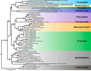
Our BI and ML concatenated phylogenetic trees yielded similar results (fig. 5); most of the clades were highly supported (PP > 0.95; BS > 80). Our specimen clustered together with other representatives of the Fecampiida in a highly supported clade (PP = 1; BS = 100) and was closely related with N. ivanovi (PP = 1; BS = 100).
Phylogenetic relationships of Octopoxenus antarcticus gen. nov., sp. nov. based on the concatenated dataset (28S and 18S). Numbers above branches indicate Bayesian posterior probabilities, and numbers under branches indicate bootstrap values.

Discussion
In this study we recorded a parasitic ‘turbellarian’ from the intestines of the octopus. This is the first report of this kind because ‘turbellarians’ have never been registered in cephalopods before. The newly found ‘turbellarian’, O. antarcticus sp. nov. gen. nov., belongs to the family Notenteridae from the order Fecampiida. We provided the description of the new species and established a new genus to accommodate it. Also, at the end of the discussion, we provide an updated diagnosis of the family Notenteridae.
Octopoxenus antarcticus gen. nov., sp. nov. clearly belongs to the family Notenteridae (Fecampiida) based on the morphology. The genetic data also show the close relationships of O. antarcticus gen. nov., sp. nov. and N. ivanovi, although only a few fecampiid taxa in the GenBank were available for the analysis. Before our study, this family contained only one species, N. ivanovi, reported from the gut of the polychaete M. minuta at the White Sea. In general, O. antarcticus gen. nov., sp. nov. is morphologically similar to N. ivanovi. Both these ‘turbellarians’ have an oval or egg-shaped body with two morphologically different poles and a genital atrium (caudal cavity). In N. ivanovi an unpaired vitelline (yolk) gland occupies almost the entire space of the body, and so it does, apparently, in O. antarcticus gen. nov., sp. nov.
The fine structure and topology of the protonephridial excretory system in N. ivanovi is not known, though the work of the excretory cells in its parenchyma was observed in vivo by Joffe et al. (Reference Joffe, Selivanova and Kornakova1997). We do not have any data on the excretory system of O. antarcticus gen. nov., sp. nov., either.
Morphological differences between the two species are evident. In N. ivanovi, the dorsal and the ventral epidermis are significantly different in height (2–6 μm vs. 120 μm, respectively) (Joffe et al., Reference Joffe, Selivanova and Kornakova1997; Joffe & Kornakova, Reference Joffe and Kornakova1998). It has been hypothesized that the dorsal epidermis, facing the intestinal lumen of the polychaete host, functionally replaces the digestive system. The concave ventral epidermis faces the intestinal wall and the entire ventral side of the ‘turbellarian’ forms a kind of sucker owing to the longitudinal muscles. No such differences in the structure of the ciliary cover were noted in O. antarcticus gen. nov., sp. nov. Notentera ivanovi has two types of frontal glands while in O. antarcticus gen. nov., sp. nov. we distinguished only one type.
There are other differences between N. ivanovi and O. antarcticus gen. nov., sp. nov. In the former, vitelline cells occupy most of the body, while in the latter, they are arranged in grape-shaped clusters in the anterior region of the middle third of the body. The epithelial lining of the posterior wall of the common genital atrium is thickened in N. ivanovi and regular in O. antarcticus gen. nov., sp. nov. On the one hand, based on the location of the ovary and the testes, and the size of their cells in N. ivanovi, which was ascertained with the help of electron microscopy (Joffe & Kornakova, Reference Joffe and Kornakova1998; Kornakova & Joffe, Reference Kornakova and Joffe1999), we can assume that rg1 of O. antarcticus gen. nov., sp. nov. is the ovary, while rg2 is the testis; however, it could be some other type of glands associated with the reproductive system. Electron-microscopic studies of O. antarcticus gen. nov., sp. nov. are necessary for clarification.
The brain of N. ivanovi looks like a star with six rays. Three pairs of long brain roots extend from the brain (Raikova et al., Reference Raikova, Kotikova and Frolova2017) and pass into the paired ventral, dorsal and lateral longitudinal cords. The brain of O. antarcticus gen. nov., sp. nov. is also star-shaped, and there are four nerve cords passing towards the posterior body end, but we did not find any nerve cords directed anteriorly. Nerve cells in O. antarcticus gen. nov., sp. nov. are accumulated under the frontal organ.
To sum up, O. antarcticus gen. nov., sp. nov., while being clearly distinct from N. ivanovi, is similar to it in general morphology. Moreover, these two species clustered together on the phylogenetic tree (fig. 5). Therefore, we see no need in establishing a new family and assign the new species to the Notenteridae. However, the two species that now comprise this family use different hosts inhabiting different geographical areas and have different localization within the host. The taxonomic position of O. antarcticus gen. nov., sp. nov. might probably have to be reconsidered after the emergence of new morphological and molecular–genetic evidence.
As for the differences between these two species and other representatives of Fecampiida, the most significant is the absence of a cocoon, the site of infection, and typical hosts. Kronborgia spp. form a cocoon and infect haemocoel of isopods and amphipods (Shinn & Christensen, Reference Shinn and Christensen1985; Williams, Reference Williams1988). Urastoma cyprinae (Graff, 1882) have a cocoon, and inhabit mantle cavity of mussels (Robledo et al., Reference Robledo, Caceres-Martinez, Sluys and Figueras Huerta1994; Noury-Sraïri et al., Reference Noury-Sraïri, Justine and Euzet1990). Fecampia spp. have a cocoon and infects isopods, decapods and cirripedids (Kuris et al., Reference Kuris, Torchin and Lafferty2002). Four known species of Genostoma Dörler, 1900 live under the carapace of leptostracan crustaceans of the genus Nebalia Leach, 1814 and possess an intestine (Hyra, Reference Hyra1993).
Octopoxenus antarcticus gen. nov., sp. nov. was found in the giant Antarctic octopus, Megaleledone setebos, in the Ross Sea, while N. ivanovi inhabits the polychaete Micronephthys minuta in the White Sea. These striking differences set one thinking about the evolutionary paths of notenterids and their coevolution with their hosts. However, information on this topic is scarce, and so far little can be said on this matter. We can only note that the polychaete host of N. ivanovi has a circumpolar distribution in the Arctic, while the cephalopod host of O. antarcticus gen. nov., sp. nov. has a circumpolar distribution in the Antarctic. Whatever pathways of evolution have led this clade of ‘turbellarians’ to their divided range (fig. 4), we can suggest that cold water could provide an advantage.
We found specimens of O. antarcticus gen. nov., sp. nov. in two sites within M. setebos: the intestine and the liver. This finding implies that these ‘turbellarians’ can move within the host. How they do it is unclear. In general, an organism with a rounded body has fewer possibilities for movement by changing the mechanical pressure on some footing such as the host tissue. However, the morphology of the newly found worm (fig. 4) suggests that it might use the frontal glands and the muscular folds at the caudal end for temporary attachment.
The description of the O. antarcticus gen. nov., sp. nov. requires an update of the family Notenteridae.
Family: Notenteridae Joffe & Kornakova, Reference Joffe and Kornakova1998 emend.
Diagnosis (based on Joffe & Kornakova, Reference Joffe and Kornakova1998 with changes):
The integument of the worms is represented by a ciliary epithelium underlain by an easily visible basal lamina. The cilia cover the body in a dense layer. The ducts of the frontal glands open terminally to form the frontal organ. A genital pore is present at the caudal body end. The pore opens into the common genital atrium, which is connected to the copulatory organ. The central portion of the body is occupied by a large unpaired vitellarium. Parasite of the digestive system of the polychetes and cephalopods.
Acknowledgements
We thank the crew of the FV Yantar-31 (ORION Ltd., Khabarovsk) and Alexander Terentiev (Russian Federal Research Institute of Fisheries and Oceanography, Kerch) for help with the sampling. We are grateful to Dr Yaroslav Zabotin (Kazan Federal University, Kazan), Dr Sc Professor Alexei Tchesunov (Lomonosov Moscow State University, Moscow), Chingiz Nigmatullin (Russian Federal Research Institute of Fisheries and Oceanography, Kaliningrad) and Dr Sergey Sokolov (A.N. Severtsov Institute of Ecology and Evolution, Moscow) for valuable advice. We are also grateful to Natalia Lentsman (St Petersburg State University, St Petersburg) for help with the preparation of the manuscript.
Financial support
This research received no specific grant from any funding agency in the public, commercial, or not-for-profit sectors
Conflicts of interest
None.
Availability of data and material
Data available within the article or its supplementary materials.
Authors’ contributions
I.G. collected the worms, contributed to genetics and manuscript preparation; N.B. contributed to text preparation and description of histological sections; I.E. contributed to phylogenetic analysis; and K.Z. made histological sections. All authors read and approved the final manuscript.
Ethics approval
Not applicable.
Consent to participate
Not applicable.
Consent for publication
Not applicable.
Compliance with ethical standards
Not applicable.