INTRODUCTION
Animals belonging to the order Polycladida are free-living platyhelminthes, with most species known from tropical seas. About 900 species have been described around the world (Newman & Cannon, Reference Newman and Cannon2003). Among them, about 125 were reported from the Tropical Western Atlantic, 70 occur in Brazil, 46 being endemic (Quiroga et al., Reference Quiroga, Bolaños and Litvaitis2004a; Bahia et al., Reference Bahia, Padula, Passeri Lavrado and Quiroga2014). From the Brazilian coast, the existing knowledge is mostly the result of works conducted between 1950 and 1970, based on specimens collected at São Paulo region (Marcus, Reference Marcus1947, Reference Marcus1948, Reference Marcus1949, Reference Marcus1950, Reference Marcus1952). Recent contributions, including new local and country records were made by Bahia & Padula (Reference Bahia and Padula2009), Bahia et al. (Reference Bahia, Padula and Delgado2012, Reference Bahia, Padula, Passeri Lavrado and Quiroga2014) and Queiroz et al. (Reference Queiroz, Sales, Neves and Johnsson2013).
On the north-eastern Brazilian coast, such as in the state of Alagoas, reef ecosystems are abundant and have a rich biological diversity (Correia & Sovierzoski, Reference Correia and Sovierzoski2009). These reefs include a benthic fauna, which provide various natural substrates for the Polycladida, composed mainly of sponges (Cedro et al., Reference Cedro, Hajdu, Sovierzoski, Correia, Custódio, Lobo-Hajdu, Hajdu and Muricy2007, Reference Cedro, Hajdu and Correia2011, Reference Cedro, Hajdu and Correia2013; Bispo et al., Reference Bispo, Correia and Hajdu2014), corals (Correia, Reference Correia2011) and bryozoans (Vieira et al., Reference Vieira, Gordon and Correia2007, Reference Vieira, Migotto and Winston2008, Reference Vieira, Migotto and Winston2010). A variety of algae also provides habitats for numerous associated organisms (Santos & Correia, Reference Santos and Correia1994, Reference Santos and Correia1995, Reference Santos and Correia2001), which are important environments for flatworms and other small invertebrates such as opisthobranch molluscs (Padula et al., Reference Padula, Bahia, Correia and Sovierzosky2012) and echinoderm brittle stars (Lima et al., Reference Lima, Correia, Sovierzoski and Manso2011, Reference Lima, Sovierzoski and Correia2013). However, there is a significant lack of information, particularly about marine platyhelminthes in this region, with no record of the order Polycladida from Alagoas reef ecosystems. Herein we aim to present the first records of Polycladida from Alagoas coast, including the description of a new species.
MATERIALS AND METHODS
Alagoas coast is approximately 230 km long, and it is limited by the Persinunga River on the north and by the São Francisco River on the south (8°54′S–35°9′W and 10°30′S–36°23′W), The main ecosystems that can be found are coral and sandstone reefs, lagoons, rivers and mangroves. The coral reefs were formed on calcareous sedimentary rock, composed of an aggregation of dead organisms, including skeletons of corals and hydrocorals combined with crusts of calcareous algae and other invertebrates (Correia & Sovierzoski, Reference Correia and Sovierzoski2009). Many of these fringing reefs are located near the beach line, where the top of the reef platform is exposed during low tides. The sandstone reefs were formed by old sandbanks solidified through sedimentation, starting from chemical reactions with calcium carbonate from the Quaternary Period, and are generally arranged in rows parallel to the coastline and near the outlets of rivers and estuaries (Correia & Sovierzoski, Reference Correia and Sovierzoski2009; Correia, Reference Correia2011).
All specimens were manually collected under rocks at the intertidal zone or in the sublittoral by snorkelling or scuba diving along the edge of the reef platforms during low tide. Collections were conducted in January 2008 and January 2012, and were carried out in reef ecosystems along the central coast of Alagoas State, Brazil (Figure 1). Seven reef ecosystems were sampled: coral reefs of Ponta Verde (9°39′57″S–35°41′32″W), Jatiúca (9°39′12″S–35°41′46″W), Piscina dos Amores (9°40′39″S–35°42′10″W), Pajuçara (9°41′06″S–35°43′22″W) and Riacho Doce (9°34′55″S–35°39′25″), and sandstone reefs of Francês (9°46′03″S–35°50′13″W) and Saco da Pedra (9°44′26″S–35°48′59″W) (Figure 1).
Map of reef environments studied at Alagoas State, north-eastern Brazil.

In the laboratory, specimens were photographed alive with a digital camera. Afterwards, they were fixed in 10% frozen formalin, following a modified methodology (Newman & Cannon, Reference Newman and Cannon2003), and transferred to 70% ethanol for histological preparation. Specimens were measured after fixation (length mm × width mm). The identification was based on morphological characteristics, colouration pattern, ocelli position and slides of the reproductive structures stained by haematoxylin-eosin method (Bolaños et al., Reference Bolaños, Quiroga and Litvaitis2007). Specimens were compared with original descriptions and previous publications (Marcus, Reference Marcus1949, Reference Marcus1950, Reference Marcus1952; Marcus & Marcus, Reference Marcus and Marcus1968; Bolaños et al., Reference Bolaños, Quiroga and Litvaitis2007). Collected material was deposited on the Platyhelminthes collection in the Museu Nacional/Universidade Federal do Rio de Janeiro, Brazil (MNRJ-PLAT).
RESULTS
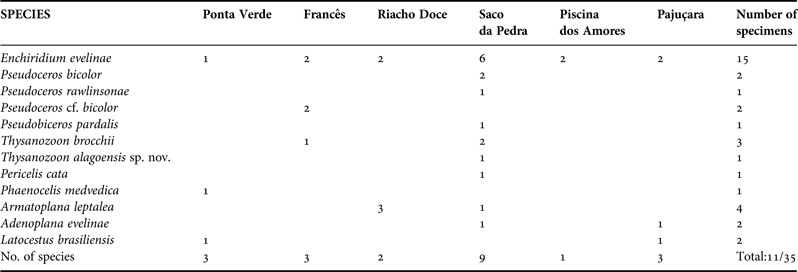
There were 35 specimens found belonging to 11 species of the order Polycladida. Pericelis cata, Enchiridium evelinae, Pseudoceros bicolor, Pseudoceros rawlinsonae, Pseudobiceros pardalis, Thysanozoon brocchii, Thysanozoon alagoensis sp. nov., Armatoplana leptalea, Adenoplana evelinae, Latocestus brasiliensis and Phaenocelis medvedica. The species Enchiridium evelinae was the most common, with 15 specimens collected, and found in all sampled reefs. Greater species richness was observed at Saco da Pedra reef. The lowest richness was found at Piscina dos Amores coral reef. All occurrences reported here are the first records of Polycladida from Alagoas ecosystems (Table 1). As some of the species found in this study were treated on previous papers by the authors, just the material examined, with measurements, distribution and remarks are included.
Species and number of specimens found at each reef ecosystem on the Alagoas coast.
SYSTEMATICS
Order POLYCLADIDA Lang, Reference Lang1884
Suborder COTYLEA Lang, Reference Lang1884
Family PERICELIDAE Laidlaw, Reference Laidlaw1902
Genus Pericelis Laidlaw, Reference Laidlaw1902
Pericelis cata Marcus & Marcus, Reference Marcus and Marcus1968
(Figure 2B)
(A) Enchiridium evelinae (MNRJ-PLAT 80); (B) Pericelis cata (MNRJ-PLAT 96); (C) Pseudoceros bicolor (MNRJ-PLAT 85); (D) Pseudoceros rawlinsonae (MNRJ-PLAT 87); (E) Pseudobiceros pardalis (MNRJ-PLAT 92).
EXAMINED MATERIAL
One mature specimen (18 × 16 mm) as sagittal sections of reproductive structures (MNRJ-PLAT 96, 13 slides). Collected 26 January 2012 at Saco da Pedra sandstone reef, Marechal Deodoro, Alagoas, Brazil.
GEOGRAPHIC DISTRIBUTION
This species was recorded from Curaçao (type locality; Marcus & Marcus, Reference Marcus and Marcus1968), Colombian Caribbean (Quiroga et al., Reference Quiroga, Bolaños and Litvaitis2004b), Cabo Frio, south-eastern Brazil (Bahia & Padula, Reference Bahia and Padula2009) and Salvador, north-eastern Brazil (Queiroz et al., Reference Queiroz, Sales, Neves and Johnsson2013). This is the first record of this species from Alagoas State.
REMARKS
The specimen herein studied was smaller than those found at Cabo Frio, south-eastern Brazil (Bahia & Padula, Reference Bahia and Padula2009) and those from Bahia State, north-eastern Brazil (Queiroz et al., Reference Queiroz, Sales, Neves and Johnsson2013). During the collections two specimens of P. cata were placed in the same container as some specimens of the opisthobranch mollusc Micromelo undatus. When freed to be photographed the Pericelis were much damaged and one was not useful for study. This could have happened because of some fighting with the molluscs or some toxic chemical compound they might present.
Family PROSTHIOSTOMIDAE Lang, Reference Lang1884
Genus Enchiridium Bock, Reference Bock1913
Enchiridium evelinae Marcus, Reference Marcus1949
(Figure 2A)
EXAMINED MATERIAL AND LOCALITY
One mature specimen (MNRJ-PLAT 77, 32 × 9 mm) collected 6 January 2008 at Brazil, Alagoas, Maceió, Ponta Verde coral reef. One mature specimen (MNRJ-PLAT 78, 29 × 8 mm) collected 7 January 2008 at Pajuçara coral reef, Maceió, Alagoas, Brazil. Two specimens (MNRJ-PLAT 79, 26 × 6 mm and 30 × 8 mm) collected 9 January 2008 at Francês sandstone reef, Maceió, Alagoas, Brazil. Two specimens (MNRJ-PLAT 80, 21 × 7 mm and 32 × 10 mm). One as sagittal sections of reproductive structures (21 slides). Collected 10 January 2008 at Riacho Doce coral reef, Maceió, Alagoas, Brazil. Five specimens (MNRJ-PLAT 81, 6 × 3 mm, 12 × 5 mm, 16 × 6 mm, 21 × 7 mm and 23 × 8 mm) collected 11 January 2008 at Saco da Pedra sandstone reef, Marechal Deodoro, Alagoas, Brazil. Two specimens (MNRJ-PLAT 82, 21 × 7 mm and 22 × 7 mm) collected 13 January 2008 at Piscina dos Amores coral reef, Maceió, Alagoas, Brazil. One specimen (MNRJ-PLAT 83, 18 × 7 mm) collected 7 February 2008 at Pajuçara coral reef, Maceió, Alagoas, Brazil. One specimen (MNRJ-PLAT 84, 26 × 11 mm) collected 27 January 2012 at Saco da Pedra sandstone reef, Maceió, Alagoas, Brazil. All specimens preserved in 70% ethanol.
GEOGRAPHIC DISTRIBUTION
Originally described from São Paulo State, south-eastern Brazil (Marcus, Reference Marcus1949) reported to Rio Grande do Norte State (Bahia et al., Reference Bahia, Padula and Delgado2012), Rio de Janeiro State (Bahia et al., Reference Bahia, Padula, Passeri Lavrado and Quiroga2014) and now Alagoas State, north-eastern Brazil. It is also known from Curaçao (Marcus & Marcus, Reference Marcus and Marcus1968). This species is considered by Rawlinson (Reference Rawlinson2008) as representative of seagrass habitat. We found our specimens both in sandstone and coral reefs and E. evelinae was the most common species in our samplings. This is the first record of this species from Alagoas State.
REMARKS
This species was observed copulating (video in Supplementary material). It presented reciprocal insemination and a somewhat violent penis-fencing behaviour. Unfortunately the following eggmass laying could not be observed. Also one specimen of Enchiridium evelinae ate a specimen of Adenoplana evelinae during a fortnight in the same Petri dish. We noticed differences in colouration between the specimens found in Alagoas and those from Rio de Janeiro, the latter sometimes has a different distribution of the dorsal brown spots, with them more densely disposed in the median line (personal observation).
Family PSEUDOCEROTIDAE Lang, Reference Lang1884
Genus Pseudoceros Lang, Reference Lang1884
Pseudoceros bicolor Verrill, Reference Verrill1901
(Figures 2C & 3)
Pseudoceros cf. bicolor (A) in vivo; (B) and (C) detail of the anterior region; (D) ventral view; (E) sagittal section of male reproductive structures; (F) sagittal section of male and female reproductive structures. ce, cerebral eyespots; cg, cement glands; cp, cement pouch; fg, female gonopore; ma, male atrium; mg, male gonopore; pe, penis; ph, pharynx; pv, prostatic vesicle; su, sucker; sv, seminal vesicle; te, tentacular eyespots; ut, uteri; va, vagina.
EXAMINED MATERIAL
One mature specimen (12 × 8 mm) as sagittal sections of reproductive structures (MNRJ-PLAT 85, 9 slides). One mature specimen (MNRJ-PLAT 86, 10 × 8 mm) collected 26 January 2012. Both collected at Saco da Pedra sandstone reef, Marechal Deodoro, Alagoas, Brazil and preserved in 70% ethanol. One mature specimen (11 × 10 mm) as sagittal section of reproductive structures (MNRJ-PLAT 90, 16 slides). One mature specimen (15 × 10 mm) as sagittal section of reproductive structures (MNRJ-PLAT 91, 15 slides). Both collected 25 January 2012 at Francês sandstone reef, Marechal Deodoro, Alagoas, Brazil.
GEOGRAPHIC DISTRIBUTION
Described from Bermudas (type locality; Verrill, Reference Verrill1901), reported from Curacao (Marcus & Marcus, Reference Marcus and Marcus1968), Caribbean coast of Colombia (Quiroga et al., Reference Quiroga, Bolaños and Litvaitis2004b), Florida, Virgin Islands, Jamaica, Belize, Honduras, Caribbean coast of Panama (Rawlinson, Reference Rawlinson2008) and south-eastern Brazil (Bahia & Padula, Reference Bahia and Padula2009). This is the first record of this species from north-eastern Brazil.
REMARKS
The specimens MNRJ-PLAT 85 and 86 are in accordance with the description and posterior amendment. Specimens MNRJ-PLAT 90 and MNRJ-PLAT 91 (at Table 1 counted separately as Pseudoceros cf. bicolor) were somewhat different from the former specimens. Their background colour was yellowish orange with scattered white and dark spots (Figure 3A); whitish translucent marginal band with a thin light yellow outermost line, only seen in live specimens. Seminal vesicle muscularized and elongated. Prostatic vesicle rounded and small, located above the penial papillae (Figure 3E). They resemble Pseudoceros bicolor in its background coloration pattern, but it lacks the white marginal band with black languettes characteristic of this species. Also the colour has hints of orange that are absent in specimens from the same locality and from other regions of the Brazilian coast (Bahia & Padula, Reference Bahia and Padula2009), and the seminal vesicle of these specimens is more elongated than rounded as usually found in P. bicolor. The material differs from the recently described P. juani in body proportion length × width, this species has a more elongated body (Bahia et al., Reference Bahia, Padula, Passeri Lavrado and Quiroga2014) than the Pseudoceros specimens found in Alagoas. Also, the seminal vesicle of P. juani is proportionally much larger than in P. bicolor and in the two specimens studied. However both P. juani and the two studied specimens have both scattered white and dark spots and there is a hint of orange in Alagoas’ specimens, which is the background colour of P. juani, therefore we cannot rule out that those specimens can be some morphotype between P. bicolor and P. juani, despite the latter having no record in Alagoas so far. These two specimens are here, for now, identified as P. bicolor; this should be confirmed through a future molecular analysis.
Pseudoceros rawlinsonae Bolaños, Quiroga & Litvaitsi, Reference Bolaños, Quiroga and Litvaitis2007
(Figure 2D)
EXAMINED MATERIAL
One mature specimen (10 × 6 mm) as sagittal sections of reproductive structures (MNRJ-PLAT 87, 18 slides). Collected 11 January 2008 at Saco da Pedra sandstone reef, Marechal Deodoro, Alagoas, Brazil.
GEOGRAPHIC DISTRIBUTION
Originally described from the American Virgin Islands and Bonaire (Bolaños et al., Reference Bolaños, Quiroga and Litvaitis2007). After the revision of Pseudoceros bicolor complex, P. rawlinsonae was also reported from Florida, Honduras, Jamaica, Bahamas, Curaçao (Litvaitis et al., Reference Litvaitis, Bolaños and Quiroga2010) and south-eastern Brazil (Bahia & Padula, Reference Bahia and Padula2009, as P. bicolor; Bahia et al., Reference Bahia, Padula, Passeri Lavrado and Quiroga2014). This is the first record of this species from north-eastern Brazil.
Genus Pseudobiceros Faubel, Reference Faubel1984
Pseudobiceros pardalis (Verrill, Reference Verrill1900)
(Figure 2E)
EXAMINED MATERIAL
One specimen (45 × 37 mm) as sagittal sections of reproductive structures (MNRJ-PLAT 92, 35 slides). Collected 27 January 2012 at Saco da Pedra sandstone reef, Marechal Deodoro, Alagoas, Brazil.
GEOGRAPHIC DISTRIBUTION
Pseudobiceros pardalis was described from Bermudas (Verrill, Reference Verrill1900) and reported from Bahamas, south Florida and Panama (Bolaños et al., Reference Bolaños, Quiroga and Litvaitis2007). It was recently reported for the first time from Brazil, but south from Alagoas (Bahia et al., Reference Bahia, Padula, Passeri Lavrado and Quiroga2014). This is the first record of this species from north-eastern Brazil.
REMARKS
Our specimens have lighter colouration than the ones in the original description (Verrill, Reference Verrill1900) and re-description (Bolaños et al., Reference Bolaños, Quiroga and Litvaitis2007), but darker than reported from south-eastern Brazil (Bahia et al., Reference Bahia, Padula, Passeri Lavrado and Quiroga2014), probably due to differences in size and the nutritional conditions of the animals. The specimens found in the Caribbean (Bolaños et al., Reference Bolaños, Quiroga and Litvaitis2007) have a concentration of white dots near the margin that is not so clear in Brazilian specimens (Figure 2E), both from Alagoas and from Rio de Janeiro (Bahia et al., Reference Bahia, Padula, Passeri Lavrado and Quiroga2014).
Genus Thysanozoon Grube, Reference Grube1840
Thysanozoon brocchii (Risso, Reference Risso1818)
(Figure 4)
Thysanozoon brocchii (A) in vivo; (B) detail of the ventral surface; (C) sagittal section of the male reproductive structures; (D) sagittal section of the female reproductive structures. cg, cement glands; fg, female gonopore; it, intestine; ma, male atrium; mg, male gonopore; mo, mouth; pe, penis; ph, pharynx; pv, prostatic vesicle; st, stylet; su, sucker; ut, uteri; va, vagina.
EXAMINED MATERIAL
Two specimens (22 × 18 mm and 6 × 4 mm), the mature as sagittal sections of reproductive structures (MNRJ-PLAT 93, 29 slides). Collected 11 January 2008 at Saco da Pedra sandstone reef, Marechal Deodoro, Alagoas, Brazil. One specimen (17 × 20 mm) (MNRJ-PLAT 94) collected 25 January 2012 at Francês sandstone reef, Marechal Deodoro, Alagoas, Brazil.
GEOGRAPHIC DISTRIBUTION
Cosmopolitan species described from Naples, Italy (type locality; Risso, Reference Risso1818) and other parts of the Mediterranean Sea, UK, south and west from Africa, Florida, Colombian Caribbean, Brazil, Japan and New Zealand (Prudhoe, Reference Prudhoe1985; Quiroga et al., Reference Quiroga, Bolaños and Litvaitis2004b). It was also reported from Canary Islands (Vera et al., Reference Vera, Moro, Bacallado and Hernández2008), Argentina (Brusa et al., Reference Brusa, Damborenea and Quiroga2009), from north-eastern (Bahia et al., Reference Bahia, Padula and Delgado2012) and south-eastern Brazil (Bahia et al., Reference Bahia, Padula, Passeri Lavrado and Quiroga2014). This is the first record from Alagoas State.
REMARKS
The specimens found at Alagoas all have rounded papillae and brownish colouration (Figure 4A). This is not the case in some south-eastern Brazil and in the Mediterranean Sea specimens (Bahia et al., Reference Bahia, Padula, Passeri Lavrado and Quiroga2014). Specimens from those areas can have slender papillae and black to greyish colouration, and European specimens present a red marginal band (Bahia pers. obs.).
Thysanozoon alagoensis sp. nov.
(Figures 5 & 6)
Thysanozoon alagoensis sp. nov. (A) in vivo; (B) ventral view; (C) detail of the anterior margin, showing pseudotentacles; (D) detail of cerebral and tentacular eyespots; (E) sagittal section of female reproductive structures; (F) and (G) sagittal sections of male reproductive structures. ce, cerebral eyespots; cg, cement glands; fg, female gonopore; ma, male atrium; mg, male gonopore; pa, penis papilla; pe, pseudotentacular eyespots; ph, pharynx; pt, pseudotentacles; pv, prostatic vesicle; su, sucker; sv, seminal vesicle; ut, uteri; va, vagina.
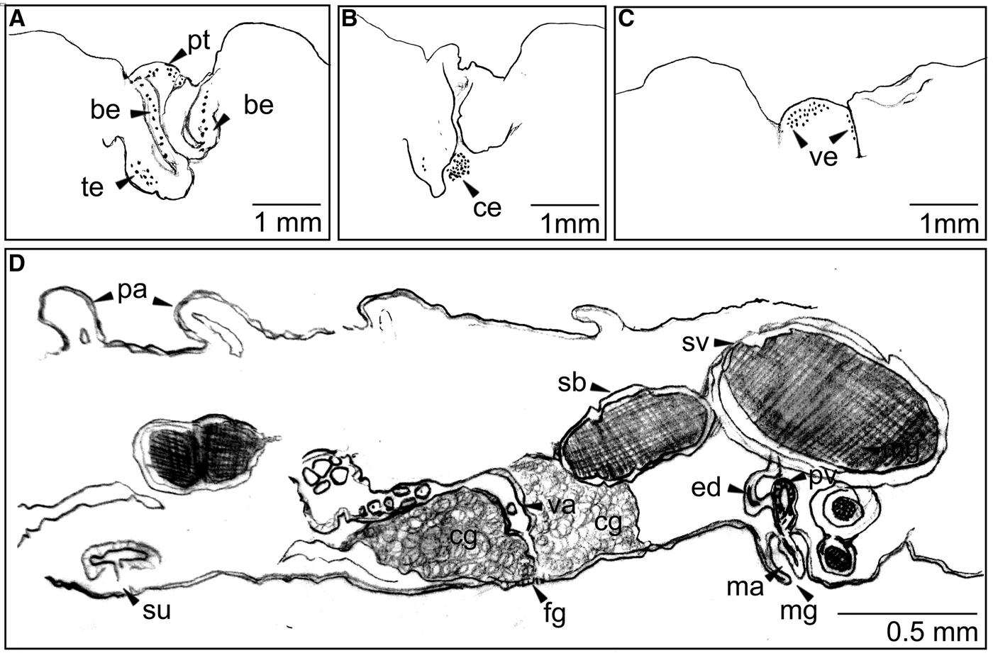
Thysanozoon alagoensis sp. nov. (A) detail of pseudotentacular eyespots; (B) cerebral eyespots; (C) ventral eyespots; (D) tentative reconstruction of serial sections. be, pseudotentacle border eyespots; ce, cerebral eyespots; cg, cement glands; ed, ejaculatory duct; fg, female gonopore; ma, male atrium; mg, male gonopore; pa, papilla; pt, eyespots between pseudotentacles; pv, prostatic vesicle; sb, spermiducal bulbs; su, sucker; sv, seminal vesicle; te, pseudotentacles tips eyespots; va, vagina; ve, ventral eyespots.
TYPE MATERIAL
Holotype: one specimen (19 × 16 mm), as sagittal sections of reproductive structures (MNRJ-PLAT 95, 26 slides). Collected 27 January 2012 at Saco da Pedra sandstone reef, Marechal Deodoro, Alagoas, Brazil, 4 m deep.
GEOGRAPHIC DISTRIBUTION
The species is only known from the type locality.
ETYMOLOGY
The specific name alagoensis makes reference to the geopolitical division of Brazil, Alagoas State, where the holotype specimen was found.
DIAGNOSIS
Greyish background colour with reddish brown papillae, reddish brown margin and dark almost black tentacles with a hint of yellowish pigmentation on the border. Unpigmented line present at the median line of the body. Tentacular eyespots arranged in a line at the border and a group of eyespots at each tentacular tip. Pharynx ruffled with seven simple folds. Seminal vesicle elongated and huge. Its extension reaches the front of the penial papillae and the seminal duct come in a curve to join the penis. Prostatic vesicle small and comma shaped. Male atrium simple. Greatly developed spermiducal vesicles.
DESCRIPTION
Colour: Greyish background colour with reddish brown papillae, reddish brown margin and dark almost black tentacles (Figure 5A). Some papillae are more lightly coloured, more like the background colour. Unpigmented line present at the median line of the body from the tentacles until the last 1/6 of the body length (Figure 5A).
Form: Elongated with papillated dorsal surface (Figure 5B) with slender whitish tipped papillae.
Tentacles: Dark almost black tentacles with a hint of yellowish pigmentation on the border. More rounded than pointed, reaches 1 mm.
Eyes: Cerebral eyes numerous and arranged in a horseshoe shaped group (Figure 6B). Tentacular eyespots arranged in a line at the border (Figure 6A) and a group of eyespots at each tentacular tip (about 20). Frontally, in between psedotentacles, there are also clusters of eyespots, it is not clear if as an extension of the border eyespots groups or distinctive cluster (Figure 6A). Ventral eyespots groups (Figure 6C) with about 25 eyespots.
Digestive system: Pharynx ruffled with seven simple folds (Figure 5B). Reaches 6 mm. Mouth opens at 6 mm from the anterior margin.
Epidermis and body wall: Thin epidermis (Figure 5D) and body wall (0.04 mm), even thinner ventrally (0.01). Sucker with 0.7 mm diameter is 2 mm behind the female pore.
Gonopores: Two male and one female gonopores present, 1.5 mm apart (Figure 5B). Male pores at 7 mm from the anterior margin, both measure 0.5 mm and the female pore 1 mm.
Male reproductive system: Seminal vesicle elongated and huge (Figure 5E). Its extension reaches the front of the penial papillae and the seminal duct comes in a curve to join the penis. Penis papillae (Figure 5D) 0.18 mm. Prostatic vesicle small (0.09 mm) and comma shaped (Figure 5E). Its duct joins the ejaculatory duct and enters the penis papillae (Figure 6D). Seminal vesicle elongated and spermiducal vesicles greatly developed and reaching the region right above the male gonopore (Figure 5E), maybe dislodging the seminal vesicle. Male atrium simple and 0.2 mm deep (Figure 5E).
Female reproductive system: Uteri well developed and full of eggs (0.089 mm diameter). Cement glands also well developed (Figure 5C).
TAXONOMIC REMARKS
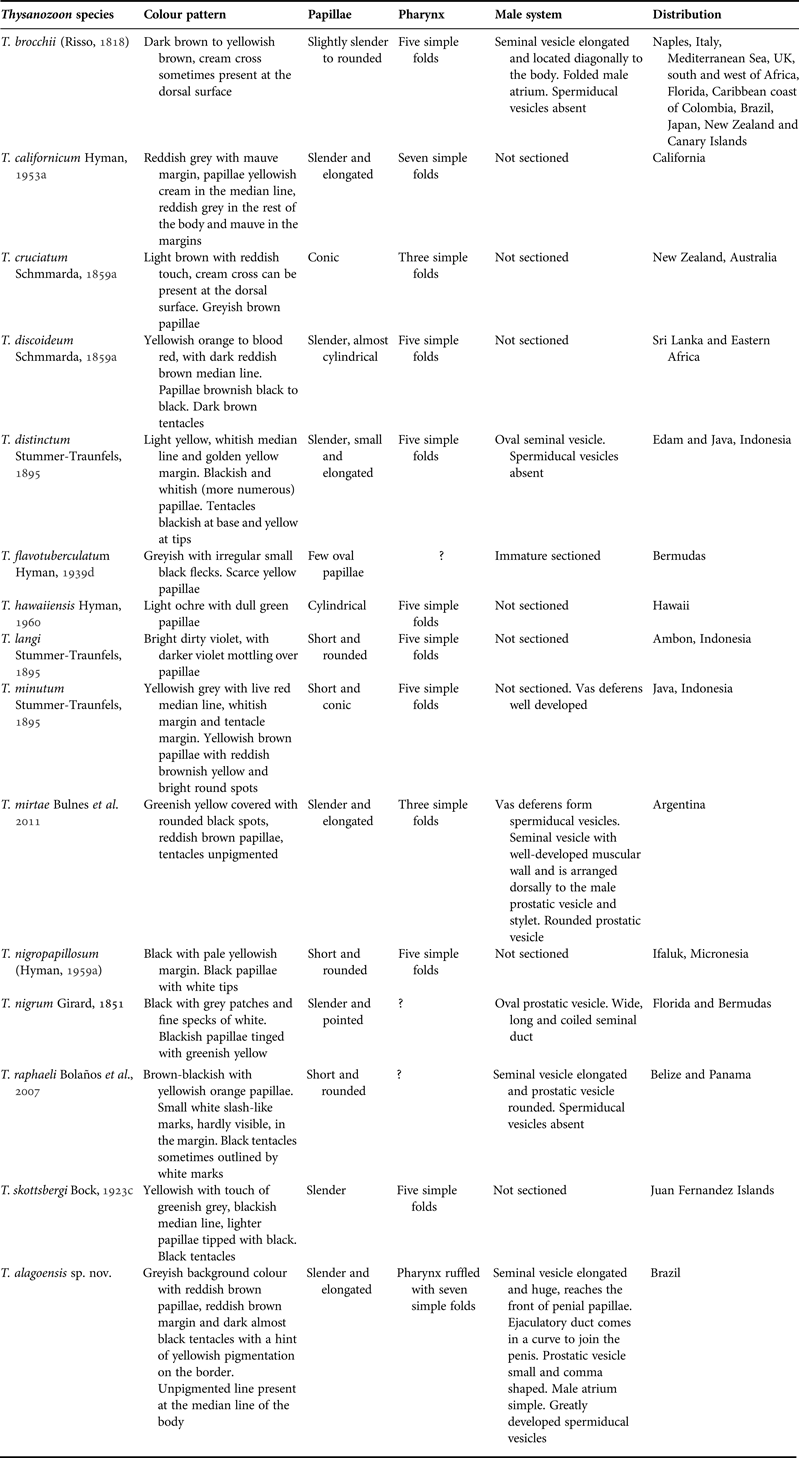
Our specimen has a simple male atrium, different from the folded atrium found in Thysanozoon brocchii. Also the T. alagoensis has a different colouration pattern, and the dorsal papillae of this species are slender and Thysanozoon brocchii have rounder ones (Table 2). Other similar species such as Thysanozoon californicum (Hyman, Reference Hyman1953a) also has slender papillae and elongated pharynx with seven simple folds, but the colouration pattern differs from the Brazilian species (Table 2). The most similar species found in the literature is Thysanozoon mirtae, recently described from Argentina (Bulnes et al., Reference Bulnes, Albano, Obenat and Cazzaniga2011). However, there are marked differences between them, T. mirtae has a smaller pharynx (5.5 mm) and with fewer folds. This is especially distinctive as the specimen from Argentina is much larger, almost double the size of ours, and it would be expected that a larger specimen would have a larger pharynx. Also, the pseudotentacles of the Argentinian species are colourless and the ones in T. alagoensis are dark with yellowish tips. The Argentinian species also present rounded black spots in the dorsal surface that are absent in our specimen (Bulnes et al., Reference Bulnes, Albano, Obenat and Cazzaniga2011). Both T. alagoensis and T. mirtae have slender, elongated papillae, which are also found in Thysanozoon skottsbergi (Bock, Reference Bock1913) and T. distinctum (Stummer-Traunfels, Reference Stummer-Traunfels1895), but the former has darker pigmentation on the papillae and the latter has a golden yellow margin which is markedly different from the reddish brown margin of T. alagoensis sp. nov. and T. mirtae. Both also have apparent spermiducal vesicles, but in T. alagoensis sp. nov. they are greatly developed. Yet another difference between these two South American species is the parenchymatic musculature, developed in T. alagoensis sp. nov. and not in T. mirtae. Other valid species of the genus are compared in Table 2. It is repeatedly stated in the literature that the female system is very uniform throughout the genus, therefore it was not included in the comparative table.
Suborder ACOTYLEA Lang, Reference Lang1884
Family CRYPTOCELIDAE Laidlaw, Reference Laidlaw1902
Genus Phaenocelis von Stummer-Traunfels, Reference Stummer-Traunfels and Bronn1933
Phaenocelis medvedica Marcus, Reference Marcus1952
(Figure 7)
Phaenocelis medvedica (A) in vivo; (B) sagittal section showing muscular layers; (C) fixed worm; (D) anterior region detail showing eyespots. ce, cerebral eyespots; cl, circular layer; dl, diagonal layer; es, eyespots; ll, longitudinal layer; lv, Lang's vesicle; me, marginal eyespots; te, tentacular eyespots.
Thysanozoon species characteristics comparison.
EXAMINED MATERIAL
One specimen (19 × 5 mm) as sagittal sections of reproductive structures (MNRJ-PLAT 97, 9 slides). Collected 6 January 2008 at Ponta Verde coral reef, Maceió, Alagoas, Brazil.
GEOGRAPHIC DISTRIBUTION
This species was described from São Paulo State, Brazil (Marcus, Reference Marcus1952), and is also known from the Caribbean coast of Colombia (Quiroga et al., Reference Quiroga, Bolaños and Litvaitis2004b). Here it is reported for the first time from north-eastern Brazil.
DIAGNOSIS
Rosaceous colour with brown pigment spots in two rows longitudinal to the body, parallel to the body axis (Figure 7A). Marginal eyespots small and present all over the body margin, disposed in a line (Figure 7D). Cerebral eyespots scarce and disposed right in front of the brain; tentacular eyespots scarce in two small groups (Figure 7C, D). Pharynx 1/3 of the body size and centrally disposed. Muscular layers disposed in the following order: longitudinal, circular, diagonal and longitudinal (Figure 7B).
REMARKS
Our specimen was immature and wounded at the level of reproductive structures, therefore on sagittal section it is possible only to identify the Lang's vesicle (Figure 6C, D).
Family STYLOCHOPLANIDAE Faubel, Reference Faubel1983
Genus Armatoplana Faubel, Reference Faubel1983
Armatoplana leptalea (Marcus, Reference Marcus1947)
(Figure 8)
Armatoplana leptalea (A) in vivo; (B) sagittal section of the male reproductive structures; (C) and (D) sagittal sections of the female reproductive structures. cud, common uterine duct; fg, female gonopore; gv, granular vesicle; lv, Lang's vesicle; lvd, Lang's vesicle duct; mg, male gonopore; pe, penis; sv, seminal vesicle; va, vagina.
EXAMINED MATERIAL
Three specimens (16 × 5 mm, 10 × 3 mm and 10 × 3 mm), one as sagittal sections of reproductive structures (MNRJ-PLAT 98). Collected 10 January 2008 at Riacho Doce coral reef, Maceió, Alagoas, Brazil. One specimen (11 × 4 mm) (MNRJ-PLAT 99, 5 slides). Collected 11 January 2008 at Saco da Pedra sandstone reef, Marechal Deodoro, Alagoas, Brazil.
GEOGRAPHIC DISTRIBUTION
This species was described from São Paulo State, Brazil (Marcus, Reference Marcus1947), and also known from Bahia State and localities in the Caribbean, such as Antigua, Barbuda, Curaçao and Florida (Marcus & Marcus, Reference Marcus and Marcus1968). This is the first record of this species from Alagoas State.
DIAGNOSIS
Light brown colouration, mostly transparent (Figure 8A). Few eyespots posteriorly to tentacular region; tentacular eyespots in a densely disposed group. Cerebral and pre-cerebral eyespots disposed in long parallel groups. Pharynx in the anterior half of the body. Vagina wall ciliated and muscular (Figure 8C, D). Long penis with stylet, seminal vesicle highly muscularized (Figure 8B), Lang's vesicle present, granular vesicle elongated and directed backwards.
REMARKS
Our specimens slightly differ in the fact that the seminal vesicle is located under the granular vesicle and not behind it as illustrated in Marcus (Reference Marcus1947: Figure 32), which can be due to the size of the specimens or fixation contraction. It is the first time that this species is illustrated with coloured photos of internal structures and live specimens.
Family LATOCESTIDAE Laidlaw, Reference Laidlaw1902
Genus Latocestus Plehn, 1896
Latocestus brasiliensis Hyman, Reference Hyman1955
(Figure 9)
Latocestus brasiliensis (A) in vivo; (B) detail of anterior region showing eyespots; (C) detail of the ventral posterior region showing mouth and gonopores; (D), (E) and (F) sagittal sections of reproductive structures. asv, accessory seminal vesicle; cud, common uterine duct; fg, female gonopore; gp, gonopores; gv, granular vesicle; lv, Lang's vesicle; lvd, Lang's vesicle duct; ma, male atrium; me, marginal eyespots; mg, male gonopore; mo, mouth; pce, precerebral eyespots; va, vagina.
EXAMINED MATERIAL
One specimen (7 × 1.5 mm) as sagittal sections of reproductive structures (MNRJ-PLAT 101, 6 slides). Collected 7 February 2008 at Pajuçara coral reef, Maceió, Alagoas, Brazil. One specimen (10 × 2 mm) as sagittal sections of reproductive structures (MNRJ-PLAT 107, 8 slides). Collected 28 January 2012 in algae at Ponta Verde coral reef, Maceió, Alagoas, Brazil.
GEOGRAPHIC DISTRIBUTION
In the original description Hyman (Reference Hyman1955) did not mention the exact type locality and only writes the unspecific term ‘off São Francisco’. However, in a later paper (Schmitt, Reference Schmitt1926, p. 89), she states that the collector (Waldo L. Schmitt) travelled in southern Brazil in 1925, including Santa Catarina State. Off the coast of this region there is an island called São Francisco do Sul, and, most probably, Hyman referred to it in the original description. It is the first time it is reported after almost 60 years after the original description. This is the first record of this species in north-eastern Brazil.
DIAGNOSIS
Colour greyish beige (Figure 9A). Body form elongated, anterior part pointed, tentacles absent, marginal eyespots around all body, precerebral eyespots in a fan-like arrangement (Figure 9B). Pharynx at the last third of the body. Mouth opens at posterior part of the body right in front of reproductive structures. Male aparat directed forward (Figure 9D). Male and female gonopores separated (Figure 9D, F). Female atrium and vagina ciliated, vagina turned backwards and connects to Lang's vesicle by a repeatedly expanded and constricted duct (Figure 9F). Granular vesicle and accessory seminal vesicle very muscularized (Figure 9E), elongated and directed forward.
REMARKS
Our specimens fit the original description and the redefinition of the genus by Faubel (Reference Faubel1983). Other species of the genus, like Latocestos callizona Marcus, Reference Marcus1947 have different body form, eyespots arrangement and differences in reproductive structures both masculine and feminine. The species Latocestus atlanticus Plehn, 1896 also has a different eyespots arrangement. The species Prolatocestus ocellatus (Marcus, Reference Marcus1947) is mostly similar, but it has a common genital aperture and therefore is located in another genus. The material here studied fits the original description both internally and in eyespots arrangement.
Family DISCOCELIDAE Laidlaw, Reference Laidlaw1902
Genus Adenoplana Stummer-Traunfels, Reference Stummer-Traunfels and Bronn1933
Adenoplana evelinae Marcus, Reference Marcus1950
(Figure 10)
Adenoplana evelinae (A) in vivo; (B) in vivo, ventral view; (C) detail of the anterior margin; (D) ventral view; (E) and (F) sagittal sections of reproductive structures. br, brain; ce, cerebral eyespots; fg, female gonopore; lvd, Lang's vesicle duct; me, marginal eyespots; mfg, male and female gonopores; mg, male gonopore; mo, mouth; ph, pharynx; po, prostatoids; spv, spermiductal vesicle; te, tentacular eyespots; va, vagina; vd, vas deferens.
EXAMINED MATERIAL
One specimen (14 × 10 mm) as sagittal sections of reproductive structures (MNRJ-PLAT 102, 8 slides). Collected 18 January 2007 at Pajuçara, Maceió, Alagoas, Brazil. One specimen (12 × 6 mm), collected 9 January 2008 at Brazil, Alagoas, Maceió, Recife do Francês (eaten by a Enchiridium evelinae). One specimen (21 × 12 mm) (MNRJ-PLAT 103, 6 slides) collected 26 January 2012 at Saco da Pedra sandstone reef, Marechal Deodoro, Alagoas, Brazil, under rocks.
GEOGRAPHIC DISTRIBUTION
This species was described from São Paulo State, Brazil (Marcus, Reference Marcus1950) and is endemic from Brazil. This is the first record of this species from north-eastern Brazil and it is the second and northernmost record in the Brazilian coast.
DIAGNOSIS
Transparent body makes it possible to see gut contents (Figure 10A, B). Marginal eyespots (Figure 10C) surround all body, are arranged in a scattered broad line and become scarcer after the first third of the body. Tentacular eyespots located before the brain level (Figure 10C). Cerebral eyespots groups extend towards the margin and begin before the brain level (Figure 10C). Male and female gonopores separated. Prostatoid organs present in the penis tissue (Figure 10F) and granular vesicle; male atrium spacious, vagina, common uterine duct and Lang's vesicle duct ciliated (Figure 10E). External vagina directed forward and female ducts directed backwards, towards the Lang's vesicle.
REMARKS
The other Tropical Western Atlantic Adenoplana species, A. obovata (Schmmarda, Reference Schmmarda1859a), does not have marginal eyespots surrounding the entire body margin (Hyman, Reference Hyman1955), as Adenoplana evelinae has. The South American species A. platae also has marginal eyespots all over the body, however it presents a rather elongated cerebral eyespots group and it begins at the brain level (Hyman, Reference Hyman1955).
DISCUSSION
The north-eastern Brazilian coast is mostly unexplored concerning polyclad biodiversity and most works about Brazilian Polycladida have been made on the south-eastern coast (Marcus, Reference Marcus1949, Reference Marcus1950, Reference Marcus1952). Until recently, only two species were reported from this region: Stylochoplana walsergia and Armatoplana leptalea, both from Bahia State, south of Alagoas State (Marcus & Marcus, Reference Marcus and Marcus1968). Bahia et al. (Reference Bahia, Padula and Delgado2012), based on material from Rio Grande do Norte State, reported six other species: Enchiridium evelinae, Phrikoceros mopsus, Pseudobiceros evelinae, Thysanozoon brocchii, Hoploplana divae. And recently, Queiroz et al. (Reference Queiroz, Sales, Neves and Johnsson2013) recorded Pericelis cata from Bahia State.
The present work adds six species to the north-eastern Brazilian coast: Pseudoceros bicolor, Pseudoceros rawlinsonae, Thysanozoon alagoensis sp. nov., Phaenocelis medvedica, Latocestus brasiliensis and Adenoplana evelinae. One of them is a new species and all 11 species are for the first time reported from Alagoas State. It is also the first time that Phaenocelis medvedica, Adenoplana evelinae, Latocestus brasiliensis and Armatoplana leptalea are illustrated by full colour photos of live specimens and histological sections. Our results emphasize that the Order Polycladida is not well known on the Brazilian coast and that the biodiversity of reef areas in Alagoas State is underestimated. More studies focused on polyclads are necessary to access their biodiversity throughout the Brazilian coast.
The different areas sampled in this study are subject to different levels of human impact (Correia & Sovierzoski, Reference Correia and Sovierzoski2010). The area with highest species richness, Saco da Pedra reef, has a good conservation status as it is within a Biological Reserve (Correia & Sovierzoski, Reference Correia and Sovierzoski2009). In contrast areas such as Piscina dos Amores coral reef, Pajuçara and Jatiúca reefs are more impacted reef areas, inside the urban perimeter of the city of Maceió and the city harbour. Apparently, polyclads can be used as bioindicators of environmental quality as they are more abundant and diverse in more pristine areas (personal observation). Since the 1980s the urban occupation in Maceió, and in the Brazilian coast in general, is growing without much planning and care, which threatens the biodiversity of reef areas. It is alarming that we are losing biodiversity before we even know its identity, and studies such as this one are relevant efforts to prevent or call attention to it.
ACKNOWLEDGEMENTS
We thank Arnaldo Campos Perez for histological slides and EcoScuba for the logistic support. We also thank Juan Lucas Cervera, Juan Carlos García-Gomes, Luis Sanchez, Manuel Malaquias, Anderson Miranda, Elizabeth Torres, Vitor Cedro, Lilian Freitas, Alvaro Borba and Leandro Vieira for help in collections and sorting material.
FINANCIAL SUPPORT
We are grateful to CAPES (Coordenação de Aperfeiçoamento de Pessoal do Ensino Superior) and CNPq (Conselho Nacional de Desenvolvimento Científico e Tecnológico) for Juliana Bahia's scholarship during the collections. Both Juliana Bahia and Vinicius Padula have PhD grants from CNPq-Brazil and DAAD-Germany.
Supplementary material and methods
To view supplementary material for this article, please visit http://dx.doi.org/10.1017/S0025315415000922.