Neonatal cyanosis often heralds a deleterious course, if not dealt with promptly. Cardiac masses, however, are seldom considered in the differential diagnosis of cyanotic CHD. The proposed mechanisms that are described in case reports include single-ventricle physiology as a consequence of an intracardiac mass filling the ventricular cavityReference Butto, Shachar and Smith 1 or pulmonary stenosis physiology resulting in tricuspid valve regurgitation and right-to-left shunting of blood through a patent foramen ovale.Reference Van der Hauwaert 2 , Reference Mesia, Forman and Donofrio 3 In this article, we present a case of “functional tricuspid stenosis” by a suspected cardiac rhabdomyoma presenting as cyanosis in a newborn.
Case presentation
The department of cardiology was alerted of a newborn, referred on the second day of life with severe cyanosis, not responding to oxygen therapy. This full-term male baby, delivered by normal vaginal delivery, was found to have cyanosis immediately after birth. Although oxygen supplementation by hood initially improved the saturation, a progressive fall was documented with the passage of time despite normal respiratory effort. He was hence referred to our tertiary-care centre to rule out congenital cyanotic heart disease. General assessment exhibited an extremely cyanotic newborn without respiratory distress and stable vital signs. The cardiovascular examination revealed a non-displaced apex with grade 3 systolic murmur in the high left parasternal area. Normal vesicular breath sounds were audible equally in both lung fields. The saturation of 70%, obtained on pulse oximetry with 100% oxygen supplementation by hood, improved to 80% after intubation and mechanical ventilation.
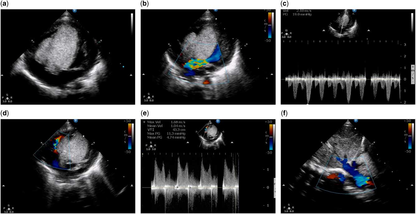
An urgent bedside transthoracic echocardiogram with colour-flow Doppler demonstrated normal segmental anatomy; two distinct echogenic masses were seen arising from the interventricular septum – a 2.2×0.8-cm homogenous hyperechoic ovoid mass in the right ventricular cavity abutting the tricuspid valve and a larger 3.3×1.8-cm mass in the interventricular septum bulging into both ventricles (Fig 1a). In the modified five-chamber view, colour Doppler showed turbulent flow in the left ventricular outflow tract with continuous wave Doppler showing a gradient of 19 mmHg, representing mild obstruction (Fig 1b and c). The parasternal short-axis view showed a morphologically normal pulmonary valve and arteries, absence of the arterial duct, and no obstruction to the right ventricular outflow; however, significant inflow obstruction was shown in the tricuspid valve Doppler due to the obstructing mass, resulting in a mean gradient of 4.7 mmHg (Fig 1d and e; Supplementary Video 1). The colour flow Doppler also revealed a 6-mm atrial septal defect shunting blood from the right atrium to the left atrium (Fig 1f; Supplementary Video 2), which was the probable cause of cyanosis. Probing into the family history, we discovered that the newborn’s father was treated for seizures with the removal of an intracranial mass in his childhood. In the presence of a possible tuberous sclerosis complex in the family, rhabdomyoma was the most probable cardiac mass to be acknowledged in our patient. Although cardiac rhabdomyomas have a high rate of spontaneous regression, in view of critically obstructed circulation, he was started on prostaglandin infusion. There was no significant improvement in the saturation, and the patient was consequently scheduled for emergency cardiac surgery involving tumour excision with closure of the septal defect. While awaiting surgery in the neonatal ICU, the patient developed resistant ventricular fibrillation from which he could not be resuscitated. This was only 3 hours after the diagnosis was made, and lactate levels had not been measured because of the family’s financial constraints. Postmortem examination was not conducted, as the family did not provide consent.
( a ) Transthoracic echocardiogram – apical four-chamber view demonstrating two hyperechoic homogeneous deformable masses, consistent with cardiac rhabdomyomata, with the larger one having dimensions of 33×18 mm in the area of the interventricular septum bulging into both ventricular cavities and a 22×8-mm smaller mass in the right ventricular cavity abutting the tricuspid valve. ( b ) Colour Doppler examination in the apical five-chamber view shows compression of the left ventricular outflow tract by the cardiac mass resulting in a turbulent flow. ( c ) A continuous wave Doppler examination reveals a maximum left ventricular outflow tract gradient of 19 mmHg. ( d ) Continuous wave Doppler at the tricuspid orifice shows a maximum gradient of 11.3 with a mean gradient of 4.74, indicating significant inflow obstruction to the right ventricle. ( e ) Modified apical four-chamber view with colour Doppler demonstrates turbulent flow at the tricuspid valve juxtaposed to the right ventricular mass. ( f ) The transthoracic echocardiogram – subcostal view colour Doppler – shows a 6-mm atrial septal defect with shunting of blood from the right atrium to the left atrium.

Discussion
Central cyanosis in a neonate is a dreaded pathophysiological sign. The aetiology of cyanosis should be promptly pursued to initiate corrective measures. When this clinical sign is associated with a cardiac murmur, the cause of decreased pulmonary blood flow or inter-circulatory mixing of blood is eagerly sought after. The diagnosis of cardiac masses resulting in cyanosis is least expected. This is compounded by the fact that primary and secondary intracardiac tumours, both benign and malignant, are extremely rare in newborns.4
Echocardiogram plays a pivotal role, not only in the identification of cardiac masses and its resultant complications but also in providing insights into possible aetiopathology. The differential diagnoses of primary intracardiac tumours in newborns include rhabdomyoma, fibroma, myxoma, teratoma, and haemangioma.
It is often difficult to differentiate solitary rhabdomyoma and fibroma. Although both are homogeneous and hyperechogenic, rhabdomyomas are deformable, occasionally multiple, and mostly involve the ventricles, whereas fibromas are non-deformable, associated with calcification, and show cystic degeneration.5–7 Myxomas are deformable, moderately echogenic masses usually located in the atrium. Teratomas are pericardial in location and are constantly associated with effusion. Haemangiomas, mostly found in the right atrium, have complex echogenicity with cystic and solid parts, with calcification.7 In view of the suggestive echocardiographic features and family history of possible tuberous sclerosis, the presenting tumour in our case is likely to be a rhabdomyoma.
The rhabdomyomas are by far the most common tumour diagnosed in utero as well as during infancy. About 80% of children with cardiac rhabdomyoma diagnosed postnatally have clinical, radiological, or a family history of tuberous sclerosis. It is an autosomal dominant multisystem disorder with variable penetrance. The majority of cases are consequent to a de novo mutation resulting in deletion at loci 9q34 and 16p13. This results in the absence of tumour suppressor gene products hamartin (TSC1) and tuberin (TSC2), respectively. The phenotypic expressions include cardiac rhabdomyomas, neurological manifestations, including cortical tubers, subependymal nodules, and seizures, renal manifestations, including angiomyolipoma, polycystic kidney disease, and carcinomas, and cutaneous manifestations, including hypomelanotic macules, facial angiofibroma, and shagreen patches.7 Cardiac rhabdomyomas may precede all the aforementioned features, and hence careful evaluation and long-term follow-up are indispensable to exclude the development of a tuberous sclerosis complex.
Although the majority are asymptomatic, clinical findings depend on the number, position, and size of the tumour. Most have a benign course with spontaneous regression noted in about 80% of patients.8 The mechanism proposed for spontaneous regression is the degradation of cytofilaments by the ubiquitin proteasome-dependent pathway along with progressive cytoplasmic vacuolisation of spider cells.9 Early complete regression is often seen before 6 years of age.10 Hence, surgical removal of the rhabdomyoma is needed only when it is associated with complications, which include intractable arrhythmias and haemodynamically significant obstruction causing heart failure; however, congenital cyanosis as a consequence of rhabdomyoma has rarely been reported. The mechanism of cyanosis in our patient is that of severe tricuspid stenosis, resulting in higher right atrial pressure and consequent shunt of deoxygenated systemic venous blood through an atrial septal defect to the left atrium. The resulting admixture physiology causes cyanosis, which is further compounded by the fact that blood flow reaching the lungs for oxygenation further declines because of the right-to-left shunt. The cardiac mass filling the ventricular cavity may also have contributed to the haemodynamic deterioration. The severity of cyanosis is directly proportional to tricuspid inflow obstruction. Hence, management in such cases should aim at relief from obstruction to prevent further desaturation. Although this can be attained in severe tricuspid stenosis with percutaneous dilatation of the tricuspid valve providing immediate relief, for cardiac masses resulting in functional tricuspid stenosis, early surgery is the only management option.
Conclusion
Routine prenatal cardiac assessment of the foetus by ultrasonogram has an upstream advantage of planning the delivery in a well-equipped tertiary-care centre. It is imperative to consider cardiac masses among the differential diagnosis of neonatal cyanosis as early surgical management is the key for survival. Although complete removal of the cardiac rhabdomyoma is impractical as they are embedded deep in the myocardium, surgery should be performed aiming at relief of the obstruction. Knowledge of outcome in affected individuals is vital for counselling after an early prenatal diagnosis, as well as for planning therapeutic strategies.
Acknowledgements
None.
Financial Support
This research received no specific grant from any funding agency, commercial, or not-for-profit sectors.
Conflicts of Interest
None.
Ethical Standards
The authors assert that all procedures contributing to this work comply with the ethical standards of the Institutional Research Committee, Government Medical College, Kozhikode and with the Helsinki declaration of 1975, as revised in 2008, or comparable ethical standards.
Supplementary material
To view supplementary material for this article, please visit https://doi.org/10.1017/S1047951116002110