Introduction
Trichinella spiralis, the aetiologic agent of trichinosis, infects a wide range of mammalian hosts and is one of the most widespread parasitic diseases worldwide. The parasite can persist in muscle tissues for many years (Froscher et al., Reference Froscher, Gullotta, Saathoff and Tackmann1988) through its ability to produce a variety of biologically active proteins that suppress the host immune response, notably macrophage migration inhibitory factor (MIF) and cysteine protease inhibitors.
Trichinella spiralis MIF (TsMIF) (Pennock et al., Reference Pennock, Behnke, Bickle, Devaney, Grencis, Isaac, Joshua, Selkirk, Zhang and Meyer1998), an orthologue of mammalian migration inhibitory factor (MIF), is expressed by all stages of the parasite, including adult worms, newborn larvae, precyst muscle larvae and postcyst muscle larvae, although not all stages express at the same level (Wu et al., Reference Wu, Boonmars, Nagano, Nakada and Takahashi2003). Previous reports showed that TsMIF can induce chemotaxis and, as with recombinant human MIF, prevent the random migration of human monocytes, and modulates the host immune defence in infected tissue (Tan et al., Reference Tan, Edgerton, Kumari, McAlister, Roe, Nagl, Pearl, Selkirk, Bianco, Totty, Engwerda, Gray and Meyer2001; Wu et al., Reference Wu, Boonmars, Nagano, Nakada and Takahashi2003). TsMCD-1, a novel multi-cystatin-like protein (MCD-1) secreted from T. spiralis, was identified as a new member of the non-inhibitory cystatin-related proteins (Robinson et al., Reference Robinson, Greig, Beattie, Lamont and Connolly2007). Expression of TsMCD-1 is largely restricted to intracellular stages, predominantly in the adult worm, and acts as a proinflammatory factor (Nagano et al., Reference Nagano, Wu and Takahashi2009). We recently demonstrated that co-expression of TsMIF and TsMCD-1 in a single vaccine can induce a Th1 immune response and confers partial protection against T. spiralis infection in BALB/c mice, although individual TsMIF and TsMCD-1 DNA vaccines were unable to protect BALB/c mice against challenge infection with T. spiralis (Tang et al., unpublished data).
Over the past decade a promising approach to DNA vaccination has been developed through fusion constructs co-expressing ubiquitin (Ub). Ubiquitin, a 76-amino-acid cytoplasmic peptide of eukaryotic cells, is a central component of the ubiquitin–proteasome system (UPS) (Tanaka & Kasahara, Reference Tanaka and Kasahara1998). Ubiquitin-tagged endogenous antigens are processed by the proteasome after being polyubiquitinated (Pickart, Reference Pickart2001), and this can enhance presentation of antigenic peptides in association with major histocompatibility complex (MHC) class I molecules and stimulate the CD8+ T-cell response. Such constructs have been tested in mice, rabbits and piglets, and are reported to heighten CD8+ T-cell responses against several viruses and parasites, shifting the immune response towards a Th1 type and enhancing cytotoxic T-lymphocyte responses (Rodriguez et al., Reference Rodriguez, Zhang and Whitton1997; Delogu et al., Reference Delogu, Howard, Collins and Morris2000; Hou et al., Reference Hou, Chen, Tong, Tian, Zhou, Li, Li, Peng, An and Yang2008; Wang et al., Reference Wang, Kang and Wang2009; Chou et al., Reference Chou, Hiromatsu, Hisaeda, Duan, Imai, Murata, Tanaka and Himeno2010).
Trichinella spiralis infection induces a polarization of the Th2 response that is thought to facilitate chronic infection (Kolodziej-Sobocinska et al., Reference Kolodziej-Sobocinska, Dvoroznakova and Dziemian2006; Fabre et al., Reference Fabre, Beiting, Bliss and Appleton2009), whereas Th1 responses are suppressed (Wakelin et al., Reference Wakelin, Goyal, Dehlawi and Hermanek1994; Hogaboam et al., Reference Hogaboam, Collins and Blennerhassett1996; Ishikawa et al., Reference Ishikawa, Goyal, Mahida, Li and Wakelin1998; Wan et al., Reference Wan, Wang, He, Liu and Zhang2006). A study on H2-compatible mouse strains revealed that resistance to T. spiralis infection and accelerated worm expulsion are associated with Th1 responses; in contrast, susceptible B10.BR mice have a lower rate of worm expulsion and primarily mount a Th2 response (Pond et al., Reference Pond, Wassom and Hayes1989). Moreover, it has been suggested that neither B cells nor a specific antibody are required for T. spiralis expulsion (Finkelman et al., 2004), and worm elimination from the intestine is also independent of interleukin (IL)-4/IL-13 activation of CD4+ T cells, macrophages and neutrophils (Michels et al., Reference Michels, Scales, Saunders, McGowan, Brombracher, Alexander and Lawrence2009). We therefore speculated that a CD8+ T-cell-mediated immune response is central to resistance to T. spiralis infection, and that an enhanced Th1 response could therefore counter the dominant Th2 response and favour protection against infection.
In the present study we aimed to investigate whether co-expression with ubiquitin might improve the protective immunity induced by the TsMIF–TsMCD-1 DNA vaccine. A plasmid co-expressing three coding sequences as a triple fusion polypeptide (mouse Ub, TsMIF and TsMCD-1) was constructed and used to evaluate protective immunity against T. spiralis infection of BALB/c mice.
Materials and methods
Proteins, antiserum, animals and parasites
Recombinant TsMIF–TsMCD-1 protein expressed in Escherichia coli BL21 (DE3) and mouse antiserum against TsMIF–TsMCD-1 were provided by the Laboratory of Veterinary Molecular and Immunological Parasitology, Nanjing Agricultural University, China. Female 6-week-old BALB/c mice were purchased from the Experimental Animal Centre of Nanjing Medical University and were maintained in plastic micro-isolator cages before and after immunization. Trichinella spiralis strain ISS 534 was maintained in BALB/c mice.
Construction of expression plasmids
Ub cDNA sequences were amplified from total RNA obtained from mouse testes using sense, 5′-GACAAGCTTGCCACCATGCAGATCTTCGTGAAGACC-3′, and antisense, 5′-TATGGTACCGCTGCCTCCCCCGCCGCTggcACCTCTCAGGCGAAGGACCAGG-3′, primers designed according to the sequence from GenBank, accession number EF580991. An artificial Kozak sequence (GCCACC) was introduced into the sense primer to promote efficient initiation of translation (Kozak, Reference Kozak1987), and a substitution of Gly76 by Ala76 was included in the antisense primer (ggc) to block removal of the ubiquitin monomer from the fusion protein, thus providing a base for polyubiquitination (Johnson et al., Reference Johnson, Ma, Ota and Varshavsky1995). The Ub sequence was amplified to include restriction sites for HindIII (AAGCTT) and KpnI (GGTACC) without translation stop codons, ligated into the expression vector pVAX1 (Invitrogen, Carlsbad, California, USA), and transformed into E. coli DH5α (Invitrogen), generating recombinant plasmid pVAX1-Ub. For co-expression of Ub with TsMIF and TsMCD-1, plasmid pVAX1-Ub-Tsmif-Tsmcd-1 was constructed from recombinant plasmids pVAX1-Ub and pVAX1-Tsmif-Tsmcd-1 using the HindIII and KpnI restriction sites. A flexible peptide linker (SerGly4Ser) was introduced into the fusion protein between the two coding sequences to facilitate folding of the proteins in their native conformations (Frigerio et al., Reference Frigerio, Jolliffe, Di Cola, Felipe, Paris, Neuhaus, Lord, Ceriotti and Roberts2001; Hu et al., Reference Hu, Li, Yang, Li, Xia, Li, Wang and Zhang2004). Clones containing inserts of the expected size were selected and subjected to DNA sequencing using T7 and BGH primers by Invitrogen (Invitrogen Co. Ltd., Shanghai, China). DNA and predicted amino-acid sequences were analysed using Lasergene 7.1 software (DNASTAR Inc., Madison, Wisconsin, USA).
Detection of target gene expression in vitro by Western blotting
Baby hamster kidney (BHK) cells were transfected with 2 μg of pVAX1, pVAX1-Tsmif-Tsmcd-1 or pVAX1-Ub-Tsmif-Tsmcd-1 using LipofectamineTM 2000 Reagent (Invitrogen, Carlsbad, California, USA) according to the manufacturer's instructions. Twenty-four hours after transfection the cells were collected and target gene expression was analysed by Western blotting. In some experiments the transfected cells were cultured with a specific proteasome inhibitor, MG-132 (Sigma-Aldrich, St. Louis, Missouri, USA), added 12 h after transfection at a concentration of 10 μm. Western blotting used a 1:40 dilution of mouse antiserum against recombinant TsMIF–TsMCD-1 protein as the first antibody and horseradish peroxidase (HRP)-conjugated goat anti-mouse IgG as the second antibody (Sigma-Aldrich). Blots were developed using diaminobenzidine HRP substrate and a chromogenic reagent kit (Boster, WuHan, China).
Immunization and challenge of mice
Six-week-old female BALB/c mice were divided into groups of 20 animals. Two groups were immunized with recombinant plasmids pVAX1-Tsmif-Tsmcd-1 and pVAX1-Ub-Tsmif-Tsmcd-1, respectively, and three control groups were immunized with pVAX1 plasmid, pVAX1-Ub plasmid, or phosphate-buffered saline (PBS; pH 7.4). Plasmid DNA for immunization was prepared using Classic Endo-free Plasmid DNAout (Tiandz, Beijing, China) and quantified using a Hitachi U-2800 spectrophotometer. Plasmid DNA was diluted to 1 μg/μl in PBS, and 100 μg aliquots were injected into the quadriceps femoris muscle of mice twice at an interval of 2 weeks. Two weeks after the second immunization, challenge was carried out by oral infection with 200 L1 infectious T. spiralis larvae per animal.
Analysis of antibody responses
Blood was drawn from the ophthalmic venous plexus of ten mice in each group at 0, 10, 20 and 28 days following the first vaccination. Serum was isolated by centrifugation and stored at − 20°C until use. Serum antibody (IgG, IgG1, IgG2a, IgG2b, IgA, IgM and IgE isotypes) levels were measured in triplicate by indirect enzyme-linked immunosorbent assay (ELISA). Briefly, 96-well plates were coated with 5 μg/ml recombinant TsMIF–TsMCD-1 protein (100 μl/well) and incubated for 18–24 h at 4°C. Serum samples were serially diluted (twofold dilutions) in buffer (PBS with 0.05% Tween20), added at 100 μl/well, and incubated for 1.5 h at 37°C. Sera from non-immunized mice were used as a negative control. After washing three times, plates were incubated with a 5000-fold dilution of HRP-conjugated goat anti-mouse IgG, IgG1, IgG2a, IgG2b, IgA, IgM or IgE (Santa Cruz Biotechnology, Santa Cruz, California, USA) for 1 h at 37°C, respectively. After final washing, 100 μl of substrate solution (3,3′,5,5′-tetramethylbenzidine, TMB; Amresco, Solon, Ohio, USA) was added and incubated for 10 min at 37°C. Reactions were halted by the addition of 50 μl 2 m sulphuric acid, and optical densities at 450 nm (OD450) and 570 nm (OD570) were determined using an automated microplate reader (Rayto, Shenzhen, China). Data were analysed after subtraction of the background OD570 reading from the OD450 value (OD450–570). The cutoff titre was defined as the lowest antibody concentration at which the OD450–570 of sample (S) was equal to that of the negative control (N) (P< 0.05).
Analysis of serum cytokines
Sandwich ELISA (Boster, WuHan, China), performed according to the manufacturer's instructions, was used to determine the concentrations of five cytokines (interferon (IFN)-γ, IL-4, IL-5, transforming growth factor (TGF)-β1, and IL-17). Serum samples were isolated from ten mice of each group at 0, 10, 20 and 28 days following the first vaccination, respectively. Briefly, biotin-conjugated anti-cytokine, avidin–HRP conjugate and TMB substrate solution were successively employed; plates were read at 450 nm.
Cytotoxic T lymphocyte (CTL) assay
To determine whether immunization with the recombinant plasmids could induce strong CTL responses, the lactate dehydrogenase (LDH) release cytotoxic assay was performed according to the protocol of the Cyto Tox 96® Non-Radioactive Cytotoxicity Assay (Promega, Madison, Wisconsin, USA).
Spleen cells were isolated from five mice of each group 14 days following the second immunization and used as effector cells. BHK cells transfected with pVAX1-Tsmif-Tsmcd-1 (BHK-TsMIF-TsMCD-1) were used as target cells, and expression of TsMIF–TsMCD-1 was confirmed by immunocytochemistry with anti-TsMIF-TsMCD-1 serum and fluorescein isothiocyanate (FITC)-labelled anti-mouse IgG (Sigma-Aldrich). BHK cells transfected with pVAX1 were used as a negative control.
Effector cells (E) were incubated with target cells (T) in 96-well round-bottom plates at different E/T ratios for 4 h in phenol-red-free RPMI (Roswell Park Memorial Institute) 1640 medium containing 2% bovine serum albumin (BSA), 2 mm glutamine and 1% penicillin and streptomycin at 37°C in 5% CO2. Supernatants (100 μl/well) were transferred to new 96-well plates, reconstituted substrate mix (100 μl/well) was added, and the mixture was incubated at room temperature for 30 min. OD492 values of the supernatants were recorded on an ELISA microplate reader. Percent lysis was calculated as follows: [(experimental release − spontaneous release)/(maximum release − spontaneous release)] × 100%.
Flow cytometric analysis of CD4+/CD8+ T cells
Two weeks after the last immunization, whole blood was drawn from five mice of each group for analysis by flow cytometry. Peripheral blood lymphocytes were analysed using FITC-labelled mouse monoclonal anti-CD3 antibody or phycoerythrin (PE)-labelled mouse monoclonal anti-CD4 or -CD8 antibodies (MultiSciences, HangZhou, China). Labelled cells were analysed using a FACScan flow cytometer with CellQuest software (BD Biosciences, Franklin Lakes, New Jersey, USA). Results were analysed using FCS Express V3 software (version 3.00.0316; De Novo Software, Los Angeles, California, USA). The percentages of CD4+ T cells and CD8+ T cells in peripheral blood lymphocytes were determined and the CD4+/CD8+ ratio was calculated.
Detection of larvae in muscle
Five weeks after challenge infection, ten mice in each group were sacrificed. Trichinella spiralis muscle larva burden was evaluated as larvae per gram (LPG) in preferred muscles (muscles of diaphragm, anterior limb and neck) following treatment with acid-pepsin digestion solution (Zhu et al., Reference Zhu, Long, Xu and Gui1993) containing 0.6% w/v pepsin (800–2500 units/mg; Sigma-Aldrich); 37% w/v HCl was added to give a pH of between 1.5 and 2. Muscles were mixed with the digestion solution (25 ml/g tissue) and incubated at 38°C for 6 h. Larvae in each digested sample were counted using light microscopy at 40 × magnification. The reduction of muscle larval burden was calculated using the following formula: reduction rate of muscle larvae = [(mean of LPG in PBS control − mean of LPG in DNA vaccination)/mean of LPG in PBS control] × 100%.
Statistical analysis
Statistical significances of inter-group differences were determined by analysis of variance (ANOVA) using SPSS Statistical Software (version 16.0; SPSS Inc., Chicago, Illinois, USA); P< 0.05 was taken to indicate statistical significance.
Results
Detection of in vitro target gene expression by Western blotting
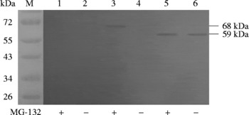
As shown in fig. 1, Western blotting analysis revealed a specific immunoreactive band at 59 kDa in BHK cells transfected with pVAX1-Tsmif-Tsmcd-1, indicating the expression of the Tsmif-Tsmcd-1 fusion gene. No significant immunoreactive bands were found in BHK cells transfected with pVAX1-Ub-Tsmif-Tsmcd-1. However, a band at 68 kDa was detected in the presence of the specific proteasome inhibitor MG-132, indicating that the Ub–TsMIF–TsMCD-1 fusion protein is generated from the plasmid but is targeted for degradation. No protein band was observed in cells transfected with the pVAX1 vector.
Western blotting of BHK cells transfected with recombinant plasmids. M, Protein ladder; BHK cells transfected with pVAX1 were cultured with (lane 1) or without MG-132 (lane 2); BHK cells transfected with pVAX1-Ub-Tsmif-Tsmcd-1 were cultured with (lane 3) or without MG-132 (lane 4); BHK cells transfected with pVAX1-Tsmif-Tsmcd-1 were cultured with (lane 5) or without MG-132 (lane 6).

Antibody response induced by DNA vaccination
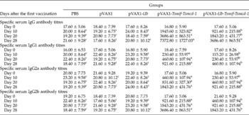
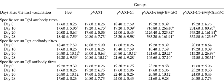
Sera collected 0, 10, 20 and 28 days following the first vaccination were assayed for the presence of specific antibodies against recombinant TsMIF–TsMCD-1 protein. As shown in tables 1 and 2, no antibody (IgM, IgG, IgA or IgE) response was detected in the pVAX1 control group or pVAX1-Ub control group. A clear IgM, IgG and IgA antibody response was observed in both pVAX1-Tsmif-Tsmcd-1 and pVAX1-Ub-Tsmif-Tsmcd-1 immunized animals (P< 0.05 compared to PBS, pVAX1 or pVAX1-Ub control groups in both cases), whereas IgE antibody was not significantly induced in any group.
ELISA titres of specific serum IgG, IgG1, IgG2a and IgG2b antibodies against the recombinant TsMIF–TsMCD-1 protein in mice.

Blood was drawn from ten mice in each group at 0, 10, 20 and 28 days following the first vaccination. Serum was isolated and production of specific antibodies (IgG, IgG1, IgG2a and IgG2b isotypes) was measured in triplicate by indirect enzyme-linked immunosorbent assay (ELISA). Values are shown as mean ± standard deviation (n= 10 mice/group). Data bearing a different superscript letter (a–c) within a line differ significantly from one another (P< 0.05).
ELISA titres of specific serum IgM, IgA and IgE antibodies against the recombinant TsMIF–TsMCD-1 protein in mice.
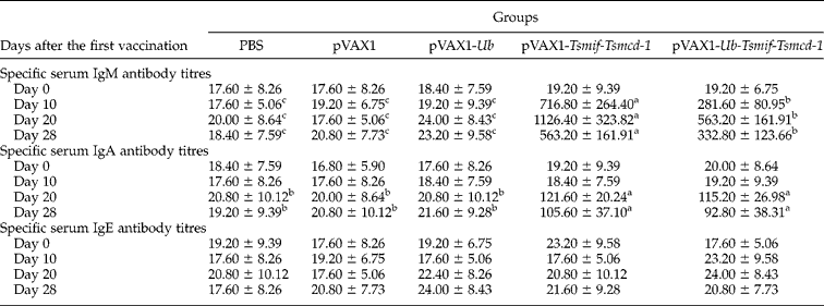
Blood was drawn from ten mice in each group at 0, 10, 20 and 28 days following the first vaccination. Serum was isolated and production of specific antibodies (IgM, IgA and IgE isotypes) was measured in triplicate by indirect enzyme-linked immunosorbent assay (ELISA). Values are shown as mean ± standard deviation (n= 10 mice/group). Data bearing a different superscript letter (a–c) within a line differ significantly from one another (P< 0.05).
Compared to the pVAX1-Tsmif-Tsmcd-1 vaccine, levels of IgG antibody induced in mice vaccinated with pVAX1-Ub-Tsmif-Tsmcd-1 were significantly lower (P< 0.05). IgG antibodies were detected 10 days following the first immunization and increased progressively to titres of 7372.80 ± 1727.03 (pVAX1-Tsmif-Tsmcd-1) and 3686.40 ± 863.51 (pVAX1-Ub-Tsmif-Tsmcd-1) before challenge (28 days following the first immunization). Further analysis of IgG isotypes indicated that both IgG2a and IgG2b had higher titres than IgG1 in these two groups (P< 0.05 in all cases). We also observed that IgM antibody levels in mice vaccinated with pVAX1-Ub-Tsmif-Tsmcd-1 were lower than those in animals receiving pVAX1-Tsmif-Tsmcd-1 (P< 0.05). IgM antibodies were detected 10 days following the first immunization and reached peak titres of 1126.40 ± 323.82 (pVAX1-Tsmif-Tsmcd-1) and 563.20 ± 161.91 (pVAX1-Ub-Tsmif-Tsmcd-1) 20 days following the first immunization. IgA antibody levels in mice vaccinated with pVAX1-Ub-Tsmif-Tsmcd-1 were not significantly different from those in mice receiving pVAX1-Tsmif-Tsmcd-1 (P>0.05). IgA antibodies were detected at lower titres (121.60 ± 20.24 for pVAX1-Tsmif-Tsmcd-1 and 115.20 ± 26.98 for pVAX1-Ub-Tsmif-Tsmcd-1) 20 days following the first immunization and levels decreased thereafter.
Cytokine production
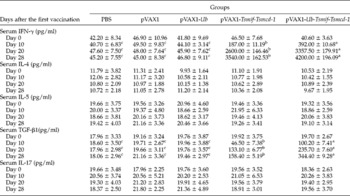
Concentrations of five cytokines (IFN-γ, IL-4, IL-5, TGF-β1 and IL-17) were measured in sera collected 0, 10, 20 and 28 days following the first vaccination. As shown in table 3, serum IL-4, IL-5 and IL-17 levels in vaccine immunized groups were not significantly different from those in control groups (P< 0.05 versus controls in all cases).
Concentrations of serum cytokines in mice.

Blood was drawn from ten mice in each group at 0, 10, 20 and 28 days following the first vaccination. Serum was isolated and cytokine (IFN-γ, TGF-β1, IL-4, IL-5 and IL-17) concentrations were measured in triplicate by ELISA kits. Values represent the mean ± standard deviation (n= 10 mice/ group). Data bearing a different superscript letter (a–c) within a line differ significantly from one another (P< 0.05).
Compared to the PBS control, the same amount of serum IFN-γ was observed with a lower level than the pVAX1 control group (P>0.05) or pVAX1-Ub control group (P>0.05). More serum IFN-γ was detected in animals vaccinated with recombinant plasmids pVAX1-Tsmif-Tsmcd-1 or pVAX1-Ub-Tsmif-Tsmcd-1 10 days following the first immunization (P< 0.05 versus controls in all cases) and reached levels of 3540.00 ± 162.53 pg/ml (pVAX1-Tsmif-Tsmcd-1) and 4200.00 ± 196.09 pg/ml (pVAX1-Ub-Tsmif-Tsmcd-1) before challenge (28 days following the first immunization). The elevation of serum IFN-γ following vaccination with pVAX1-Ub-Tsmif-Tsmcd-1 was significantly greater than the level obtained by vaccination with pVAX1-Tsmif-Tsmcd-1 (P< 0.05).
Similarly, the same amount of serum TGF-β1 was observed with a lower level than the pVAX1 or pVAX1-Ub groups (P>0.05 versus PBS controls in both cases). More serum TGF-β1 was detected in both the pVAX1-Tsmif-Tsmcd-1 and the pVAX1-Ub-Tsmif-Tsmcd-1 immunized groups (P< 0.05 versus controls) 10 days following the first immunization and reached 158.40 ± 5.19 pg/ml (pVAX1-Tsmif-Tsmcd-1) and 344.40 ± 9.28 pg/ml (pVAX1-Ub-Tsmif-Tsmcd-1) before challenge. Serum TGF-β1 levels in the pVAX1-Ub-Tsmif-Tsmcd-1 immunization group were significantly higher than in animals immunized with pVAX1-Tsmif-Tsmcd-1 (P< 0.05).
Improved CTL response by Ub-co-expressing DNA vaccines
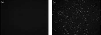
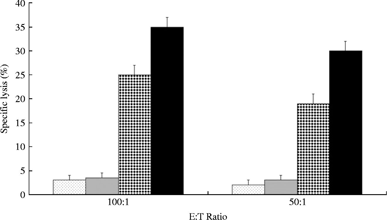
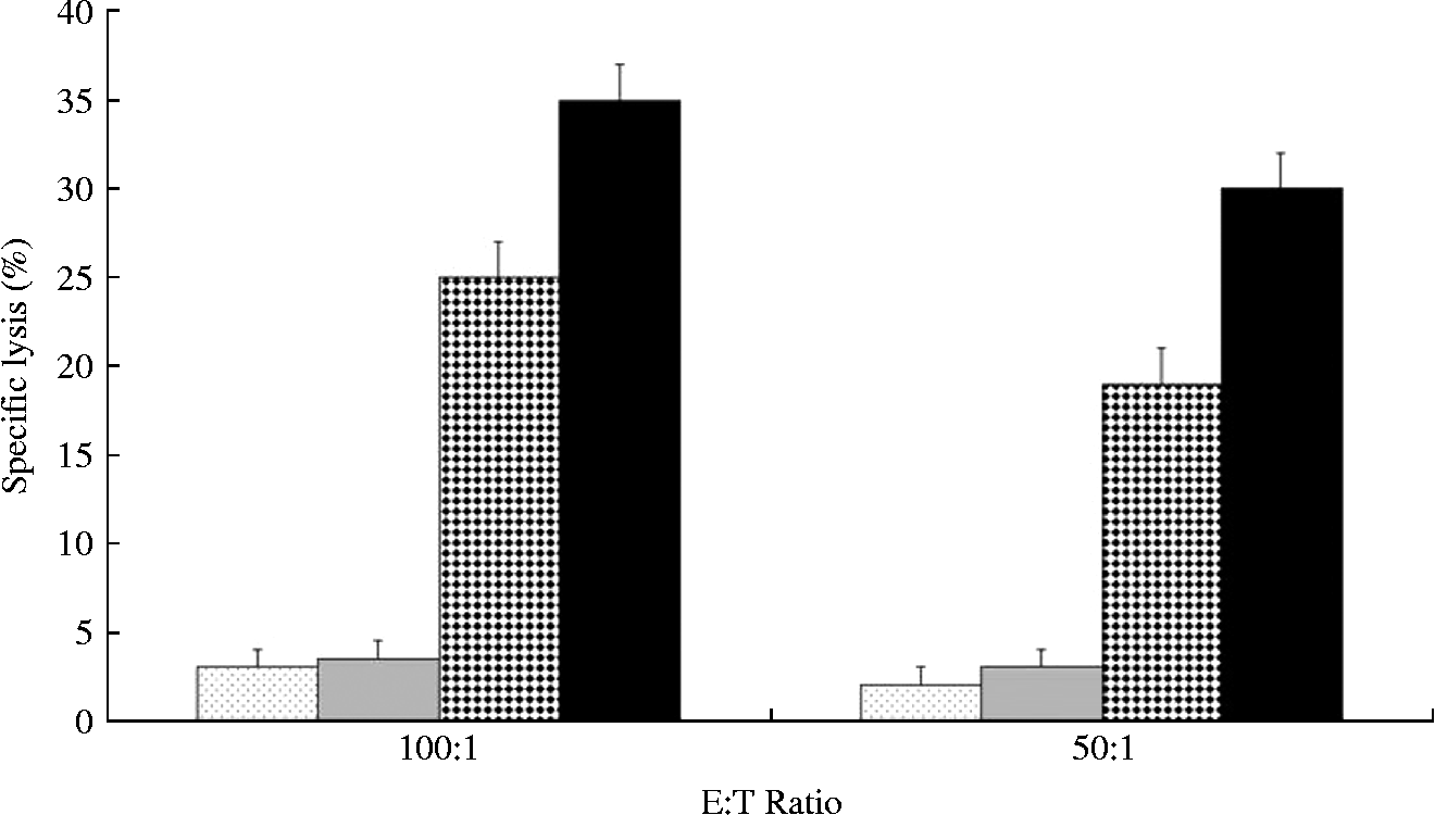
CTL responses were evaluated using BHK-TsMIF-TsMCD-1 target cells; expression of the TsMIF–TsMCD-1 protein was confirmed by immunocytochemistry (fig. 2). Following in vitro restimulation, CTL responses were determined using the LDH release assay. BHK cells transfected with pVAX1 provided the negative control. As shown in fig. 3, blank vector or pVAX1-Ub immunization failed to induce a CTL response, indicating that the spontaneous release rate was below 10%. Vaccination with pVAX1-Tsmif-Tsmcd-1 resulted in a strong CTL response which was significantly above that generated by immunization with blank vector or pVAX1-Ub (P< 0.05). Treatment with the ubiquitin-co-expressing DNA vaccine pVAX1-Ub-Tsmif-Tsmcd-1 further enhanced the CTL response compared to pVAX-TsMIF-TsMCD-1 (P< 0.05).
Immunocytochemistry of BHK cells transfected with recombinant plasmids. Expression of TsMIF–TsMCD-1 in BHK cells was confirmed by immunocytochemistry with anti-TsMIF-TsMCD-1 serum and FITC-labelled anti-mouse IgG: (a) BHK cells were transfected with pVAX1 vector plasmid; (b) BHK cells were transfected with pVAX1-Tsmif-Tsmcd-1 plasmid.

Cytotoxicity of splenocytes in mice immunized with DNA vaccines was determined with a lactate dehydrogenase (LDH) release assay. BHK cells effectively transfected with pVAX1-Tsmif-Tsmcd-1 were used as target (T) cells. Spleen cells isolated from vaccinated mice 14 days following the second immunization were used as effector (E) cells. The columns with dotted shading indicate vaccination with pVAX1, the grey columns indicate vaccination with pVAX1-Ub, the columns with diamond shading indicate vaccination with pVAX1-Tsmif-Tsmcd-1, and the black columns indicate vaccination with pVAX1-Ub-Tsmif-Tsmcd-1. Values represent the mean ± standard deviation (n= 5 mice/ group).
Flow cytometric analysis of CD4+/CD8+ T cells
Two weeks after the last immunization, the percentages of CD4+ and CD8+ T cells in peripheral blood lymphocytes were determined and the CD4+/CD8+ ratio was calculated. As shown in table 4, there were no significant differences among the three controls (PBS, pVAX1 and pVAX1-Ub) in CD4+%, CD8+% or CD4+/CD8+ ratio (P>0.05 in all cases). Compared to the PBS, pVAX1 and pVAX1-Ub control groups, a higher percentage of CD4+ cells was observed in animals receiving pVAX1-Tsmif-Tsmcd-1 (P< 0.01) or pVAX1-Ub-Tsmif-Tsmcd-1 (P< 0.05). The level of CD4+ cells in animals treated with pVAX1-Tsmif-Tsmcd-1 was significantly higher than in animals receiving pVAX1-Ub-Tsmif-Tsmcd-1 (P< 0.05).
Flow cytometric analysis of CD4+ T cells and CD8+ T cells in peripheral blood lymphocytes of vaccinated mice 14 days following the second immunization.

Values are shown as mean ± standard deviation. Data bearing a different superscript letter (a–c) within a line differ significantly from one another (P< 0.05).
Vaccination with either pVAX1-Tsmif-Tsmcd-1 (P< 0.05) or pVAX1-Ub-Tsmif-Tsmcd-1 (P< 0.01) gave rise to increased levels of CD8+ cells compared to controls. Moreover, levels of CD8+ cells were significantly higher in animals vaccinated with pVAX1-Ub-Tsmif-Tsmcd-1 than in animals receiving pVAX1-Tsmif-Tsmcd-1 (P>0.05).
CD4+/CD8+ ratios were then calculated. The CD4+/CD8+ ratio in animals immunized with pVAX1-Tsmif-Tsmcd-1 was significantly elevated versus controls (P< 0.05), whereas the CD4+/CD8+ ratio was significantly reduced in animals receiving the pVAX1-Ub-Tsmif-Tsmcd-1 vaccine versus controls (P< 0.05).
Worm burden after challenge infection
Five weeks after challenge infection, T. spiralis muscle larva burden was evaluated as larvae per gram (LPG) in each group. There were no significant LPG differences between the three control groups: PBS (2398.09 ± 187.57), pVAX1 (2375.98 ± 131.91) and pVAX1-Ub (2362.46 ± 138.54). In contrast, LPG was significantly reduced in mice vaccinated with either pVAX1-Tsmif-Tsmcd-1 (1842.47 ± 234.93) or pVAX1-Ub-Tsmif-Tsmcd-1 (1488.06 ± 277.61) compared to controls (P< 0.05 in all cases), representing reduced muscle larval burdens of 23.17% and 37.95%, respectively. Vaccination with pVAX1-Ub-Tsmif-Tsmcd-1 resulted in a significantly greater reduction in worm burden than did vaccination with pVAX1-Tsmif-Tsmcd-1 (P< 0.05).
Discussion
Trichinella spiralis produces two important modulators of the host immune response: TsMIF, a cytokine homologue, and TsMCD-1, a protease inhibitor. We found previously that co-expression of the two coding sequences in a single DNA vaccine induced Th1 immune responses and conferred partial protection against T. spiralis infection in BALB/c mice (Tang et al., unpublished data). To improve the protective efficacy of the vaccine we have explored the incorporation of Ub coding sequences into the vaccine. Mouse cDNA encoding Ub was cloned and inserted into the TsMIF–TsMCD-1 plasmid to generate a vaccine encoding a triple fusion protein, Ub–TsMIF–TsMCD-1. The new DNA vaccine, pVAX1-Ub-Tsmif-Tsmcd-1, was evaluated for its ability to induce cellular and humoral immunity and protection against T. spiralis challenge of BALB/c mice. We report that immunization with the triple fusion vaccine led to significantly increased levels of IFN-γ and TGF-β1 versus the parental vaccine expressing only the TsMIF–TsMCD-1 fusion protein, but reduced levels of IgG and IgM. The Ub-containing triple fusion vaccine also induced a differential CTL response, with a significantly reduced CD4+/CD8+ ratio compared to the parental vaccine. Importantly, worm burden following T. spiralis challenge of mice immunized with the pVAX1-Ub-Tsmif-Tsmcd-1 DNA vaccine was significantly lower than in animals receiving pVAX1-Tsmif-Tsmcd-1, indicating that Ub co-expression improves protective immunity.
Ubiquitin and the proteasome are the two central players of the UPS (ubiquitin–proteasome system), and ubiquitin-tagged endogenous antigens are rapidly degraded by the proteasome after being polyubiquitinated (Hershko & Ciechanover, Reference Hershko and Ciechanover1998; Pickart, Reference Pickart2001; Finley, Reference Finley2009). To provide a base for polyubiquitination (Johnson et al., Reference Johnson, Ma, Ota and Varshavsky1995), Gly76 at the C-terminus of ubiquitin was substituted by Ala76, thereby generating a stable ubiquitin polypeptide that can be polyubiquitinated and processed by the proteasome in the cytoplasm of eukaryotic cells (Dantuma et al., Reference Dantuma, Lindsten, Glas, Jellne and Masucci2000; Hou et al., Reference Hou, Chen, Tong, Tian, Zhou, Li, Li, Peng, An and Yang2008; Wang et al., Reference Wang, Kang and Wang2009). Using an in vitro system, we have confirmed that the UPS plays a crucial role in processing of the hybrid Ub–TsMIF–TsMCD-1 polypeptide. The hybrid polypeptide was largely undetectable in BHK cells transfected with pVAX1-Ub-Tsmif-Tsmcd-1, whereas untagged TsMIF–TsMCD-1 protein was readily detected. Treatment with the proteasome inhibitor MG-132 led to efficient detection of the Ub-fusion polypeptide, indicating that the Ub–TsMIF–TsMCD-1 is efficiently recognized and targeted for degradation by the UPS.
Immunization with DNA vaccines leads to the production of foreign proteins within host cells, thereby permitting presentation of foreign antigens by host MHC class I molecules (Whitton et al., Reference Whitton, Rodriguez, Zhang and Hassett1999). Targeting of exogenous peptides to the endoplasmic reticulum (ER) is thought to be a limiting factor in the generation of the mature MHC class I complex (Ortmann et al., Reference Ortmann, Androlewicz and Cresswell1994), and it has been suggested that ubiquitination can enhance polypeptide delivery to the ER and thereby potentiate antigen presentation by MHC class I molecules (Rodriguez et al., Reference Rodriguez, Zhang and Whitton1997; Wang et al., Reference Wang, Kang and Wang2009). It has also been demonstrated that ubiquitin plays a crucial role in the degradation of target proteins (Dantuma et al., 2000; Gravier et al., 2007; Hou et al., Reference Hou, Chen, Tong, Tian, Zhou, Li, Li, Peng, An and Yang2008; Wang et al., Reference Wang, Kang and Wang2009; Chou et al., Reference Chou, Hiromatsu, Hisaeda, Duan, Imai, Murata, Tanaka and Himeno2010), and could thereby improve the delivery of antigenic peptides to the MHC, thus improving Th1 responses and enhancing CTL responses. In the present study we observed that the Ub–TsMIF–TsMCD-1 vaccine co-expressing Ub resulted in reduced antibody (IgG, IgM) responses but elevated IFN-γ levels, increased peripheral blood CD8+ T cells, and enhanced CTL activity compared to the parental TsMIF–TsMCD-1 vaccine. This indicates that Ub co-expression is likely to target these T. spiralis antigens to the UPS and thereby improve their presentation in conjunction with host MHC class I molecules. However, IgA antibody levels in mice vaccinated with pVAX1-Ub-Tsmif-Tsmcd-1 were not significantly different from those in mice receiving pVAX1-Tsmif-Tsmcd-1 (P>0.05), which differs from the reduction of IgG and IgM antibody responses resulting from Ub co-expression with TsMIF–TsMCD-1. More research will be needed to explain the diversity of the influence on IgA and IgG/IgM.
The mechanism by which DNA immunization induces an antibody response against the encoded polypeptide remains controversial, although the release and reuptake of intact protein appears to be required (Whitton et al., Reference Whitton, Rodriguez, Zhang and Hassett1999). It has been reported that ubiquitinated DNA vaccines induce CTL responses but not antibodies (Rodriguez et al., Reference Rodriguez, Zhang and Whitton1997; Hou et al., Reference Hou, Chen, Tong, Tian, Zhou, Li, Li, Peng, An and Yang2008), or that ubiquitin fusion reduces the levels of antibodies directed against target proteins (Wang et al., Reference Wang, Kang and Wang2009). In the present study we report that vaccination with the Ub-co-expressing plasmid pVAX1-Ub-Tsmif-Tsmcd-1 resulted in a lower humoral response than did vaccination with the construct expressing TsMIF–TsMCD-1, suggesting that the Ub–TsMIF–TsMCD-1 triple fusion polypeptide is rapidly degraded intracellularly, reducing the levels of intact protein able to interact with the B cells.
IFN-γ is widely regarded as the major macrophage-activating cytokine (Nathan et al., Reference Nathan, Murray, Wiebe and Rubin1983; Suzuki et al., Reference Suzuki, Orellana, Schreiber and Remington1988) and stimulates antigen presentation by enhancing the expression of MHC genes (Gaczynska et al., 1993). It has been suggested that IFN-γ might be important for host protection against T. spiralis infection (Pond et al., Reference Pond, Wassom and Hayes1989; Bautista-Garfias et al., Reference Bautista-Garfias, Ixta, Orduna, Martinez, Aguilar and Cortes1999) because T. spiralis worm expulsion in mice is thought to take place largely as a result of inflammatory responses (Robinson et al., Reference Robinson, Bellaby and Wakelin1995). In the present study, immunization with the Ub-co-expressing Ub–TsMIF–TsMCD-1 construct induced a greater level of serum IFN-γ than did the vaccine expressing TsMIF–TsMCD-1; a finding also seen in other studies on ubiquitin-expressing DNA vaccines (Gravier et al., 2007; Hou et al., Reference Hou, Chen, Tong, Tian, Zhou, Li, Li, Peng, An and Yang2008; Wang et al., Reference Wang, Kang and Wang2009).
The increased level of IFN-γ might be related to the increase in CD8+ T cells. The role of CD8+ T cells in expulsion of T. spiralis is unclear (Khan & Collins Reference Khan and Collins2004; Dea-Ayuela et al., Reference Dea-Ayuela, Rama-Iniguez and Bolas-Fernandez2006). In our DNA vaccination experiments the Ub fusion vaccine induced a greater CD8+ T-cell (CTL) response than did the untagged TsMIF–TsMCD-1 DNA vaccine, implying that a CD8+ T-cell-mediated immune response contributes to protective immunity against T. spiralis infection.
Following challenge infection there was a significantly greater reduction in the burden of muscle larvae in animals immunized with pVAX1-Ub-Tsmif-Tsmcd-1 versus pVAX1-Tsmif-Tsmcd-1. This indicates that Ub co-expression with TsMIF–TsMCD-1 enhances resistance to T. spiralis infection.
In conclusion, immunization with the pVAX1-Ub-Tsmif-Tsmcd-1 DNA vaccine, in which TsMIF and TsMCD-1 are expressed as a fusion protein with mouse Ub, led to a reduced humoral immune response but gave enhanced Th1 cytokine levels and CD8+ T-cell (CTL) responses compared to vaccination with the untagged pVAX1-Tsmif-Tsmcd-1 vaccine. Importantly, the modification significantly improved protection against T. spiralis infection. The mechanisms underlying the improvement of protective immunity against T. spiralis warrant further research.