Left ventricular non-compaction is a rare cardiomyopathy, resulting from failure of the myocardial compaction in embryogenesis of the myocardium.Reference Zambrano, Marshalko, Jaffe and Hui1 Left ventricular non-compaction can be sometimes associated with other cardiac anomalies and neuromuscular disorders.Reference Stöllberger, Finsterer and Blazek2 Clinical manifestations of this cardiomyopathy range from no symptoms to heart failure, systemic thromboemboli, and arrhythmias (supraventricular or ventricular).Reference Kohli, Pantazis and Shah3 The diagnosis of this entity is achieved by echocardiography and magnetic resonance imaging (MRI).Reference Petersen, Selvanayagam and Wiesmann4 Echocardiographic criteria of left ventricular non-compaction have been established by Jenni et al,Reference Jenni, Oechslin and van der Loo5 and Stöllberger et alReference Stöllberger, Finsterer and Blazek2 have suggested left ventricular non-compaction as two layers of myocardium with a ratio of non-compacted to compacted myocardium >2, measured at the end-systole, coexisting cardiac anomalies should be absent and, typical localisation of the pathology was mid-lateral, apical and mid-inferior zones and colour-Doppler displayed blood flow within intertrabecular recesses. ST-segment elevation myocardial infarction is an uncommon clinical manifestation of left ventricular non-compaction and rarely reported, whereas cardiac ischaemia and chest pain are not uncommon and generally are related to diminishing coronary flow reserve and microvascular dysfunction.Reference Maron, Towbin and Thiene6
In this report, we described a 55-year-old male diagnosed with anterior myocardial infarction and isolated left ventricular non-compaction.
Case presentation
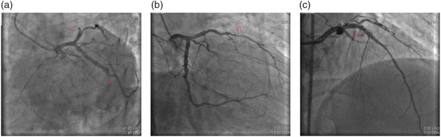
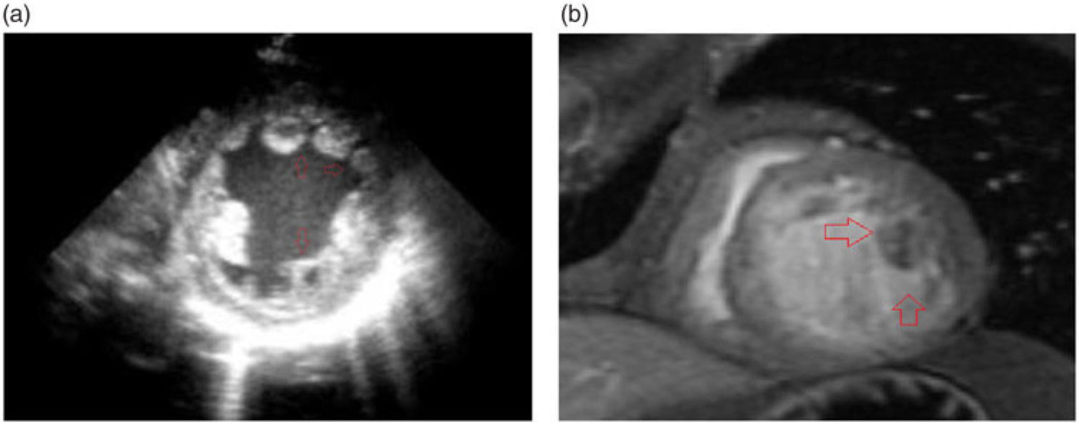
A 55-year-old male patient was admitted to our emergency department of our hospital complaining with typical chest pain and exertional dyspnoea (NYHA class 2) for two days. He was a smoker, had no history of diabetes mellitus, hypertension, hyperlipidemia, or obesity, and had no family history of ischaemic heart disease. On physical examination, his blood pressure was 125/80 mmHg and heart rate was 88 beats/min. Cardiac auscultation revealed an apical systolic murmur and the other system examinations were normal. The 12-lead electrocardiogram showed sinus rhythm, ST-segment elevation in leads V1–4, poor R-wave progression in right precordial leads, and ST-segment depression in leads I – aVL. The levels of creatinine kinase, creatine kinase-MB, and troponin I were elevated at presentation (65 U/L, 681 U/L, and 17.7 ng/ML, respectively). He was hospitalised with the diagnosis of ongoing anterior myocardial infarction. In the coronary care unit, he received anti-ischaemic and anticoagulant treatment. The patient did not suffer from any malignant ventricular arrhythmias during follow-up. Because of complaining recurrent chest pain, he has been transferred to the catheterisation laboratory, and coronary angiography revealed a thrombotic total occlusion of the mid-left anterior descending artery, significant tight lesions in left circumflex, and diagonal artery (Fig 1a and b). Percutaneous transluminal coronary angioplasty and bare metal stent implantation were successfully performed in left anterior descending artery lesion after obtaining his consent (Fig 1c). The two-dimensional echocardiography showed left ventricular hypertrabeculation and deep intertrabecular recesses in the anterior and lateral walls of the left ventricle (Fig 2a). Colour-Doppler blood flow displayed flow within the intertrabecular areas, and the ratio of non-compacted to compacted myocardium was 2:6 in the parasternal short axis view of the left ventricle. The echocardiography demonstrated severe left ventricle systolic dysfunction (left ventricle ejection friction was 23%) with akinesis of apex, anterior, and anterolateral walls of the left ventricle. After the echocardiographic findings, we considered isolated left ventricular non-compaction, and then we performed cardiac MRI for supporting the diagnosis. The cardiac MRI confirmed the diagnosis of isolated left ventricular non-compaction (Fig 2b). Because of familial pattern, we screened first-degree relatives by echocardiography who were negative for left ventricular non-compaction. The patient was discharged on the fifth hospital day with dual antiplatelet, beta-blocker, angiotensin-converting enzyme inhibitor, and a high-intensity statin therapy. His control visit after 40 days of ST-segment elevation myocardial infarction revealed that severe left ventricle systolic dysfunction has been continuing. The patient was instructed about implantation of implantable cardioverter defibrillator for primary prophylaxis, but he refused.
Coronary angiography demonstrating a thrombotic total occlusion of the mid-left anterior descending artery, significant tight lesions in left circumflex, and diagonal artery (a and b). Percutaneous transluminal coronary angioplasty and bare metal stent implantation were successfully performed in left anterior descending artery lesion (c).

Echocardiography (a) and cardiac MRI (b) showing left ventricular hypertrabeculation and deep intertrabecular recesses in the anterior and lateral walls of the left ventricle.

Discussion
Isolated left ventricular non-compaction is an uncommon cardiomyopathy, which is a characterised hypertrabeculation of the left ventricle and deep recesses in the affected segments.Reference Freedom, Yoo, Perrin, Taylor, Petersen and Anderson7 Left ventricular non-compaction can be associated with a wide spectrum of clinical findings. Its main clinical manifestations are systemic thromboembolism, heart failure, and atrial or ventricular arrhythmias. For the detection of left ventricular non-compaction, the first diagnostic tool is transthoracic echocardiography. When the echocardiographic assessment is not definitive, cardiac MRI confirms the diagnosis. When we look at the management of left ventricular non-compaction, it is treated with traditional heart failure treatment as other cardiomyopathies and device therapy should be considered due to malignant arrhythmias.
In this disorder, ischaemic symptoms are not uncommon that is particularly revealed with chest pain. The angina is usually related to microvascular dysfunction and decreased coronary flow reserve in patients with normal coronary arteries.Reference Jenni, Wyss, Oechslin and Kaufmann8 Although one of the major reasons of the chest pain is coronary artery disease, it is said that association of left ventricular non-compaction with coronary artery disease is rarely seen.Reference Panduranga and Mukhaini9 There are few reports in the literature which are associated with left ventricular non-compaction and ST-segment elevation myocardial infarction, but it is suggested that increasing number of cases reported association between left ventricular non-compaction and ST-segment elevation myocardial infarction, and such association may be undiagnosed.Reference Yavuzgil, Gürgün, SoydasCinar and Yüksel10 Even if the main clinical features of left ventricular non-compaction are heart failure, arrhythmias, and thromboembolism, association of left ventricular non-compaction and coronary artery disease should be considered carefully. In case of ST-segment elevation, myocardial infarction may occur in a patient with left ventricular non-compaction, and it may affect the left ventricle remodelling negatively; hence, the prognosis of left ventricular non-compaction may be worst.
Conclusion
To conclude, this case is an example of association between left ventricular non-compaction and coronary artery disease which is known as unexpected manifestation and it is also necessary to investigate the possibility of this combination.
Acknowledgement
None.
Financial Support
This research received no specific grant from any funding agency, commercial, or not-for-profit sectors.
Conflicts of Interest
None.