Introduction
Many microorganisms possess strategies to overcome unfavourable environmental conditions, as they might occur regularly in their habitat, as cold, drought, high salinity, seasonal differences, or during space flight. Some bacteria and many algae can desiccate and form durable resting states (Kaprelyants et al. Reference Kaprelyants, Gottschal and Kell1993). In the vegetative state, various green algae (Chlorophyceae) and other algae groups live in haploid configuration and perform asexual reproduction (only mitosis). When conditions become unfavourable, these algae produce gametes. Two gametes fuse to form a diploid zygote, which often becomes a resistant thick-walled dormant life form, called hypnozygote (Sandgren Reference Sandgren1988). After ‘hatching’, it rapidly produces haploid vegetative cells. In the event that this zygote directly develops into a diploid alga (in many algae haploid as well as diploid vegetative generations exist), it is called a zygospore (spores in general develop directly to a new organism, gametes in contrast fuse to a zygote). A variety of vegetative cells can become dormant as well. In many cyanobacteria, which are prokaryotes and in filamentous green algae certain cells of the filaments become thick-walled in unfavourable environmental conditions and serve as dormant, often desiccation-tolerant resting stages (Scherer & Potts Reference Scherer and Potts1989; Hill et al. Reference Hill, Peat and Potts1994).
Not every resting state is related to a generation cycle: the unicellular fresh water flagellate Euglena gracilis is capable of transforming into resistant cysts in order to survive adverse conditions (Rosowski Reference Rosowski1977). These cells excrete a mucilaginous protective coat and in addition Euglena replaces intracellular water with the disaccharide trehalose (α-α-1-1 glucose) to a considerable extent (Takenaga et al. Reference Takenaga, Kondo, Nazeri, Tamura, Tokunaga, Tsuyama, Miyatake and Nakano1997). Similarly, in other desiccation-tolerant organisms (e.g. mosses), trehalose and other sugars as well as certain proteins preserve cellular structures upon desiccation (Ingram & Bartels Reference Ingram and Bartels1996; Alpert Reference Alpert2006).
In the laboratory, resting states do not need sophisticated environmental control compared with the respective vegetative cell cultures. This allows for simpler and less expensive storage of inocula for biotechnological, ecotoxicological, or space applications. Malik tested various genera of microorganisms to determine best way to induce long-term resting stages (Malik Reference Malik1990) including Euglena (Malik Reference Malik1993).
To clarify our methodology in comparison with already established methods, we quickly report the conditions to be tested for viability: in an upcoming experiment, Euglena will be part of a closed ecological life support system in a space flight experiment aboard a satellite. The cells need to survive several stages of the mission prior to the experiment. First they must survive 3 months travel to the USA from Germany within the environmentally uncontrolled satellite followed by integration of the satellite into the launch vehicle. After launch, the cells have to be brought out of the resting state to the vegetative state autonomously in the simplest and most reliable manner possible: the storage vessel will be integrated into an existing water loop in the experiment, where Euglena is pumped into a gas-exchange compartment; this excludes storage under vacuum. Euglena did not respond well to freeze-drying in previous experiments, so we choose this approach.
In preliminary experiments (data unpublished), we found that Euglena cysts are quite desiccation-tolerant, but a certain degree of moisture is necessary in order to ensure survival. In this report we present a method to preserve resting stages of Euglena for at least 9 months and provide physiological data as well as results of gene expression analysis in the course of recovery of the cells after storage. Additionally we show primary approaches to minimize the energy needed to keep the resting states alive, allowing us to understand how this organism may perform during and in preparation for space flight experiments.
Material and methods
Cell culture and growth condition
Euglena gracilis Z Klebs cells were grown in mineral medium as described previously (Starr Reference Starr1964; Checcucci Reference Checcucci1976) under continuous light of 80 µmol photons m−2 s−1 (about 21 W m−2) from mixed cool white and warm tone fluorescent lamps at a temperature of about 20°C. Cells were grown in 1 l Schott bottles for about 3 weeks.
Cell counts
Cells were counted after having been fixed with 1 ml 70% ethanol 9 ml−1 cell culture by means of a Thoma chamber and a standard light microscope.
Preparation of the cells for dormancy
The cell density of the cultures in the Schott bottles was determined after around 3 weeks when stationary phase was achieved. They were harvested by centrifugation at 800 g for 5 min and the supernatant discarded. The pellet was resuspended with a part of the supernatant to a density of 107 cells ml−1, calculated from the previous count and re-dilution with some of the supernatant. Then 50 ml Falcon-type tubes were equipped with each 500 mg of autoclaved, pure common cotton wool. Different amounts of mineral medium (Starr Reference Starr1964) were added to the tubes: 1, 2, 3, 4, 5, 6, 8, 10, 20 ml. A control was kept without the addition of any mineral medium. Then 400 µl of concentrated cell suspension was added to each tube (a total of 106 cells). The tubes were hermetically sealed and placed in a rack under low light (about 43 µmol photons m−2 s−1 or 9 W m−2 from mixed cool white and warm tone fluorescent lamps) in a culture chamber at 20°C for 9 months (‘culture chamber samples’). All steps were performed under sterile conditions. For technical reasons, we could not set up replicates. In order to test a way to use much less illumination (and power) to keep the cells alive, a second experiment was set up using 4 ml of mineral medium placed into 50 ml Falcon-type tubes with 500 mg autoclaved cotton wool. Later, 400 µl of a concentrated Euglena cell culture was added (about 4 × 10⁶ cells in total) per falcon tube. Two tubes were illuminated with identical joule thieves (‘joule thief samples’) while three tubes served as dark controls (‘dark samples’). The samples were kept in light-tight boxes at 20°C for 9 months.
In a third experiment, four independent cell samples were stored on cotton wool with 4 ml of mineral medium (as described for the first experiment). The storage time was the same as in the first experiment (9 months). These cells were checked for gene expression changes by means of multiplex polymerase chain reaction (PCR) and capillary electrophoresis (see below).
Application of a joule thief device for low-power illumination
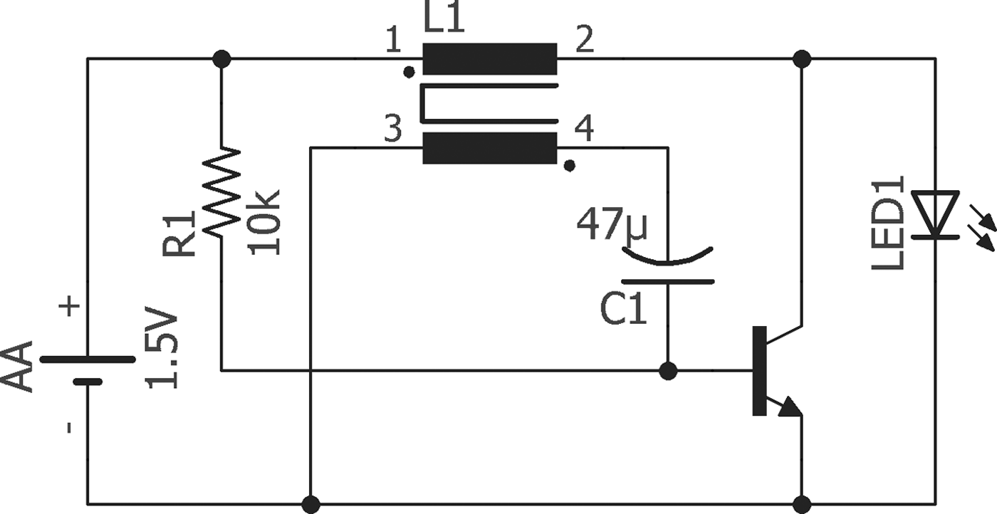
To test the suitability of very low and blinking light emitting diode (LED)-light with extremely low power consumption, a so-called joule thief was constructed to obtain a self-oscillating voltage booster for generating a driving voltage. Its utility is known for LEDs or other light sources such as neon lamps. The main difference between the joule thief and other voltage boosters is its ability to work with very low voltages down to 0.3 V (see also US Patent US4734658). It is also possible to flash an LED with a joule thief to obtain a longer lifetime. For example, with only one AA-battery, flashing joule thieves, can operate an LED for more than 1 year with a duty cycle of 30% at approximately 45 kHz.
The circuit operates as an unregulated up-converter with the self-oscillating characteristic of a blocking oscillator. The transistor connects the coil cyclically with the supply voltage, which is stored in the magnetic field. This energy induces a higher voltage in the blocking phase, causing a current flow through the LED until the stored energy is dissipated.
Thus, the function of a joule thief is based on a self-stroking/positive feedback process, which turns the transistor on, so that the capacitor is loaded until the voltage is high enough to cause the LED to flash. For this experiment we built a flashing joule thief with a warm white LED (Nichia NSPL510DS), shown in the circuit diagram depicted in Fig. 1. The coil consisted of two times 20 turns of copper wire (0.1 mm) around a ferrite toroid with a diameter of 10 mm. An electrolytic capacitor of 47 µF and a 10-kΩ resistor was connected to the base of a standard BC547 NPN transistor. A single AA battery supplies the circuit with 1.5 V.
Circuit diagram of a Joule thief: the flashing LED provides enough light to preserve Euglena gracilis cells in the state of dormancy while only drawing a minute amount of power from the battery.

Recovery of the cells
After 9 months of storage, the culture chamber samples (cells and cotton) were transferred to Schott bottles containing 1 l of mineral medium each (same medium used for regular culture growth). The Schott bottles were kept in the culture chamber at 20°C and a light intensity of about 40 µmol photons m−2 s−1 or 9 W m−2 from mixed cool white and warm tone fluorescent lamps. Cell counts as well as photosynthetic efficiency and movement analysis were performed at regular time intervals following rehydration. For analysis, 7–8 ml of each cell culture were withdrawn from the culture bottle under sterile conditions and divided for the three different analysis procedures.
Due to the bad aspect (see the section Results) of the joule thief samples and the dark samples, in the first step the tubes were filled with mineral medium with the lid slightly loosened in order to allow gas exchange with the atmosphere. At first, cell counting monitored the growth in the fluid, whereas many cells stuck in the cotton wool. After 3 days, the recovered cultures were transferred to 500 ml mineral medium in Schott bottles, which were subsequently located in a culture chamber under the same conditions as indicated above.
Image analysis
Image analysis was performed with the computer-based motion-analysis software Wintrack 2000 (Lebert & Häder Reference Lebert and Häder1999). In brief, every 40 ms the software detects the position (x- and y-coordinates) of all objects, which meet pre-adjusted expectations (e.g. size, velocity, grey level) and calculates the center of gravity (COG) for each detected object. The COG of an object in the first image and in the fifth image determines the movement vector of the object. Vector length and angle are calculated for all the recognized objects. A known frame rate (x images per second) allows for the calculation of velocity. In addition, cell size and form are calculated and all data are stored. A defined (adjustable) amount of such single movement vectors is calculated into pooled population data such as mean velocity, amount of upward swimming cells and statistical data, which indicate the precision of the orientation of a cell culture. The primary parameter used in this respect is the r-value, which ranges from 0 (no orientation of a cell culture, no preferred movement direction of the cells) to 1 (high orientation, all cells swim exactly in the same direction). The hardware used was a manual Ecotox device (Tahedl Reference Tahedl2000; Tahedl & Häder Reference Tahedl and Häder2001), that is in principle a horizontally mounted microscope with a camera attached. Infrared-LEDs serve as bright-field illumination, as the cells cannot perceive IR-light and it is not suitable for photosynthesis. Therefore, it does not induce any photo-orientation or photosynthesis. Single use cuvettes are assembled for each measurement: two microscope slides are glued together with two layers of a U-shaped cut-out of common adhesive tape. The space between the two slides is 100 µm wide. Cells are carefully and slowly filled in the cuvette from the upper side. Then, the cuvette is inserted in a slit of the microscope so that the position is vertical and enables movement of cells with respect to the vector of gravity. The microscope setup is impermeable to light after insertion of the cuvette.
Determination of photosynthetic efficiency
Photosynthetic efficiency was measured by means of a PAM (pulse-amplitude modulated) fluorometer. As long as the cell concentration in the samples was very low, photosynthetic efficiency was determined with a Water PAM (Walz, Germany). With increasing cell number, fluorescence of the cells exceeded the measurement range of that instrument. Therefore, a less sensitive PAM 2000 device was employed. Comparison of both devices revealed that they delivered identical results in a cell concentration range that both devices were able to detect. The single saturation pulse method was used for the experiments. A 20 min dark period ensured that the redox systems, especially the primary electron acceptor (QA) were all in an oxidized state. The maximum quantum yield F V/F M was determined by the following equation:
 $$F_{\rm V} /F_{\rm M} = {\rm} (F_{\rm M} -F_{\rm 0} )/F_{\rm M}, $$
where F M is the maximum Chl a fluorescence yield in the dark-adapted state and F 0 is the minimum Chl a fluorescence yield in the dark-adapted state (Kalaji et al. Reference Kalaji2014). Every data point consists of five subsequent measurements.
Multiplex PCR and capillary gel electrophoresis
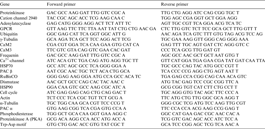
Four independent samples with dormant cells (4.2 × 10⁶ cells in 4 ml mineral medium on 0.5 g cotton wool in 50 ml falcon tubes as described above) were recovered with 5 ml of fresh mineral medium. Samples for mRNA-analysis were withdrawn directly after addition of medium and subsequently after 1 h, 2, 4, 6, 8, and 10 d, respectively. RNA was isolated with the TRIzol-method (Rio et al. Reference Rio, Ares, Hannon and Nilsen2010). In total 100 ng of mRNA were transcribed into cDNA using the QuantiTECT reverse transcription kit from QIAGEN. For the simultaneous amplification of several genes from Euglena gracilis, a multiplex PCR was carried out to detect gene expression differences. The PCR reactions were carried out in 25 µl volume containing 2 × Multiplex Master Mix (QIAGEN Multiplex PCR Plus Kit), 0.2 µM of each primer (see Table 1) and template DNA (10 ng) in a thermal cycler (C1000 Touch, Bio-Rad, USA.) The PCR profile was as follows: 95°C for 5 min followed by 42 cycles of amplification at 95°C for 30 s, 60°C for 3 min, 72°C for 90 s and with a final extension at 68°C for 30 min. Samples were analysed using the QIAxcel Advanced System from QIAGEN. Screen gel analysis took place using the OM500 Multiplex setting, with Default DNA, which was not diluted. For analysis, 1 µl of the QX DNA SizeMarker FX 174/ Hae III (QIAGEN, Lot. Nr. 148044119) was mixed with 9 µl RNAse free water.
Primer used for the dormant state multiplex PCR. The mixture of primer was resuspended in 10 mM Tris, 1 mM EDTA buffer

Data calculation and statistics
The experiments were performed as independent quadruplicates. The signals were normalized to two housekeeping genes, actin and RuBisCO (large subunit). The raw data of both genes showed a relatively stable signal with a mean deviation of about 4.6% (RuBisCO) or 3.7% (actin). After normalization, data of all time points were compared with each other. The difference of the corresponding means was calculated and a two-side student-test (p < 0.05) was performed (each set of four results with any other set) to test the significance between the datasets. Two criteria were set to regard a gene as differentially expressed: a difference of 10 and more percent and significance expressed by student t-test.
Results
Observations during incubation
When the cells were placed in the 50 ml tubes on pre-wetted cotton wool, they moved to the surface on top of the cotton wool where they formed a green layer. In two samples, the cotton chunk did not form a completely flat surface, but some fiber skeins arose from the surface. The cells climbed up the skeins, where they probably dried due to a lack of water.
Control cells without additional liquid did not move onto the surface. During the storage period, these cells continually bleached, while the other samples with additional amounts of medium kept a green colour during the entire experiment.
Cell recovery and cell growth of dormant states kept in dim light
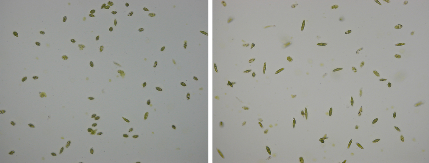
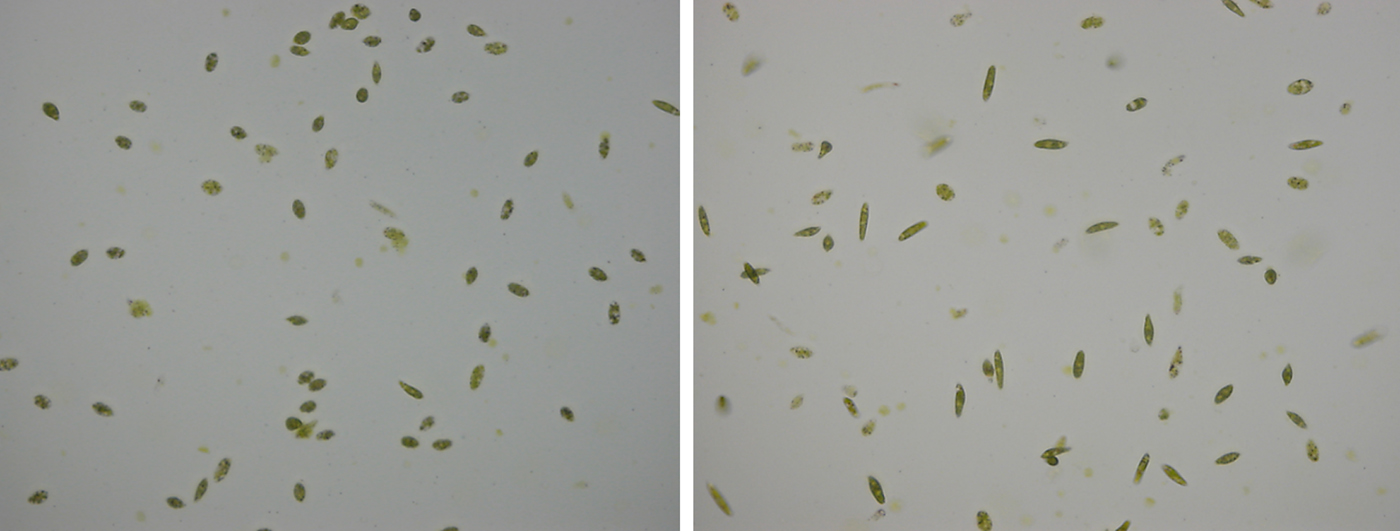
After 9 months on cotton wool in hermetically closed 50 ml falcon tubes the complete content of the tubes including immobilized cells was transferred to 1 l bottles with mineral medium. While almost all of the cells were found to be immotile and rounded (Fig. 2, left) upon sampling during the transfer to the Schott bottles, their aspect and behavior changed profoundly during the following hours (Fig. 2, right), when samples were drawn and examined under the microscope. Although the initial cell density was very low in all flasks, revitalization of the cells was observed. Only the samples without additional fluid did not recover.
Euglena gracilis cells after 9 months of storage on moist cotton wool in hermetically closed containers. Left: Cell sample drawn and observed immediately after opening the container, right picture: cell sample from the same culture after 2 h incubation in fresh medium.
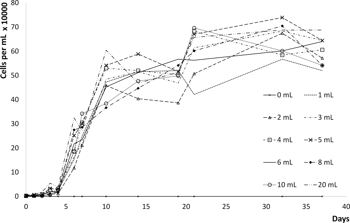
During the first 6 days, the cell number seemed to be low, but the data showed an exponential growth during this time (Fig. 3). The fluctuation of the data is most likely due to different numbers of cells in the different setups were attached to the cotton wool and the bottle. It was possible to remove the cells from the bottle wall by shaking, but in each setup a different number of cells remained attached to the cotton. Initially the 10 ml sample as well as the 2 ml sample showed a slightly lower growth rate compared with the others. The reason might be that in those samples the cotton skeins as described earlier reduced the number of surviving cells and inhibited recovery.
Increase of cell number of rehydrated dormant stages of Euglena gracilis. Cells (4 × 10⁶ cells in a total volume of 400 µl medium) were kept for 9 months in closed containers (50 ml) on 0.5 g cotton wool with 0, 1, 2, 3, 4, 5, 6, 8, 10, and 20 ml of medium. Cells were stored under low light conditions (7 W × m−2 PAR) at 20°C. In order to induce rehydration, cells in the container were transferred into 1 l of medium. Cells without medium added in the beginning of the experiment (0 ml line) did not recover.

Cell motility
After 1 day of rehydration, motile cells were found in all samples (except for the control without additional medium during dormancy, which never recovered).
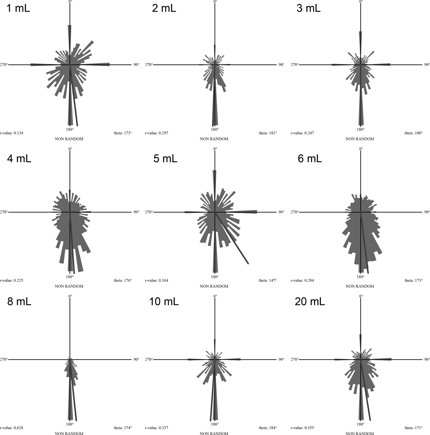
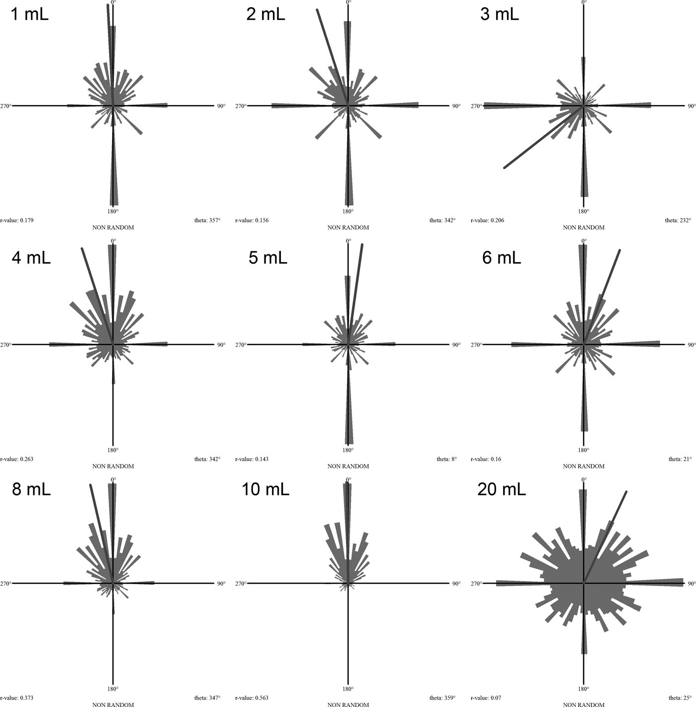
Due to the low cell density in combination with high amounts of debris, it was not possible to trace the cells by means of image analysis until day 4 after inoculation. Then the image analysis indicated positive gravitaxis in all samples, which means that the cells analysis indicated positive gravitaxis in all samples, which means that the cells moved downward in the water column (Fig. 4). After about 10 days, negative gravitaxis was detected in almost all samples (Fig. 5).
Circular histograms of the orientation of the cultures stored with dim light after 4 days of revitalization. All samples show downward orientation. Numbers indicate the volume of medium added in the beginning of the experiment. Grey line: theta, average direction of the movement.

Circular histograms of the orientation of the cultures stored with dim light after about 10 days of revitalization. Samples show mostly upward orientation. Numbers indicate the volume of medium added in the beginning of the experiment. Grey line: theta, average direction of the movement.

Photosynthetic yield
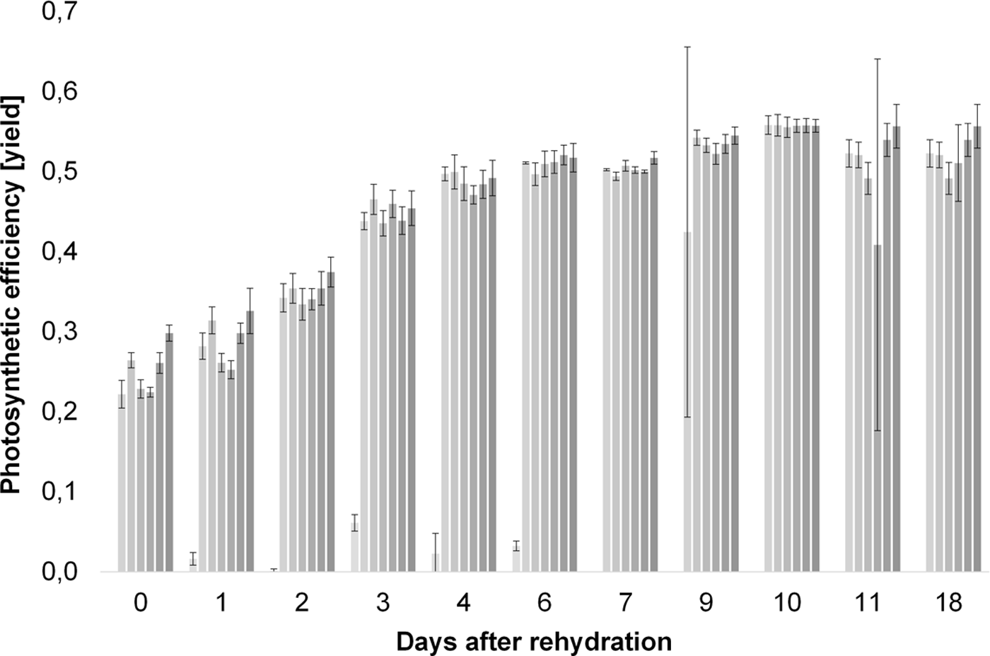
Immediately after rehydration, photosynthesis was observed. Within 3 days, it almost doubled and indicated a good development of the cell culture. After that, it remained stable and similar regardless of the amount of medium used at the beginning of the storage time (Fig. 6).
Photosynthetic efficiency of rehydrated dormant stages of Euglena gracilis. From light to dark bars: hibernation in the presence of 0, 1, 2, 3, 4, 5, 6, 8, 10 and 20 μl of medium. Each bar represents the mean of five subsequent measurements and their standard deviation. For details see Fig. 3.

Cell recovery and growth of dormancy states in darkness or illuminated with low-energy light pulses
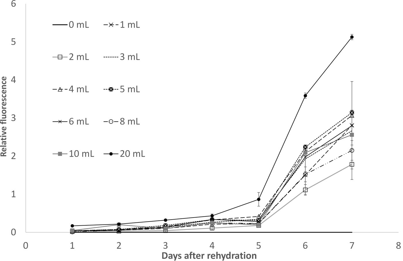
In contrast to the cells kept under dim light, the aspect of the cells that were kept in total darkness and those that were illuminated by the joule thief appeared not very promising after 9 months: all cells on the cotton wool had a blackish colour and seemed unlikely to have survived. For this reason, the containers were filled with medium to a total volume of 50 ml. Remarkably, the samples showed recovery after 4 days. With the high sensitivity water-PAM, considerable photosynthetic activity was detected in all samples (Fig. 7).
Increase of chlorophyll fluorescence of rehydrated dormant stages of Euglena gracilis in the first days of recovery determined with a water-PAM device. The fluorescence signal was normalized and indicates growth of the cell cultures. For details see Fig. 3.

After 6 days in all samples freely swimming cells were detected. After 11 days, the samples were transferred into 500 ml medium, where all cultures developed comparable with the samples, which were stored under constant light.
Transcription analysis
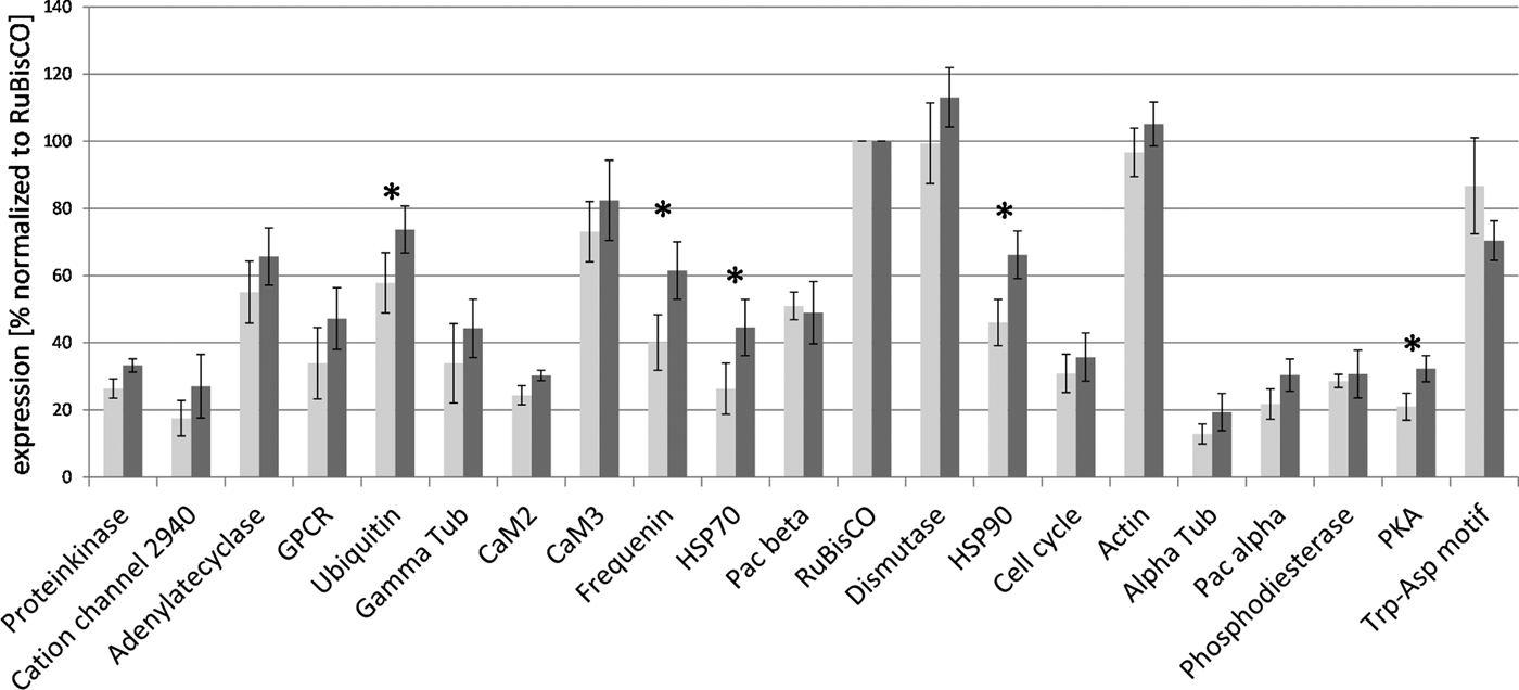
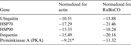
Immediately after rehydration with fresh mineral medium many of the investigated genes showed reduced transcription compared with the sample withdrawn 1 h after recovery. Many transcripts were found to be more highly expressed in the 1 h-sample (Fig. 8) compared with the sample immediately after rehydration. Only genes that met the two criteria (10% and t-test) in both normalizations against actin and RuBisCO were considered. After applying these stringent criteria, the following transcripts were significantly more highly (p < 0.05) expressed after 1 h: the heat shock proteins HSP 70 and HSP 90, the calcium-sensor protein frequenin, and ubiquitin (Table 2, Fig. 8). Further development of the cell culture from day 1 to day 2 showed a significant increase of γ-tubulin.
Changes in the expression profile normalized to RuBisCO between cells directly after rehydration (light grey) and 1 h after rehydration (dark grey). Asterisks display changes more than 10% and significance (p < 0.05).

Comparison of gene expression between Euglena cells directly after rehydration and 1 h after rehydration

The table shows only genes, which were significantly higher expressed after normalization for actin and RuBisCO, respectively. In addition only genes, which differ at least 10% were considered.
*Significant but below 10%-threshold.
Using another method to estimate the expression level (samples that deviate 15% from the mean of all experimental days), a higher number of differentially expressed genes became visible. While directly after addition of mineral medium 9 of 20 genes were down-regulated compared with the mean of all days, 8 genes were significantly up-regulated compared with the mean after 1 h (no gene was lower expressed than the mean). In the 2-day sample only two genes with lower expression were found, on the 4th day two were up-regulated. At the 6th day no investigated gene was changed compared with the mean. At day 8, strong changes occurred with 10 genes down- and 3 genes up-regulated, while at day 10 only four genes were different with a higher expression.
Discussion
Microscopic observation showed that not all Euglena gracilis cells formed thick-walled dormant states. However, the described method of cell preservation leads to reliably recoverable cell cultures of Euglena gracilis after prolonged storage. Dormant states of microorganisms are employed in a number of applications: encysted cyanobacteria are used for rice field fertilization (Vaishampayan et al. Reference Vaishampayan, Sinha, Häder, Dey, Gupta, Bhan and Rao2001). For the restauration of eroded landscapes (Hu et al. Reference Hu, Zhang, Huang and Liu2003; Xie et al. Reference Xie, Liu, Hu, Chen and Li2007; Rao et al. Reference Rao, Liu, Wang, Hu, Dunhai and Lan2009; Wang et al. Reference Wang, Liu, Li, Hu and Rao2009; Li et al. Reference Li, Rao, Wang, Shen, Li, Hu and Liu2014) huge amounts of cells are sprayed onto the soil; when humidity is available, they grow as filamentous cell strains and excrete considerable amounts of polysaccharides forming crusts, which immobilize the soil. During dry periods, the cells survive in dormancy. Field experiments resulted in a rapid recovery of the soil with a fast succession from moss to grass and even trees within about 10 years.
Also within the laboratory, dormant states offer a number of advantages: they can be stored as stock cultures under conditions less demanding in terms of power (illumination, temperature) and environmental control. They do not require as many person-hours of maintenance as serial subculturing. However, many of the standard procedures come with certain conditions, that were not compatible with our requirements: the organisms are stored under vacuum and preserving agents are added (Malik Reference Malik1990, Reference Malik1993).
Dormant states of eukaryotic algae such as the resting states of Euglena gracilis are potentially of great interest for further applications (e.g., stock culture for ecotoxicological studies). Malik determined survival of different green algae and Euglena gracilis after long term storage (6 month and 12 month) (Malik Reference Malik1995): liquid cultures (logarithmic growth phase) of the algae were kept under low light conditions (50–100 lux) at 9°C or 20°C. All green algae (Chlorella pyrenoidosa, Chlorella vulgaris, Selenastrum capricornutum, and Scenedesmus subspicatus) recovered well in all storage conditions (both temperatures, with and without additives). Euglena gracilis survived only 1 month under these conditions (then, ‘overgrowth of associated bacterial flora’ occurred), although the culture conditions had been very similar to the conditions in our experiment. Obviously, the survival of Euglena gracilis is supported by the presence of cotton wool and availability of medium, because all those samples showed good recovery after 9 months, while the sample without medium did not. Similarly, without cotton wool the survival of concentrated cells is highly unlikely over a prolonged period (as observed in previous experiments). Hence, a certain residual moisture is important. The cotton wool provides a huge surface for the cells to attach. Compared with a liquid culture of similar cell density, cotton wool substrate improves the recovery rate considerably (as tested in previous experiments, data not shown), even though the reason is not clear. About 10% of absolute water content (0.1 g H2O per gram dry mass, which corresponds to an equilibrium with air of about 50% relative humidity) is necessary to form a monolayer of water around proteins and membranes (Alpert Reference Alpert2006). This could well mean that in the presence of high amounts of medium (20 ml) not all cells may have developed into resting states and it could be argued that we dealt with starving cells rather than resting cells in this case.
The motility of the cells during recovery showed a normal pattern: young cell cultures usually show positive gravitaxis (Häder et al. Reference Häder, Richter and Lebert2006; Richter et al. Reference Richter, Schuster, Meyer, Lebert and Häder2006, Reference Richter, Schuster, Lebert, Streb and Häder2007), that changes after a couple of days to negative gravitaxis.
Photosynthetic yield indicates an initially slightly faster development of the culture with 20 ml of medium compared with the samples with less medium (Fig. 7). However, the difference decreases over time as it does with the cell numbers (Fig. 3). While in the other samples the medium was completely soaked up by the cotton wool (being a ‘water-in-cotton-habitat’), at 20 ml the substrate was completely submersed (being a ‘cotton-in-water-habitat’). In any case, addition of a total amount of 1.4 ml of medium with 4 000 000 cells (1 ml of medium and 400 µl of concentrated cells) on 0.5 g of cotton wool enables survival of Euglena gracilis-cells for at least 9 months. It is very likely that also lower amounts of humidity would be sufficient but this remains to be investigated.
The preliminary results achieved with low energy light pulses proved to be quite interesting because they show that even with a minute amount of energy, the viability of an inoculum can be maintained over a long period. The power of one AA cell was sufficient to supply light over 9 months to enable better recovery of cells compared with cells stored in darkness. In any case, these non-illuminated cells did recover as well but they needed more time to grow to a considerably dense culture. For this reason, we do not recommend storing resting stages for in darkness. After all, the dormant state is a natural adaption of E. gracilis to unfavourable natural conditions, which do not usually include months of darkness.
Changes of external parameters such as light, temperature, humidity, salinity etc., make adaptation of the cellular machinery necessary. This is achieved by means of protein-modification (e.g., phosphorylation (Toroser & Huber Reference Toroser and Huber1997), protein degradation (see below) as well as synthesis of new proteins (Kosová et al. Reference Kosová, Vítámvás, Prášil and Renaut2011). The latter is detected by a change in the transcription profile.
In this study, significant changes of the ubiquitin-transcription level was observed 1 h after rehydration, indicating accumulation of degenerated proteins during long-term storage. Ubiquitination is a sign of degradation of proteins and is crucial for regulation of proteins as well as removal of misfolded proteins (Ciechanover Reference Ciechanover1994). Among the important roles of ubiquitination in plants is the local as well as the systemic responses of phosphate starvation in Arabidopsis thaliana (Rojas-Triana et al. Reference Rojas-Triana, Bustos, Espinosa-Ruiz, Prat, Paz-Ares and Rubio2013). Involvement of desiccation/rehydration-associated changes in ubiquitin-mediated protein degradation was shown in desiccation tolerant moss (O'Mahony & Oliver Reference O'Mahony and Oliver1999). In yeast, ubiquitin was found to have a vital role in germination of ascospores (no sporulation in UBI4-deficient Saccharomyces cerevisiae clones). In addition, these cells were hypersensitive to high temperature, amino acid analogs (lead to misfolded proteins) and starvation (Finley et al. Reference Finley, Özkaynak and Varshavsky1987). The authors suggest that stress leads to the accumulation of degenerated proteins that are toxic, if not degraded.
Multiplex data indicate an over expression of the heat shock proteins 70 and 90 one hour after rehydration. Abiotic stress leads to formation of different HSPs, that support protein folding and stabilize proteins and membranes (Wang et al. Reference Wang, Vinocur, Shoseyov and Altman2004; Timperio et al. Reference Timperio, Egidi and Zolla2008). HSPs play an important role in desiccation of the resurrection plant Xerophyta viscosa (Farrant et al. Reference Farrant, Cooper, Hilgart, Abdalla, Bentley, Thomson, Dace, Peton, Mundree and Rafudeen2015). An important signal for HSP-activation are abnormally folded proteins (Jayakumar et al. Reference Jayakumar, Hwang, Fabiny, Chinault and Barnes1989). Depletion of essential elements is also accompanied by increased HSP-production: Fe-depletion was described to increase, among many other effects, the expression of HSPs and calcium-sensor proteins in Phaeodactylum tricornutum (Allen & Crane Reference Allen and Crane1976; Allen et al. Reference Allen, LaRoche, Maheswari, Lommer, Schauer, Lopez, Finazzi, Fernie and Bowler2008).
The gene expression levels off after 2 days, leading to the assumption that the differences in expression is a transient effect on the cells. Microgravity-induced stress response in Euglena gracilis was observed during a space flight experiment, where expression of certain genes was determined after about 40 min of microgravity (Nasir et al. Reference Nasir2014). It was found that many stress-related genes were up-regulated (HSPs, oxidative stress and repair proteins). Although the number of analysed genes was limited, the data indicate transient stress response of the cells after rehydration.
Long-term storage of Euglena gracilis cells probably resulted in accumulation of misfolded proteins, which made their degradation via an ubiquitin-proteasome system necessary. Increase of heat-shock proteins transcription indicates a transient stress response after rehydration in fresh medium. The restart of metabolism and preparation of cell division is most likely accompanied with an increase in protein translation. The HSPs increase the likelihood of proper protein folding.
The calcium-sensor protein frequenin was found to become up-regulated 1 h after rehydration of the cells. An increase of a calcium-related protein is in agreement with other results in stress research. Calcium-dependent signaling in mediation of abiotic stress is an intensively studied research field (Knight Reference Knight2005; Vinocur & Altman Reference Vinocur and Altman2005). Stress induced calcium increase regulates signal transduction chains such as CaM, CDPK/CCaMK, phosphatases, etc. (Poovaiah & Reddy Reference Poovaiah and Reddy1990; Reddy et al. Reference Reddy, Ali, Celesnik and Day2012).
Application of microarrays would deliver a more complete picture of the gene-families involved in recovery of long-term stored Euglena gracilis cells. Under water stress (20% remaining humidity) in Rhodococcus jostii more than 700 differentially expressed genes were detected by microarray analysis (LeBlanc et al. Reference LeBlanc, Goncalves and Mohn2008).
The results of this study clearly demonstrates that the method described enables long-term storage of Euglena gracilis cells with reliable recovery rates. In recent tests, dormant cells were recovered after 20 months of storage (unpublished results) and some of our samples still wait to be awoken.
As Euglena is a candidate organism for bio-regenerative life support systems (BRLSS) and micro-gravity studies in general, this simple storage method can be used to increase the reliability of such a system for long-term space flights, however, one of the potential risks for BRLSS is contamination. With a stock of dormant states stowed away without much need of energy and space, it would be possible to re-inoculate the system after proper cleansing if contamination occurred. Once grown to an appropriate cell density (and still axenic), a sample could easily been drawn to replace the spent inoculum, thus decreasing the need of additional backups.
This study clearly demonstrates that Euglena can survive the long and sometimes inhospitable conditions necessary for space flight research and for its applied use in space habitats.
Acknowledgments
This work was funded by the German Aerospace Center (DLR, Deutsches Zentrum für Luft- und Raumfahrt) on behalf of the Federal Ministry of Education and Research (BMBF, Bundesministerium für Bildung und Forschung, grant 50WB1528). Ina Becker is a doctoral candidate of the Helmholtz Space Life Sciences Research School (SpaceLife), German Aerospace Center (DLR) Cologne, Germany, which was funded by the Helmholtz Association (Helmholtz–Gemeinschaft) over a period of 6 years (Grant No. VH-KO-300) and received additional funds from the DLR, including the Aerospace Executive Board and the Institute of Aerospace Medicine.