

Introduction
Podocarpus L'Hér. ex Pers. is the largest genus in the family Podocarpaceae, with about 100 species (Farjon, Reference Farjon2010). Podocarpus has two subgenera, Podocarpus subgenus Podocarpus and Podocarpus subgenus Foliolatus de Laub. (de Laubenfels, Reference de Laubenfels1985). In species within Podocarpus subgenus Podocarpus, the stomata possess a distinct raised rim around their openings, a structure known as a Florin ring, a term coined by Buchholz & Gray (Reference Buchholz and Gray1948: 52) and named after the morphologist and specialist on gymnosperms Rudolf Florin, who first discovered them in conifers (Florin, Reference Florin1931). In contrast, species within Podocarpus subgenus Foliolatus are characterised by the absence of Florin rings (de Laubenfels, Reference de Laubenfels1985). The Florin ring may function in reducing wettability of the leaf surface, as suggested by Pariyar et al. (Reference Pariyar, Chang, Zinsmeister, Zhou, Grantz, Hunsche and Burkhardt2017) for Chamaecyparis obtusa (Siebold & Zucc.) Endl. In addition, Mohammadian et al. (Reference Mohammadian, Hill and Watling2009) found that in Agathis robusta (C.Moore ex F.Muell.) F.M.Bailey the Florin rings had a noticeable, although not quantifiable, effect in deflecting fungal hyphae away from the stomata and so protecting them from fungal invasion. Investigation of this in Podocarpus was outside the scope of this study.
Species of Podocarpus subgenus Podocarpus are found in habitats ranging from forest to alpine heath in Southeast Australia, Tasmania, New Zealand and Chile, as well as in the tropical highlands of Africa and America (including the Caribbean Islands), and rarely in the tropical lowlands of those areas (de Laubenfels, Reference de Laubenfels1985). In contrast, species of Podocarpus subgenus Foliolatus are especially abundant in the Asian and Pacific tropics (lowlands and highlands), in subtropical areas of eastern Asia and in Australia, and are absent from Africa and the Americas (de Laubenfels, Reference de Laubenfels1985). The two subgenera overlap only in two areas: the Pacific tropics of north-eastern Australia and on the island of New Caledonia (de Laubenfels, Reference de Laubenfels1985).
The cross-section of the stomatal complex of Podocarpus has not previously been studied using either light microscopy or scanning electron microscopy (SEM). Indeed, there have been very few such studies of the stomatal complex of any gymnosperm. However, SEM can provide further detail and information on the architecture of the stomatal complex and the wax plug and how they function as a unit.
Two early studies were those by Hanover & Reicosky (Reference Hanover and Reicosky1971) and Jeffree et al. (Reference Jeffree, Johnson and Jarvis1971). Hanover & Reicosky's paper concerned surface wax deposits on the foliage of various conifers including Picea pungens Engelm., while that by Jeffree et al. studied epicuticular wax in what they and numerous other workers have termed the stomatal antechamber (e.g. Jeffree et al., Reference Jeffree, Johnson and Jarvis1971; Appleby & Davies, Reference Appleby and Davies1983; Morvan, Reference Morvan1987, Podocarpus [various organs, but not stomata in cross-section]; Karabourniotis et al., Reference Karabourniotis, Tzobanoglou, Nikolopoulos and Liakopoulos2001). The stomatal antechamber is equivalent to the space in the stomatal complex that is exterior to the subsidiary cells and below the surface of the leaf. This cavity was also termed the epistomatal chamber by Hanover & Reicosky (Reference Hanover and Reicosky1971), and that term has likewise been used by numerous later workers (e.g. Yoshie & Sara, Reference Yoshie and Sara1985; Prior et al., Reference Prior, Pritchard, Runion, Rogers and Mitchell1997; Vovides & Galicia, Reference Vovides and Galicia2016). Mill & Whiting (Reference Mill and Whiting2012) used ‘peristomatal chamber’ for the same cavity in the stomatal complex of Podocarpus orarius R.R.Mill & Whiting, but that expression has been used by no other authors and is rejected here. Instead, following Jeffree et al. (Reference Jeffree, Johnson and Jarvis1971), Morvan (Reference Morvan1987) and subsequent authors, we use ‘stomatal antechamber’.
Materials and Methods
Cross-sectioning
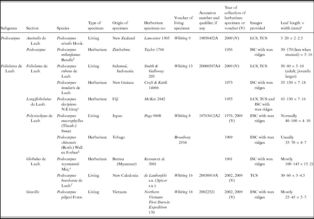
Leaf samples were taken from either herbarium or living specimens of four species of Podocarpus conserved at the Royal Botanic Garden Edinburgh (Table 1). Cross-sections of living material (Podocarpus nivalis Hook., P. rubens de Laub. and P. beecherae de Laub.) were obtained using cryofracture. A section of the leaf was cut into slices of c.5 × 7 mm and placed on to a stub that had a layer of colloidal graphite C303, allowing the segments to be placed with the longitudinal or transverse edge up. The specimen was rapidly frozen using liquid nitrogen subcooled to about −210°C and transferred to the cold stage of a Gatan Alto 2500 cryopreparation system, where the leaf segments were fractured using a blade at a temperature of about –175°C. Cross-sections of herbarium material (Podocarpus decipiens N.E.Gray) were obtained by fracture of the dried leaf, using tweezers at room temperature. These slices were mounted with the longitudinal or transverse edge up on a specimen stub with a carbon adhesive disc and Electrodag 915 high-conductivity paint.
Voucher and collection details for material of species of Podocarpus examined

ECS, external surface of the cuticle; ICS, internal surface of the cuticle; LCS, longitudinal cross-section; TCS, transverse cross-section.
a Adapted from Farjon (Reference Farjon2010) with extreme values omitted, except: Podocarpus beecherae from de Laubenfels (Reference de Laubenfels2003), P. decipiens from Gray (Reference Gray1955) and P. teysmannii from measurements of the holotype specimen of its synonym P. epiphyticus at E.
b Often synonymised with the South African Podocarpus latifolius (Thunb.) R.Br. ex Mirb. (e.g. de Laubenfels, Reference de Laubenfels1985; White et al., Reference White, Dowsett-Lemaire and Chapman2001), but retained as a separate species by Farjon (Reference Farjon2010), the view followed here.
c Podocarpus decipiens was regarded as a synonym of the widespread P. neriifolius D.Don by de Laubenfels (Reference de Laubenfels1985) and Farjon (Reference Farjon2010) but kept separate by Smith (Reference Smith and Smith1979). Cuticle micromorphologies of the two taxa show differences (Whiting, Reference Whiting2009), and in her protologue Gray (Reference Gray1955) pointed out anatomical differences. Smith (Reference Smith and Smith1979) retained Podocarpus decipiens as a distinct species, and the same view was recently accepted by de Laubenfels (Reference de Laubenfels2015), who classified it in Podocarpus section Longifoliolatus.
d Treated as Podocarpus macrophyllus var. maki Siebold & Zucc. by Farjon (Reference Farjon2010).
e The studied material was the holotype of Podocarpus epiphyticus de Laub. & Silba. This name was regarded as a synonym of Podocarpus neriifolius by Farjon (Reference Farjon2010), but cuticle micromorphology shows distinct differences from that species (Whiting, Reference Whiting2009). Most recently, Podocarpus epiphyticus was synonymised with P. teysmannii by de Laubenfels (Reference de Laubenfels2015).
f A recently described species (de Laubenfels, Reference de Laubenfels2003) that has been synonymised with Podocarpus novae-caledoniae Vieill. by Farjon (Reference Farjon2010). Cuticle micromorphologies of the two taxa are markedly different, however (Whiting, Reference Whiting2009), so Podocarpus beecherae is retained here. This is also in agreement with the most recent treatment of these two species by de Laubenfels (Reference de Laubenfels2015), who transferred Podocarpus beecherae to Podocarpus section Globulus.
Cuticle preparation
The cuticle of six other taxa of Podocarpus (P. chinensis (Roxb.) Wall. ex Forbes,Footnote 1 P. insularis de Laub., P. macrophyllus (Thunb.) Sweet, P. milanjianus Rendle, P. pilgeri Foxw., P. teysmannii Miq.Footnote 2 : Table 1) was also examined, although cross-sections of these species were not undertaken. To observe the internal surface of the cuticle (hereafter abbreviated ISC), the cuticle was isolated following the method of Alvin & Boulter (Reference Alvin and Boulter1974), with changes made to suit Podocarpaceae based on work by Kershaw (Reference Kershaw1997), Stockey et al. (Reference Stockey, Frevel and Woltz1998) and Stark Schilling & Mill (Reference Stark Schilling and Mill2011). Six leaf slices of around 0.8 cm2 were placed in glass vials with 6 mL of 20/120% (16.66%) solution of dihydroxidodioxidochromium (H2CrO4, ‘chromic acid’). The vials were sealed and kept at room temperature in a fume hood for 96 h. Isolated cuticles were removed from the chromic acid solution, washed with distilled water and left to dry on filter paper for at least 4 h. Cuticles were checked for homogeneity under the light microscope. Two cuticle slices were mounted on an aluminium stub using carbon adhesive discs. The external surface of the cuticle (ESC) was observed using untreated leaf slices. These samples were mounted on to stubs as above. The leaf segments were sputter coated with 60%:40% gold:palladium for 2 min at 12 mA in an Emscope SC500 sputter coater.
Scanning electron microscopy
Examination of all samples by SEM, whether they were cryosectioned, fractured or not, was done using a Hitachi S-4700 II field emission SEM at the University of Edinburgh. Accelerating voltage was 5 kV. Working distance varied from 11.9 to 15.2 mm for the cross-sections, and from 13.0 to 16.6 mm for the ISC and ESC. It was found that optimal images of leaf cuticles were obtained by setting LensMode to Analysis, although images of Podocarpus beecherae, P. decipiens and P. nivalis in cross-section were taken with LensMode set to Normal. Magnifications ranged from approximately 1.3 to 6 k [i.e. × 1300–6000], as given exactly in the SEM data imprinted at the bottom of each figure.
Results
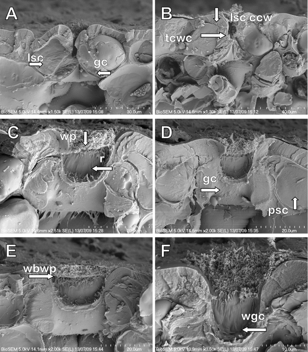
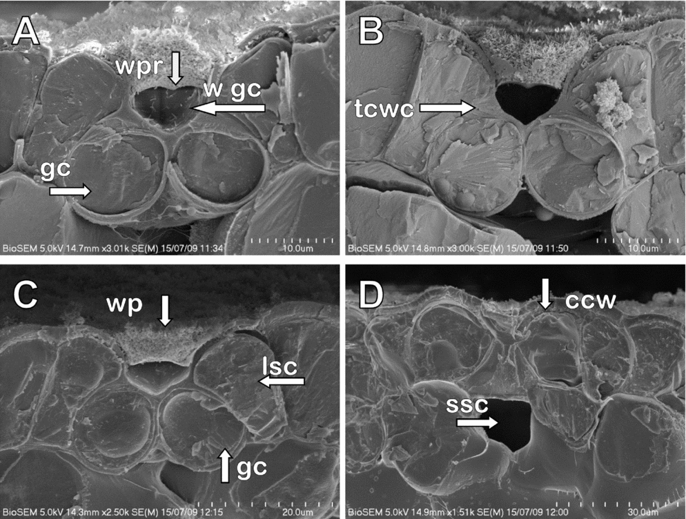
Cross-sections of the stomatal apparatus of Podocarpus rubens (Fig. 1), P. beecherae (Fig. 2) and P. decipiens (Fig. 3) revealed the inside of the stomatal antechamber with wax plug intact. The wax plug sat high in the stomatal antechamber, in contact with the subsidiary cells and with a relatively wide gap between the wax plug and the guard cells. The wax plug of Podocarpus nivalis (Fig. 4) was missing for the transverse cross-section (TCS: Fig. 4A) and appeared damaged in the longitudinal cross-section (LCS: Fig. 4B).
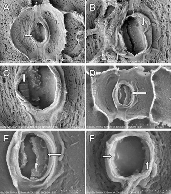
Podocarpus rubens: cross-sections of stomatal complexes under scanning electron microscopy. A, Transverse cross-section (TCS) showing lateral subsidiary cell (lsc) and guard cell (gc). B, TCS showing cuticle and cell wall of lateral subsidiary cell (lsc ccw) and triangular area of cell wall and cuticle (tcwc). C, Longitudinal cross-section (LCS) showing wax plug (wp) and ridges (r). D, LCS showing polar subsidiary cell (psc) and guard cell (gc). E, LCS showing wax below the wax plug (wbwp). F, LCS showing wax on guard cell surface (wgc). Scale bar and magnification shown in each image.

Podocarpus beecherae: cross-sections of stomatal complexes under scanning electron microscopy. A, Transverse cross-section (TCS) showing wax plug ridge (wpr), wax on the guard cell surface (w gc) and guard cell (gc). B, TCS showing triangular area of cell wall and cuticle (tcwc). C, TCS showing wax plug (wp), lateral subsidiary cell (lsc) and guard cell (gc). D, TCS showing substomatal cavity (ssc) and cuticle and cell wall (ccw). Note the elliptical guard cells in this sample. Scale bar and magnification shown in each image.

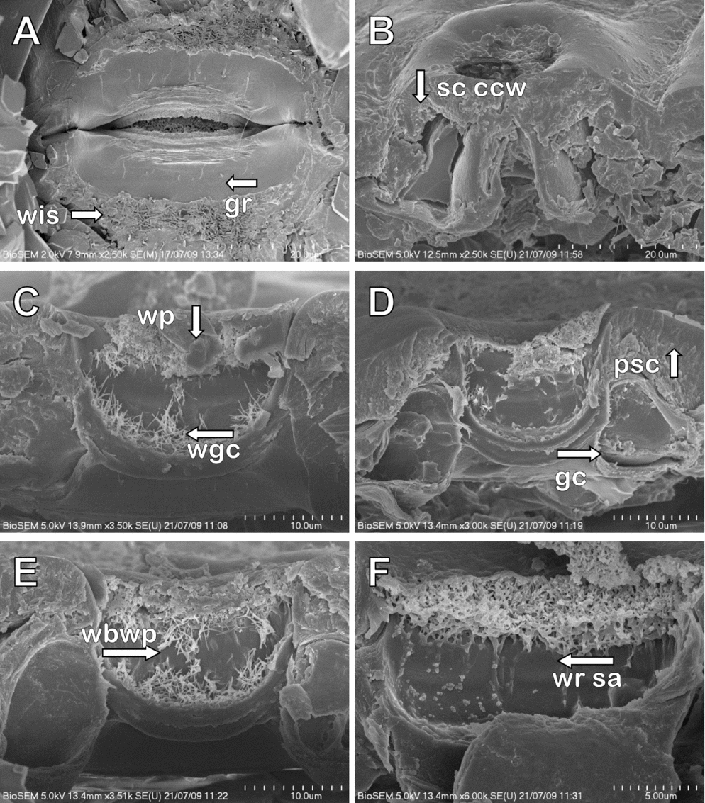
Podocarpus decipiens: stomatal complexes under scanning electron microscopy. A, Inner surface of stomatal complex, showing wax inside stomata (wis) and groove (gr). B, Outer view of stomatal complex, showing cuticle and cell wall of subsidiary cells (sc ccw). C, Longitudinal cross-section (LCS) showing wax plug (wp) and wax on guard cells (wgc). D, LCS showing polar subsidiary cell (psc) and guard cell (gc). E, LCS showing wax below wax plug (wbwp). F, LCS showing wax ridges in the stomatal antechamber (wr sa). Scale bar and magnification shown in each image.

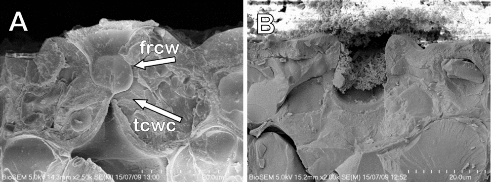
Podocarpus nivalis: cross-sections of stomatal complexes under scanning electron microscopy. A, Transverse cross-section showing Florin ring made up of cell wall material and cuticle (frcw) and triangular area of cell wall and cuticle (tcwc). B, Longitudinal cross-section. Scale bar and magnification shown in each image.

Transverse cross-section
The TCS of the stomatal apparatus is illustrated for Podocarpus rubens (Fig. 1A,B), P. beecherae (Fig. 2A–D) and P. nivalis (Fig. 4A). This view shows a cut through the two lateral subsidiary cells and through the two guard cells. All transverse sections revealed closed guard cells, a narrow stomatal antechamber and a wax plug with a central ridge on the underside (apart from in Podocarpus nivalis, in which the wax plug was not present). The wax plug of Podocarpus beecherae appeared very smooth on the underside compared with a rough upper surface consisting of protruding wax rods (Fig. 2A–C). The guard cells in Podocarpus beecherae were usually almost round in cross-section (Fig. 2A–C), although they were elliptical in the sample used for Fig. 2D; whereas those of P. rubens (Fig. 1A,B) and P. nivalis (Fig. 4A) were very elongated and elliptical, although this may vary. The lateral subsidiary cells tended to arch over the guard cells, coming to approximately halfway, where a distinct triangle of cell wall material (probably including cuticle and wax) sat on top of the guard cell (seen in all three species for which transverse sections were obtained: Figs 1B tcwc, 2A–C, 4A). The most notable difference between the species was the presence in Podocarpus nivalis of a Florin ring created by the lateral subsidiary cells and that appears to be composed of cell wall material and cuticle (Fig. 4A frcw, 4B).
Longitudinal cross-section
The LCS of the stomatal apparatus is illustrated for Podocarpus rubens (Fig. 1C–F), P. decipiens (Fig. 3C–F) and P. nivalis (Fig. 4B). This view shows a section through the two polar subsidiary cells and one mostly complete guard cell. No ridge could be seen on the underside of the wax plug when seen in this view (the ridge appearing to correspond with the area where the two guard cells meet). In this view, wax crystals could clearly be seen on the guard cells and on the stomatal antechamber walls, where wax ridges occasionally occurred in Podocarpus rubens (Fig. 1C,D,F) and P. decipiens (Fig. 3F). In Podocarpus nivalis, a layer of wax covered the stomatal complex and a wax plug appeared to sit halfway up the stomatal antechamber (Fig. 4B). In Podocarpus rubens (Fig. 1D) and P. decipiens (Fig. 3D), a portion of the polar subsidiary cells, mostly cell wall material, arched almost completely over the polar ends of the guard cells; the same was observed to a greater extent in P. nivalis (Fig. 4B). The presence of the Florin ring in Podocarpus nivalis was less obvious than in TCS, and the Florin ring appeared to consist of cell wall material. The polar ends of the guard cells tended to be oblong to reniform in LCS in all three species. An internal view of the stomatal complex of Podocarpus decipiens was seen, resulting from fracturing of the leaf during cross-sectioning. Wax was seen on the inside area of the stomata and a groove on the underside of the guard cells (Fig. 3A).
Ridges within the stomatal antechamber
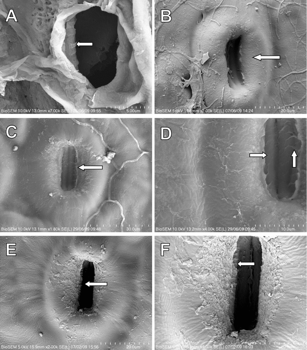
Ridges within the stomatal antechamber were observed in the cross-sections of Podocarpus rubens and P. decipiens, and also when viewing the ISC of P. decipiens (Fig. 5A–C), P. teysmannii (Fig. 5D,E), P. insularis (Fig. 5F) and P. milanjianus (Fig. 6A), as well as the ESC of P. pilgeri (Fig. 6B), P. chinensis (Fig. 6C,D), and P. macrophyllus (Fig. 6E,F). The ridges appeared to be particularly prominent and regularly spaced in Podocarpus decipiens, P. teysmannii and P. macrophyllus. In Podocarpus decipiens (Fig. 5B) and P. teysmannii (Fig. 5E), a cracked wax plug, which would have been positioned at the top of the stomatal antechamber, could also be seen. The ridges were less prominent and less regularly spaced in Podocarpus insularis, P. pilgeri, P. chinensis and P. milanjianus.
Internal surfaces of cuticle of Podocarpus species, showing ridges on the cuticle that appear to be in the stomatal antechamber: A–C, Podocarpus decipiens; D and E, P. teysmannii; F, P. insularis. Scale bar and magnification shown in each image.

Internal (A) and external (B–F) surfaces of cuticle of Podocarpus species, showing ridges on the cuticle that appear to be in the stomatal antechamber. A, Podocarpus milanjianus; B, P. pilgeri; C and D, P. chinensis; E and F, P. macrophyllus. Scale bar and magnification shown in each image.

Wax plugs
The wax plug of Podocarpus rubens seen in LCS and TCS appeared at the very top of the stomatal antechamber, in line with the top of the subsidiary cells (Fig. 1A–F). In Podocarpus decipiens, the wax plug sat slightly below the level of the subsidiary cells (Fig. 3B–E), whereas in P. nivalis it was situated halfway up the stomatal antechamber (Fig. 4B), although this may have been the result of damage. The wax plug of Podocarpus beecherae seen in TCS often had the outer edges lower than the subsidiary cell edges, although the central area or the edges of the wax plug bulged up to the height of the subsidiary cells (Fig. 2A–C). Overall, the wax plug occurred as a flat layer when viewed in LCS and was more irregular when viewed in TCS, often with a ridge on the lower surface and bulges on the upper surface.
Dimensions
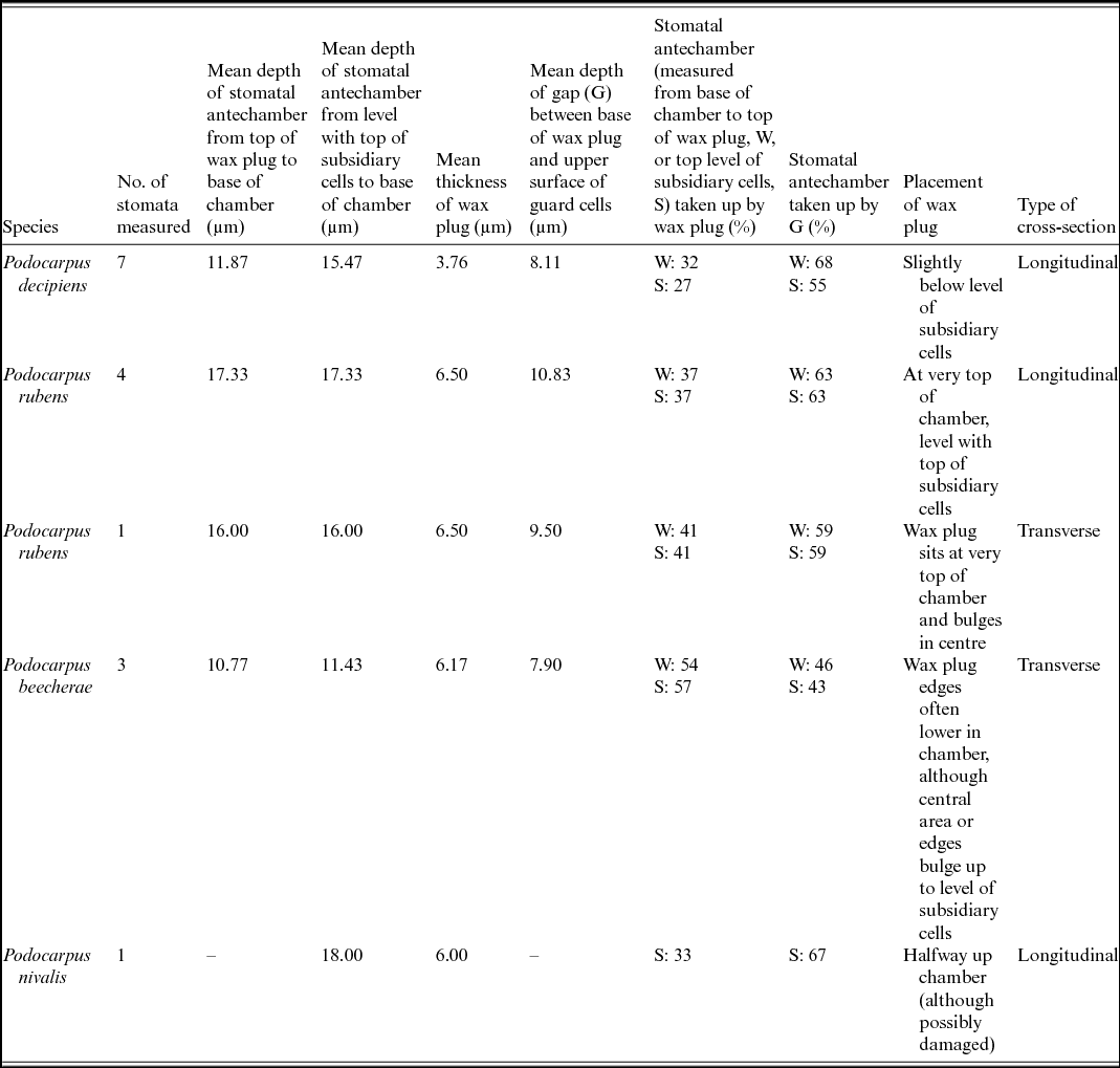
In total, measurements were taken from seven different stomata in LCS for Podocarpus decipiens, four in LCS for P. rubens, one in LCS for P. nivalis, three in TCS for P. beecherae and one in TCS for P. rubens (Table 2). Podocarpus rubens was the only species with images of undamaged stomata available in both LCS and TCS, with the main difference being the shape of the wax plug (Fig. 1B,C).
Measurements taken from longitudinal cross-sections of Podocarpus decipiens, P. rubens and P. nivalis, and transverse cross-sections of P. rubens and P. beecherae
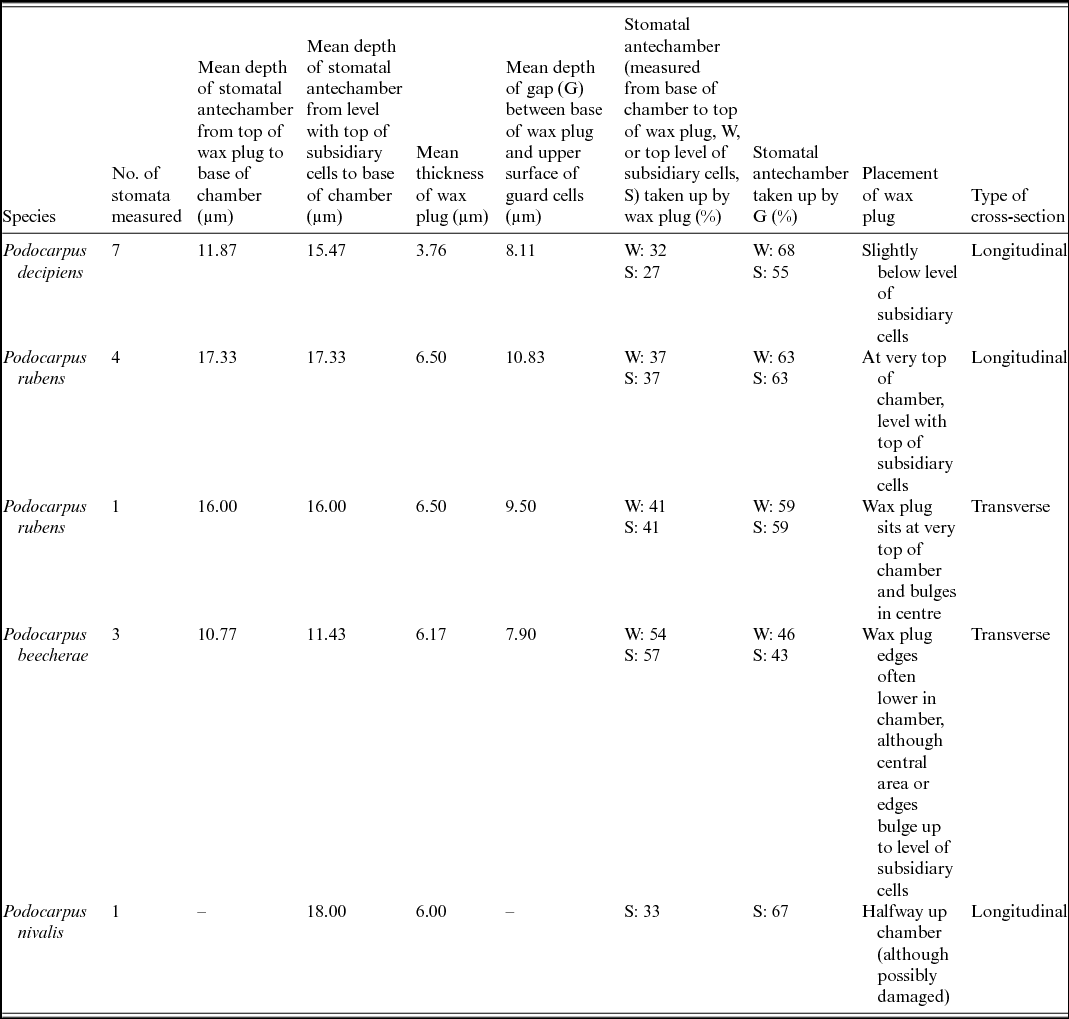
Table 2 shows that in Podocarpus subgenus Foliolatus, on average the stomatal antechamber was deeper in P. rubens (17.33 µm in LCS and 16 µm in TCS) than in any other species examined, while it was shallowest in P. beecherae (10.77 µm in TCS). The deepest stomatal antechamber, although by less than 1 µm, was that of Podocarpus nivalis (18 µm). The wax plug was similar in thickness for Podocarpus rubens (6.5 µm in both LCS and TCS), P. nivalis (6 µm) and P. beecherae (6.17 µm), although it was markedly thinner in P. decipiens (3.76 µm). The gap between the base of the wax plug and the upper surface of the guard cells was fairly similar between the studied species, varying only by up to 3 µm, being greatest in Podocarpus rubens (10.83 µm in LCS) and shallowest in P. beecherae (7.9 µm in TCS). The mean percentage of the stomatal antechamber taken up by the gap between the lower surface of the wax plug and the upper surface of the guard cells was greatest in Podocarpus decipiens (68%), if measuring depth from the top of the wax plug, or in P. nivalis (67%), if measuring to the top of the subsidiary cells, whereas it was least in P. beecherae (46% to top of wax plug, or 43% to top of subsidiary cells).
Discussion
We studied cross-sections of three species of Podocarpus subgenus Foliolatus (Podocarpus beecherae, P. decipiens and P. rubens) and one of Podocarpus subgenus Podocarpus (P. nivalis). Cross-sections of the stomatal complex provide a basis for understanding the architecture of the stomatal complex with regard to the guard cells, subsidiary cell shape, Florin ring (where present) and wax plug. Very few examples of studies on cross-sections of gymnosperm stomata seem to exist. One such study is that by Johnson & Riding (Reference Johnson and Riding1981), who used light microscopy and SEM to study the structure and ontogeny of two species of Pinus L. (Pinaceae). In addition, a study by Mohammadian et al. (Reference Mohammadian, Hill and Watling2009) used cryofracture to examine the cross-section of the stomatal complex of Agathis robusta. Their study also found that the wax plug occurred as a layer of wax that sat high in the stomatal antechamber (although no images were provided to make comparisons with the material that we studied). Our study represents the first of its kind for Podocarpus, and is also one of the first studies to use either cryofracture techniques or fracture at room temperature to examine the stomata of any gymnosperm and apply the results in a taxonomic context. Within Podocarpaceae, the same techniques have been used to study the cross-section of the stomata of the monospecific genus Sundacarpus (J.T.Buchholz & N.E.Gray) C.N.Page (Whiting, Reference Whiting2009).
More images are needed, in particular for Podocarpus subgenus Podocarpus, before generalisations can be drawn, although the images provided here do give some insight into the organisation and functioning of the stomatal complex in Podocarpus. In all species of Podocarpus subgenus Foliolatus examined in cross-section, the wax plug sat high in the stomatal antechamber and was in contact with the subsidiary cell walls, but with a relatively large gap between the wax plug and the guard cells. The underside of the wax plug had a central ridge that appeared to correspond with the crease where the guard cells meet (seen in Podocarpus beecherae and P. rubens). This is most clearly seen in Podocarpus beecherae, in which the shape of the upper surface of the guard cells and the shape of the lower surface of the wax plug (including the ridge) are similar although do not quite match. Material of both Podocarpus beecherae and P. rubens originated from young trees kept in a glasshouse (Table 1). Both these factors could have had an effect on the wax plug; for example, the ridge may disappear over time and the production of wax may vary. Had more samples, from leaves of different ages, been studied, these questions could perhaps have been addressed, but doing so was outside the scope of this study. Wax plug shape and thickness and placement within the stomatal antechamber may also vary in nature over time or depending on the seasons, as was found for Podocarpus macrophyllus by Morvan (Reference Morvan1987). A cycle of wax plug extrusion, solidification and degradation was found to occur in cultivated Agathis robusta (Mohammadian et al., Reference Mohammadian, Hill and Watling2009).
The wax plug of Podocarpus nivalis appeared to sit halfway up the stomatal antechamber, although it may have been damaged during cryofracture. A view of the external surface of the cuticle in this species revealed wax plugs with cracks, which could indicate that the wax plugs of this species are easily damaged during cryofracture or drying of specimens (Whiting, Reference Whiting2009) or that the wax plug may have been damaged because of the ageing of the leaf or changing seasons. Alternatively, it could indicate the presence, as opposed to absence, of specific fungi in the stomata, as was found in Pinus strobus L. by Deckert et al. (Reference Deckert, Melville and Peterson2001). In cultivated Agathis robusta, it was found that wax plugs were degraded in winter and that leaves over 2 years old had incomplete, irregularly shaped wax plugs (Mohammadian et al., Reference Mohammadian, Hill and Watling2009). There does appear to be more wax overall present in the stomatal antechamber of this species, which could indicate that it has a much thicker wax plug than Podocarpus decipiens, P. beecherae and P. rubens. The stomatal antechamber in Podocarpus subgenus Podocarpus potentially has more depth/height because the Florin ring, providing the possibility of having a thicker wax plug and still leaving a fairly large gap between the guard cells and wax plug. Wax plugs were found to block fungal hyphae entering the stomata in Agathis robusta (Mohammadian et al., Reference Mohammadian, Hill and Watling2009), although Deckert et al. (Reference Deckert, Melville and Peterson2001) found that in Pinus strobus the epistomatal chamber acts as a microhabitat for specific, presumably beneficial, ‘stomatal fungi’ that are protected from desiccation and the effects of ultraviolet light by the wax plug; the hyphae of these fungi never protruded into the stomatal pore or occupied the substomatal space. A thicker wax plug may also last longer and be more resistant to the seasons, which would provide protection against harmful fungi for a greater period of time. Studies of more species of Podocarpus subgenus Podocarpus are needed to test this hypothesis.
The ridges within the stomatal antechamber observed in the cross-sections of Podocarpus rubens and P. decipiens and when viewing the ISC of P. decipiens, P. teysmannii, P. insularis and P. milanjianus and the ESC of P. pilgeri, P. chinensis and P. macrophyllus were a novel discovery. They may be present in a greater number of species (in Podocarpus and in other genera), although difficulty can be encountered in observing them. In the case of the ISC, the ridges are hidden when the guard cells are closed and can be seen only when the stomatal antechamber is collapsed slightly inwards. In addition, these ridges may be hidden from view by the wax plug when observing the ESC. The process of removing the wax may also remove the ridges if they consist of wax only. These ridges were observed mainly in species of Podocarpus subgenus Foliolatus, although a similar pattern was observed on the internal cuticle of P. milanjianus from Podocarpus subgenus Podocarpus, but not in other members of that subgenus examined (Whiting, Reference Whiting2009).
Examination of young leaves of another, recently described species of Podocarpus subgenus Foliolatus, Podocarpus orarius R.R.Mill & Whiting, found that in the developing stomata the wax plug appears to lie on top of the guard cells within the stomatal antechamber (Mill & Whiting, Reference Mill and Whiting2012, ‘peristomatal chamber’). In this species, the developing lateral subsidiary cells form narrow ridges on either side of the guard cells. It may be that as the leaf expands, the subsidiary cells push the guard cells downwards. If this is so, it may cause the cuticle to be stretched in this region of the stomatal antechamber. This could cause the polymers of the cuticle to align, which in turn could make the wax crystals orient in the direction of the polymers, thereby creating ridges by a process of epitaxis. It is possible that these ridges are a result of wax crystals being produced by the guard cells and then travelling up the epistomatal wall to become incorporated into the wax plug. The cross-sections of Podocarpus decipiens and P. rubens appear to show that wax crystals are produced by the guard cells and then climb the epistomatal wall to the wax plug. It was noted that a triangular area of cell wall material (from the guard cell and the subsidiary cell) and cuticle sits on top of the guard cell. This area, as well as the lateral subsidiary cells, may become increasingly curved inwards during development of the stomatal complex and stomatal antechamber; ridges may then occur in this region's inner area. The subsidiary cells have a specific and complex shape in comparison with the surrounding epidermal cells. The subsidiary cells are likely to have a number of functions, which may include creating the Florin ring and surrounding groove (in subgenus Podocarpus), positioning of guard cells, wax production, raising the wax plug from the guard cells, and creating a greater depth for a thicker wax plug to occur (in subgenus Podocarpus). The subsidiary cells may therefore have an important role in the development of the stomatal complex as well as in the functioning of the stomata (as found by Johnson & Riding, Reference Johnson and Riding1981).
Few images were available from which to take measurements, in particular for Podocarpus nivalis, the only species of subgenus Podocarpus that was studied. Ideally, more cross-sections would need to be done, with more species and more specimens per species to obtain more accurate dimensions. However, the measurements taken give a general idea as to the proportions of the stomatal antechamber taken up by the wax plug and the gap between the wax plug and the guard cells. In Podocarpus beecherae, the younger specimen (a sapling 40 cm tall when sampled) was noticeable in having the greatest proportion of the stomatal antechamber taken up by the wax plug and therefore the least proportion taken up by the gap between the guard cells and wax plug. Possibly in a mature tree (and mature leaf) this gap may have increased in depth and/or the wax plug be lifted higher. However, images of Podocarpus beecherae were all in TCS, which could have affected the measurements, because in P. rubens the proportion of the stomatal antechamber taken up by the wax plug was slightly higher in TCS than in LCS. Interestingly, the wax plug mean thickness was fairly constant in Podocarpus rubens (TCS and LCS), P. beecherae and P. nivalis, although it was on average thinner in P. decipiens. The sample of Podocarpus decipiens was the only one that was taken from a herbarium specimen (Table 1). More work would need to be done to see if, and in what way, the wax plug changes in morphology during the process of making a herbarium specimen, which includes drying, as well as over time in the herbarium. The data, however, suggest that either such changes do take place, or that (given the similarities between the wax plug thicknesses of Podocarpus rubens, P. beecherae and P. nivalis) differences in thickness do occur between some species but these differences are not correlated with the subgeneric placement of the species.
In the LCS of Podocarpus rubens, the wax plug appears as a rectangular layer mostly flush with the upper level of the lateral subsidiary cells, although it appears slightly sunken in P. decipiens. This sunken wax plug may also be a consequence of the sample coming from a herbarium specimen (the wax plug shrinking as it dries out). More work is necessary to determine the true placement and shape of the wax plug in Podocarpus nivalis.
Conclusions
Although the results presented here must be regarded as a preliminary study, they show the potential for cross-sections of stomata obtained by cryofracture and fracture at room temperature to be used in taxonomic studies. Very little is known about the variation in stomatal cross-sectional morphology in gymnosperms, or indeed any group of plants, and it is hoped that this study will stimulate further research in this field. As well as further SEM of a wider range of leaf ages than was possible to be studied here, light microscopy of samples histochemically stained with Sudan III and IV (to show waxes and cuticles) and floroglucinol/HCl (to illustrate lignin in the stomatal complexes) would be highly desirable.
Acknowledgements
We thank Stephen Mitchell (University of Edinburgh) for help with SEM work and Frieda Christie and Ruth McGregor (both Royal Botanic Garden Edinburgh), who helped with laboratory work. We are grateful to A. P. Vovides and an anonymous reviewer for their helpful comments. The research reported here represents part of a project carried out while M.W. was in receipt of a Natural Environment Research Council studentship that allowed her to undertake the M.Sc. in the Biodiversity and Taxonomy of Plants at the Royal Botanic Garden Edinburgh. The Royal Botanic Garden Edinburgh is supported by the Scottish Government's Rural and Environment Science and Analytical Services Division.